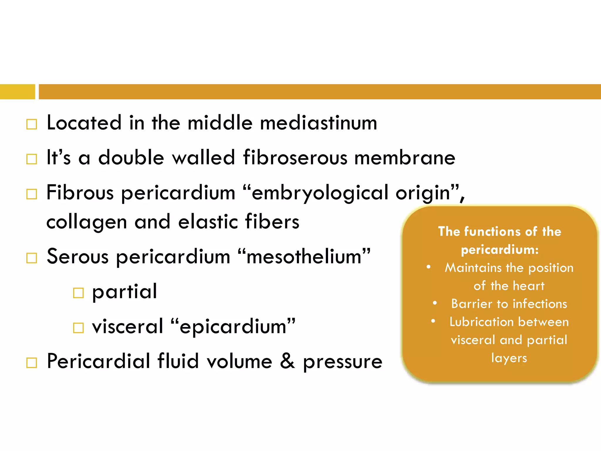
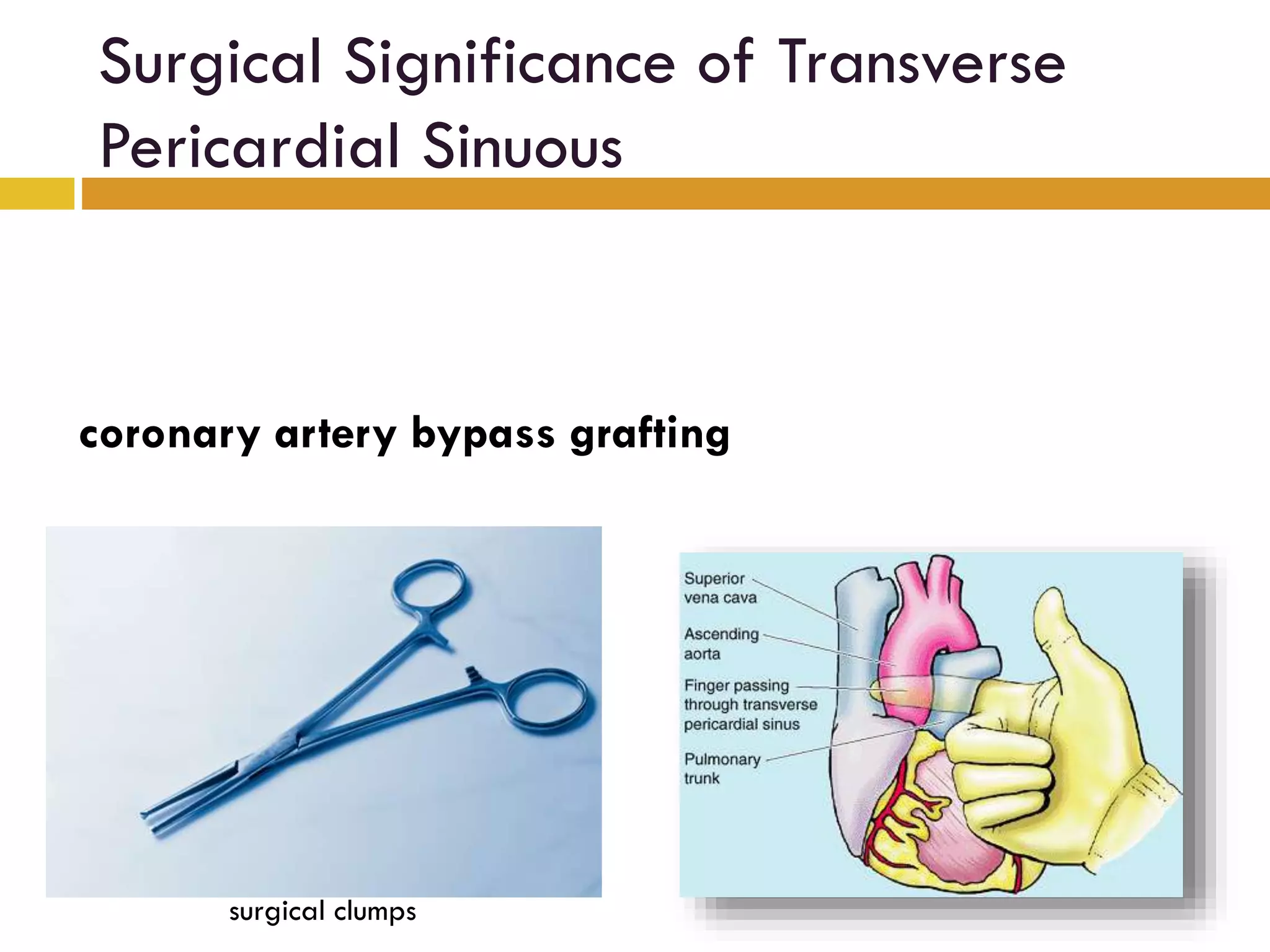
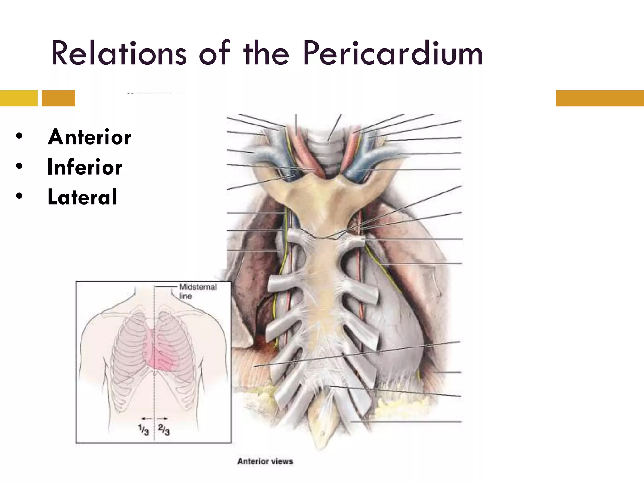
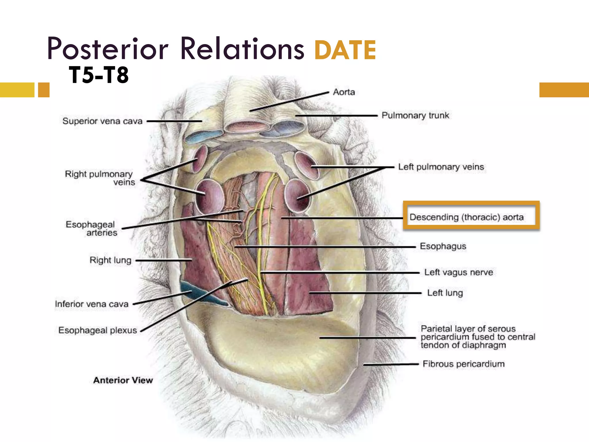
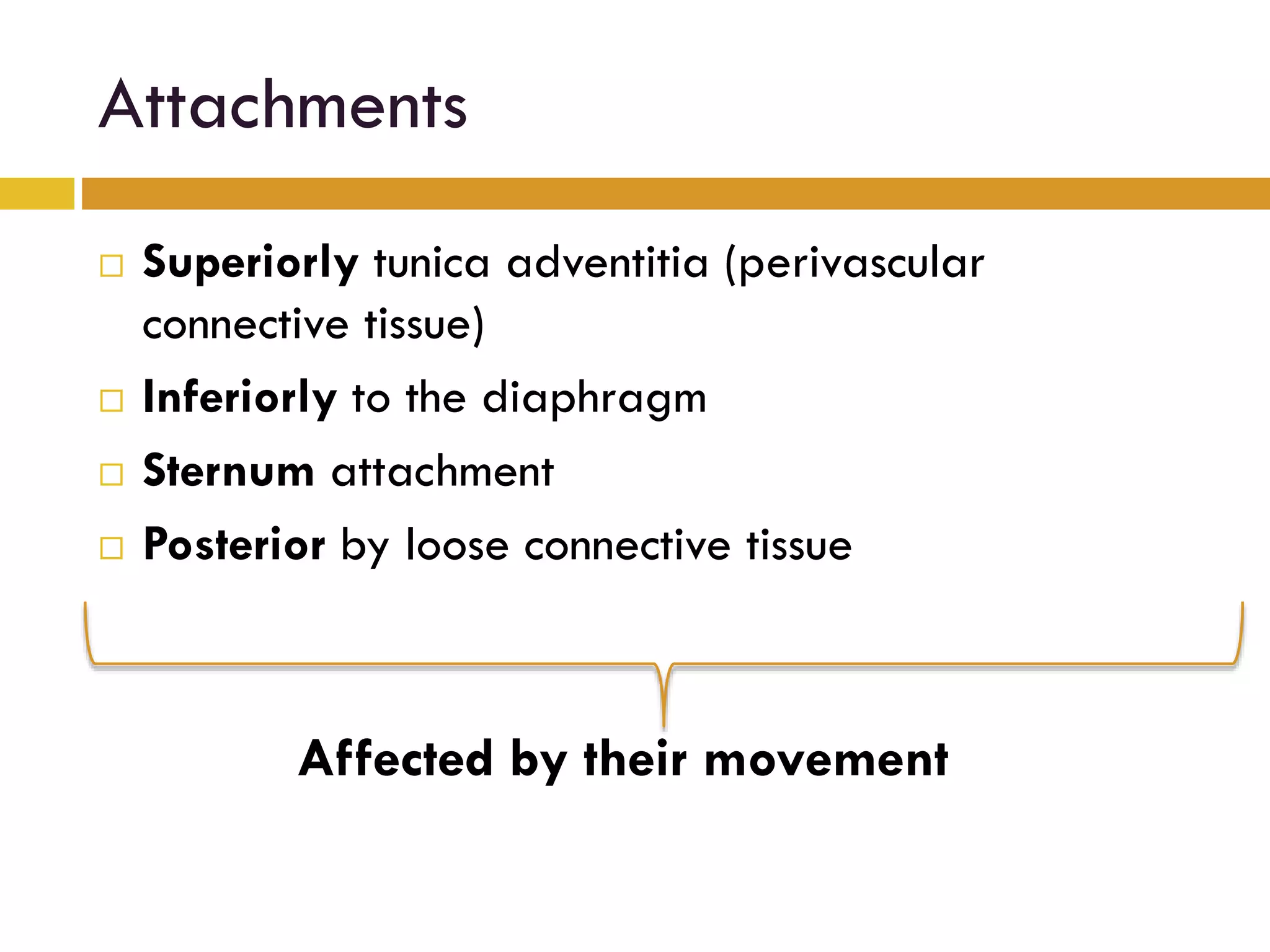
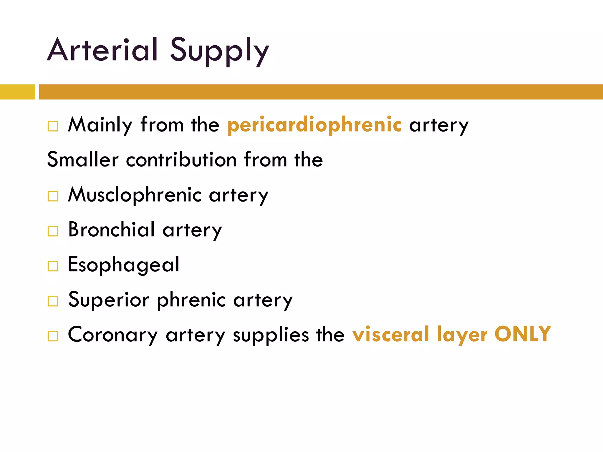
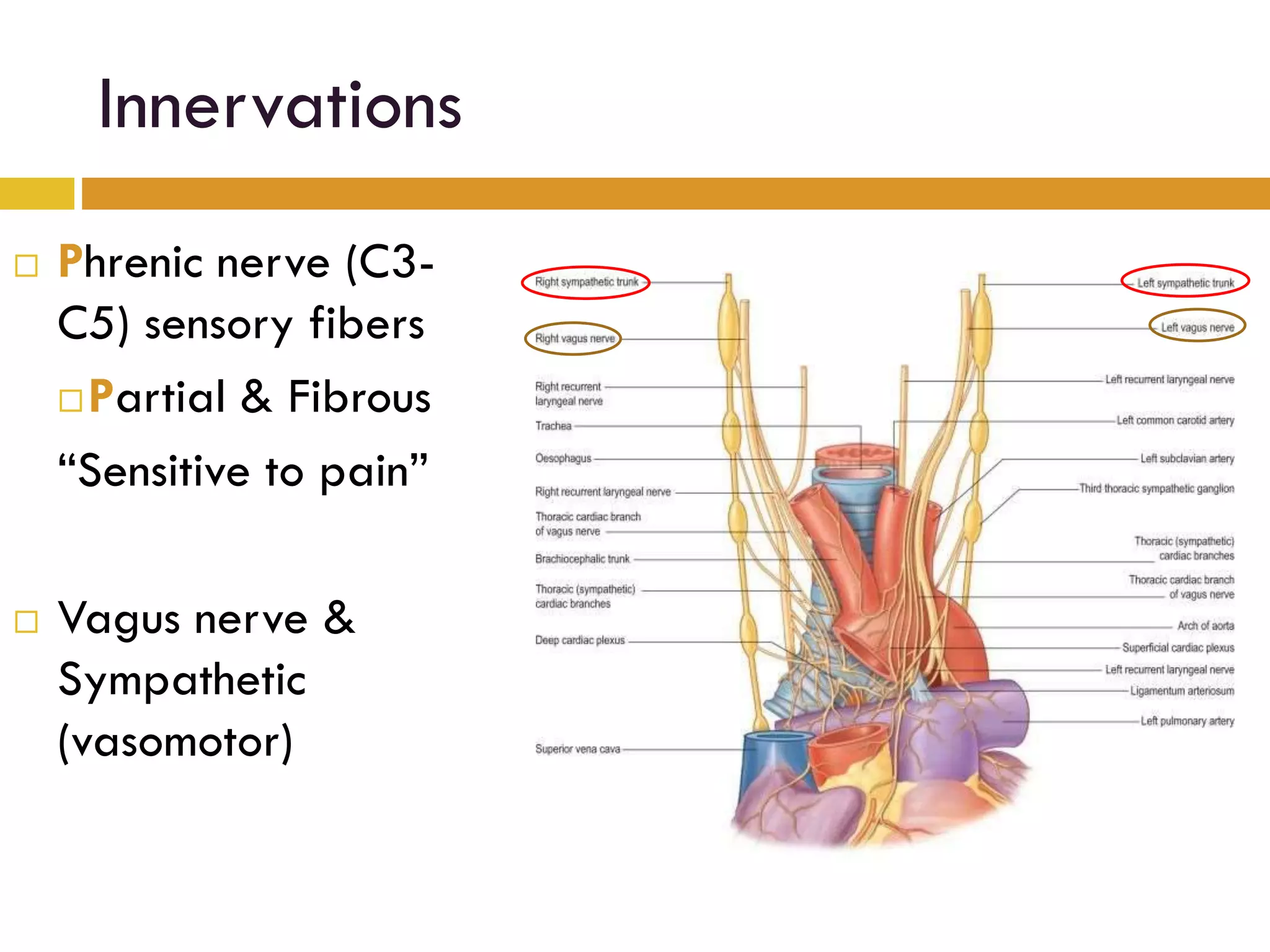
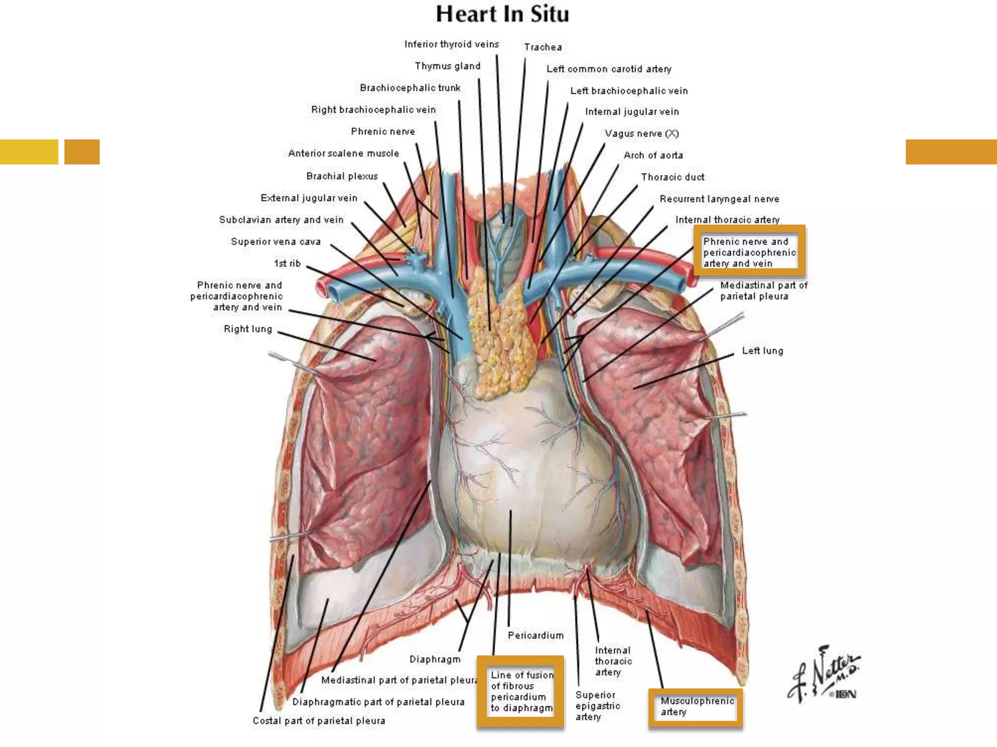
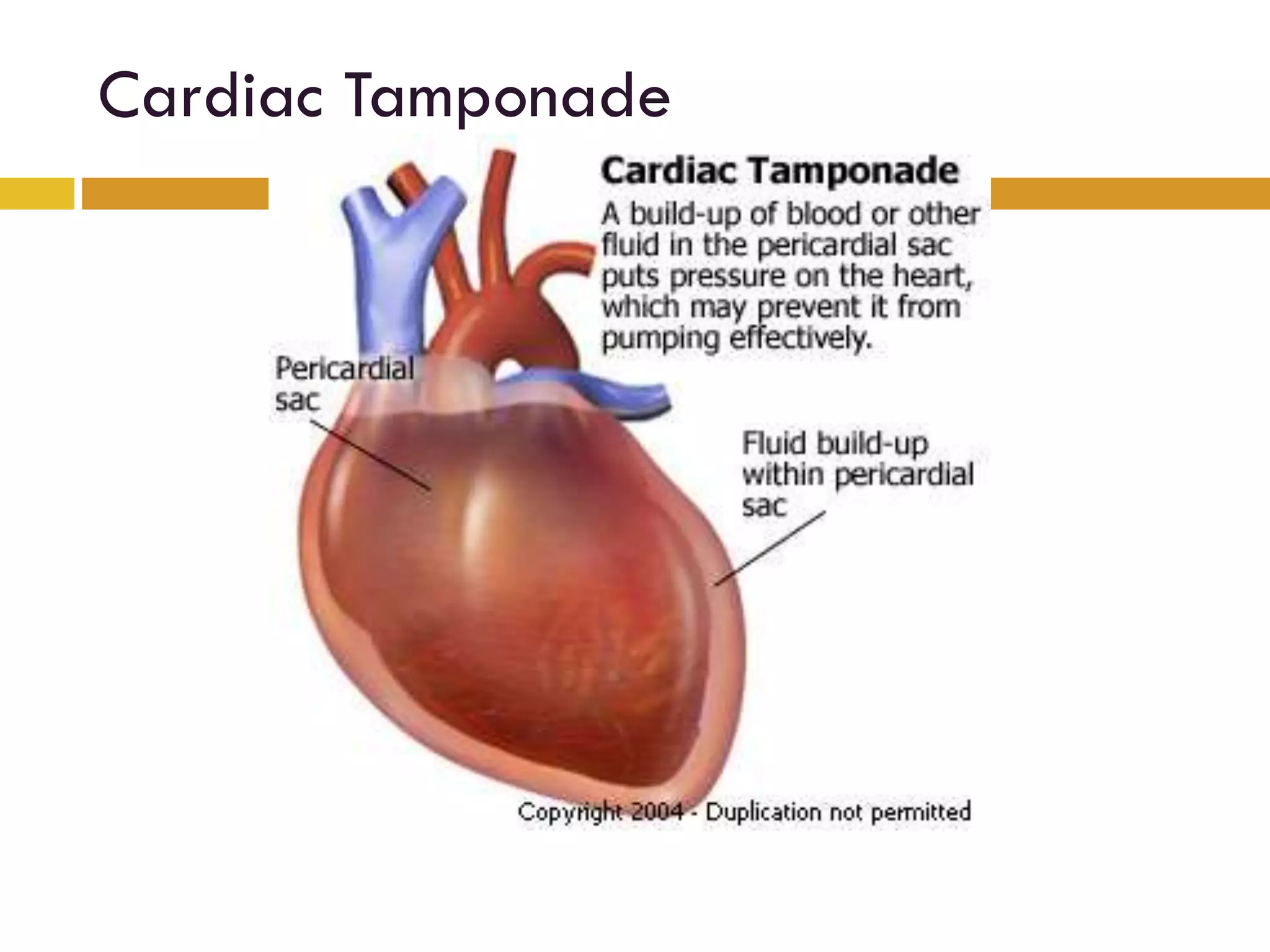
PERICARDIUM
ANATOMYShahd AlAli
 Located in the middle mediastinum
 It’s a double walled fibroserous membrane
 Fibrous pericardium “embryological origin”,
collagen and elastic fibers
 Serous pericardium “mesothelium”
 partial
 visceral “epicardium”
 Pericardial fluid volume & pressure
The functions of the
pericardium:
• Maintains the position
of the heart
• Barrier to infections
• Lubrication between
visceral and partial
layers
Pericardial Sinuses
Transverse sinuous Oblique sinuous
Development of the Pericardial Sinuous
• The oblique sinuses
separates the
esophagus from the
left atrium
Surgical Significance of Transverse
Pericardial Sinuous
coronary artery bypass grafting
surgical clumps
Relations of the Pericardium
• Anterior
• Inferior
• Lateral
Posterior Relations DATE
T5-T8
Attachments
 Superiorly tunica adventitia (perivascular
connective tissue)
 Inferiorly to the diaphragm
 Sternum attachment
 Posterior by loose connective tissue
Affected by their movement
Arterial Supply
 Mainly from the pericardiophrenic artery
Smaller contribution from the
 Musclophrenic artery
 Bronchial artery
 Esophageal
 Superior phrenic artery
 Coronary artery supplies the visceral layer ONLY
Venous drainage
 Pericardiophrenic veins
 Azygos venous system
Innervations
 Phrenic nerve (C3-
C5) sensory fibers
Partial & Fibrous
“Sensitive to pain”
 Vagus nerve &
Sympathetic
(vasomotor)
Cardiac Tamponade
Clinical ~
 Pericardiocentesis:
 Parasternal
left 5th or 6th intercostal spaces.
 Subxiphoidal approach
Thoracocentesis ???

Pericardium Anatomy