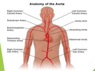
SlideShare a Scribd company logo
Hridya. P
And it’s branchesit’s
Branches
 The aorta is the main artery in the human body
originating from the left ventricle of the heart and
extending down the abdomen, where it splits into 2
smaller arteries ( The common iliac arteries )
 The aorta distributes oxygenated blood to all part of the
body through the systemic circulation
 The aorta initialy one inch wide in diameter
 The aorta can be divided into four sections:
1.The ascending aorta
2.The aortic arch Thoracic
3.The thoracic (descending)
aorta
4.The abdominal aorta.
.The first 3 parts are confined to the thoracic cavity and
together create the thoracic aorta
Function
 The Aorta Supplies blood to the entire body, except the respiratory zone of
the lungs.
 Ascending Aorta supplies blood to the heart
 Arch of Aorta- Supplies blood to the Head, neck and arms
 Descending Aorta- Supplies blood to the Chest
 Abdominal Aorta-Supplies blood to the Abdomen
 At the cellular level aorta is composed of
three layers:
 The Tunica Intima,which surrounds the
lumen & is composed of single squamous
epithelial cells.
 The Tunica Media ,composed of smooth
cell muscles & elastic fibres.
 The Tunica Adventitia,composed of loose
collagen fibres
 Ascending aorta originates from the aortic orifice from the left
ventricle, and continues as an arch of aorta at the sternal
angle.
 It is about 5cm long and its diameter is about 3cm. It
completely enclosed in the pericardium.
 Lies in the pericardium (in middle mediastinum )below the
level of sternal angle.
 Branches :
 The left and right aortic sinuses are dilations in the ascending
aorta, located at the level of the aortic valve. These give rise
to:
 The left coronary artery
 The right coronary artery
- These supply the myocardium
 Located in the superior mediastinum
 About 2.5 cm diameter.
 It is a continuation of the ascending aorta and begins at the
level of second sternocostal joint.
 The arch is directed upward, backwards to the left and then
downwards on to the left side of vertebral column.
 The aortic arch ends at the level of sternal angle
 The arch is still connected to the pulmonary trunk by the
ligamentum arteriosum(remnant of the foetal ductus
arteriosus)
 The first &largest from the aortic arch.
 It is about 4-5 cm in length.
 Branchiocephalic artery arises opposite the center of manubrium,
ascends upwards, backwrads and to the right.
 The branchiocephalic artery ends behind the right sternoclavicular joint
by dividing to,
1.Right Subclavian artery
2.Right common carotid artery
 These arteries supply the right side of the head and neck and right upper
limb.
 It found below the clavicle
 It supplies the left arm
 It runs upwards on the left mediastinal pleura and makes groove on the left
lung and enters the neck by passing behind the left sternoclavicular joint.
 At the outer border of the first rib, it furnishes by becoming axillary artery.it
extends along the upper arm as brachial artery to divided into two terminal
branches radial and ulnar arteries below the elbow.
Parts
 Each of subclavian artery is madeup of three parts defined in the relation
to the anterior scalene muscle of the neck
 First part- This part of subclavian artery is medial to the anterior scalene
muscle
Branches : Vertebral artery,Internal thoracic artery,Thyrocervical
trunk
 Second part-This part of subclavian is posterior to the anterior scalene
muscle .
Branches : Costocervical trunk
 Third part – This part of subclavian lies lateral to the anterior scalene
muscle
Branches : Dorsal scapular artery.
 There are 2 common carotid arteries, left and right.
 These are the main arteries of the head and neck.
 The left common carotid artery originates directly from the arch
of aorta in thorax.
 It ascends to the back of left sternoclavicular joint and enters the
neck.
 The left common carotid artery runs upwards from left
sternoclavicular joint to the upper border of thyroid cartilage.
 Left common carotid artery supplies the head and neck
 The descending thoracic aorta which is contained in the
posterior mediastinum
 It is the continuation of the arch of aorta .
 Descends in front of vertebral column
Thoracic aorta
Branches
1.Bronchial arteries: Paired visceral branch arising laterally to supply bronchial &
peribronchial tissue,& visceral pleura. most commonly only the paired left
bronchial artery arises directly from the aorta
2. Mediastinal arteries :Small artery that supply the lymph glands & loose areolar
tissue in the posterior mediastinum
3. Oesophageal arteries: Unpaired visceral branches arising anteriorly to supply the
oesophagus
4. Pericardial arteries: small unpaired arteries that arise anteriorly to supply the dorsal
portion of the pericardium.
5.Superior phrenic arteries:Paired parietal branches that supply the superior portion of
diaphragm
 Intercostal & Subcostal arteries : Small paired arteries that branch off
throughout the length of posterior thoracic aorta. The 9 paires of intercostal
arteries supply the Intercostal space, with the exception of first & second( They
are supplied by a branch from the subclavian artery). The subcostal arteries
supply the flat abdominal wall muscles
 Abdominal aorta is a continuation of the thoracic aorta .It is approximately 13cm
long
 It descends and terminating by divided into right and left common illiac arteries
 The branches of abdominal aorta are,
1. Inferior phrenic arteries:They supply the diaphragm
2.Coeliac artery : supplies the liver, stomach, abdominal oesophagus, spleen,
the superior duodenum & superior pancreas
3.Superior mescentric artery: it supply the distal duodenum, jejuno –ileum,
ascending colon& the part of transver
4.Middle suprarenal artery: They supply the adrenal glands
5.Renal arteries: supply the kidneys
6.Gonadal arteries:Supply the testicular artery,& ovarian artery
7.Inferior mesenteric artery: It supplies the large intestine .
8.Median Sacral artery : supply the Coccyx, lumbar vertebrae,& the
sacrum.
9.Lumbar arteries: supply the abdominal wall and spinal cord.
 Terminal branches of abdominal aorta are two common illiac arteries.
 External iliac artery& Internal illiac artery
 Branches of external illiac artery
 1.Inferior epigastric artery
 2.Deep circumflex artery
 Internal illiac artery
 1.Superior vesical artery
 2.Inferior vesical artery
 3.Middle rectal artery
 4.Inferior gluteal artery
 5.Obturator artery
 6.Uterine artery( In female)
 7.Lateral sacral artery
 It describe the dialation of the artery to more than 1.5 times its original size .
 The abdominal component of the aorta is the most common site for aneurysmal
changes.
 Patients suffering with an abdominal aortic aneurysm may experience
abdominal pulsations,abdominal pain &back pain.
 The aneurysm may also compress nerve roots causing pain/numbness in the
lower limbs.
 A patient with an aortic arch aneurysm may have a hoarse voice due to the
dilation stretching the left recurrent laryngeal nerve .Patients may not have
any symptoms at all.
 Small aortic aneurysms do not usually pose a serious immediate
threat.Diagnosis is made from an ultrasound &the weakened vessel wall can be
surgically replaced with a piece of synthetic tubing.if left untreated, a large
aneurysm can rupture. This is a medical emergency & often fatal.
 Coarctation of aorta refers to narrowing of vessel, Usually at the insertion
of ligamentum arteriosum( former ductus arteriosus)
 It is congential narrowing of the aorta just proximal or distal to the
entrance of the ductus arteriosus.
 The narrow vessel has a increased resistance to blood flow which increases
the after load for the left ventricle – leading to left ventricular
hypertrophy
 Coarctation are most common in the Arch of Aorta
 Accordingly it is named as preductal type and postductal type of
coarctalion of aorta.
 The blood supply to the head, neck & upper limbs is not compromised as
the vessel that supply them emerge proximal to the coarctation, However
yhe blood supply to the rest of the body reduced, This result in a weak,
delayed femoral pulse which prevent clinically as radio-femoral delay
 It probably take place because of hyper involution of the ductus arteriosus.
 The narrowing is proximal to the ductus arteriosus . Life threatening , if
severe;seen in Turner’s syndrome .
The narrowing occurs at the insertion of the ductus arteriosus this kind usually
appears when the ductus arteriosus closes.
The narrowing is distal to the insertion of the ductus arteriosus .Postductal
coarctation is most likely the result of the extenction of a muscular artery in to
an elastic artery .This type is most common in adult.
Aorta&its branches . pptx.pptx

More Related Content

What's hot

Blood supply of heart
Blood supply of heartBlood supply of heart
Blood supply of heart
Nikhil Vaishnav
 
Mitral valve anatomy - ppt by kunwar sidharth
Mitral valve    anatomy - ppt by kunwar sidharthMitral valve    anatomy - ppt by kunwar sidharth
Mitral valve anatomy - ppt by kunwar sidharth
kunwar sidharth
 
Cath hemodynamics vir
Cath hemodynamics virCath hemodynamics vir
Cath hemodynamics vir
Dr Virbhan Balai
 
Coronary artery anatomy
Coronary artery anatomyCoronary artery anatomy
Coronary artery anatomy
Dr. Yash Kumar Achantani
 
SEGMENTAL ANALYSIS OF CONGENITAL HEART DISEASE
SEGMENTAL ANALYSIS OF CONGENITAL HEART DISEASE SEGMENTAL ANALYSIS OF CONGENITAL HEART DISEASE
SEGMENTAL ANALYSIS OF CONGENITAL HEART DISEASE
Dr. Murtaza Kamal MD,DNB,DrNB Ped Cardiology
 
Thoracic aorta
Thoracic aortaThoracic aorta
Thoracic aorta
Aisha Sadaf
 
Dual left anterior descending coronary artery
Dual left anterior descending coronary arteryDual left anterior descending coronary artery
Dual left anterior descending coronary artery
Ramachandra Barik
 
Aortic arch anomalies
Aortic arch anomaliesAortic arch anomalies
Aortic arch anomalies
Sumit Shanker
 
Normal anatomy of tricuspid valve
Normal anatomy of tricuspid valve Normal anatomy of tricuspid valve
Normal anatomy of tricuspid valve
India CTVS
 
Pulmonary Artery Anatomy and Pulmonary Embolism
Pulmonary Artery Anatomy and Pulmonary EmbolismPulmonary Artery Anatomy and Pulmonary Embolism
Pulmonary Artery Anatomy and Pulmonary Embolism
Gamal Agmy
 
Aortic Arches
Aortic ArchesAortic Arches
Aortic Arches
DRHAFIZ07
 
Anatomy of mitral valve echo evaluation
Anatomy of mitral valve echo evaluationAnatomy of mitral valve echo evaluation
Anatomy of mitral valve echo evaluation
madhusiva03
 
Anatomy tricuspid valve DR NIKUNJ R SHEKHADA (MBBS,MS GEN SURG ,DNB CTS SR)
Anatomy tricuspid valve DR NIKUNJ R SHEKHADA (MBBS,MS GEN SURG ,DNB CTS SR)Anatomy tricuspid valve DR NIKUNJ R SHEKHADA (MBBS,MS GEN SURG ,DNB CTS SR)
Anatomy tricuspid valve DR NIKUNJ R SHEKHADA (MBBS,MS GEN SURG ,DNB CTS SR)
DR NIKUNJ SHEKHADA
 
The valves of the heart
The valves of the heartThe valves of the heart
The valves of the heart
Idris Siddiqui
 
Cardiac venous system
Cardiac venous systemCardiac venous system
Cardiac venous system
Jyotindra Singh
 
Sequential segmental analysis
Sequential segmental analysisSequential segmental analysis
Sequential segmental analysis
Ramachandra Barik
 
Mitral valve repair
Mitral valve repairMitral valve repair
Mitral valve repair
DR NIKUNJ SHEKHADA
 
DM cardiology Exam Spotter
DM cardiology Exam SpotterDM cardiology Exam Spotter
DM cardiology Exam Spotter
PRAVEEN GUPTA
 
Echo assessment of lv systolic function and swma
Echo assessment of lv systolic function and swmaEcho assessment of lv systolic function and swma
Echo assessment of lv systolic function and swmaFuad Farooq
 
Mitral valve surgical anatomy DR NIKUNJ R SHEKHADA (MBBS ,MS GRN SURG , DNB C...
Mitral valve surgical anatomy DR NIKUNJ R SHEKHADA (MBBS ,MS GRN SURG , DNB C...Mitral valve surgical anatomy DR NIKUNJ R SHEKHADA (MBBS ,MS GRN SURG , DNB C...
Mitral valve surgical anatomy DR NIKUNJ R SHEKHADA (MBBS ,MS GRN SURG , DNB C...
DR NIKUNJ SHEKHADA
 

What's hot (20)

Blood supply of heart
Blood supply of heartBlood supply of heart
Blood supply of heart
 
Mitral valve anatomy - ppt by kunwar sidharth
Mitral valve    anatomy - ppt by kunwar sidharthMitral valve    anatomy - ppt by kunwar sidharth
Mitral valve anatomy - ppt by kunwar sidharth
 
Cath hemodynamics vir
Cath hemodynamics virCath hemodynamics vir
Cath hemodynamics vir
 
Coronary artery anatomy
Coronary artery anatomyCoronary artery anatomy
Coronary artery anatomy
 
SEGMENTAL ANALYSIS OF CONGENITAL HEART DISEASE
SEGMENTAL ANALYSIS OF CONGENITAL HEART DISEASE SEGMENTAL ANALYSIS OF CONGENITAL HEART DISEASE
SEGMENTAL ANALYSIS OF CONGENITAL HEART DISEASE
 
Thoracic aorta
Thoracic aortaThoracic aorta
Thoracic aorta
 
Dual left anterior descending coronary artery
Dual left anterior descending coronary arteryDual left anterior descending coronary artery
Dual left anterior descending coronary artery
 
Aortic arch anomalies
Aortic arch anomaliesAortic arch anomalies
Aortic arch anomalies
 
Normal anatomy of tricuspid valve
Normal anatomy of tricuspid valve Normal anatomy of tricuspid valve
Normal anatomy of tricuspid valve
 
Pulmonary Artery Anatomy and Pulmonary Embolism
Pulmonary Artery Anatomy and Pulmonary EmbolismPulmonary Artery Anatomy and Pulmonary Embolism
Pulmonary Artery Anatomy and Pulmonary Embolism
 
Aortic Arches
Aortic ArchesAortic Arches
Aortic Arches
 
Anatomy of mitral valve echo evaluation
Anatomy of mitral valve echo evaluationAnatomy of mitral valve echo evaluation
Anatomy of mitral valve echo evaluation
 
Anatomy tricuspid valve DR NIKUNJ R SHEKHADA (MBBS,MS GEN SURG ,DNB CTS SR)
Anatomy tricuspid valve DR NIKUNJ R SHEKHADA (MBBS,MS GEN SURG ,DNB CTS SR)Anatomy tricuspid valve DR NIKUNJ R SHEKHADA (MBBS,MS GEN SURG ,DNB CTS SR)
Anatomy tricuspid valve DR NIKUNJ R SHEKHADA (MBBS,MS GEN SURG ,DNB CTS SR)
 
The valves of the heart
The valves of the heartThe valves of the heart
The valves of the heart
 
Cardiac venous system
Cardiac venous systemCardiac venous system
Cardiac venous system
 
Sequential segmental analysis
Sequential segmental analysisSequential segmental analysis
Sequential segmental analysis
 
Mitral valve repair
Mitral valve repairMitral valve repair
Mitral valve repair
 
DM cardiology Exam Spotter
DM cardiology Exam SpotterDM cardiology Exam Spotter
DM cardiology Exam Spotter
 
Echo assessment of lv systolic function and swma
Echo assessment of lv systolic function and swmaEcho assessment of lv systolic function and swma
Echo assessment of lv systolic function and swma
 
Mitral valve surgical anatomy DR NIKUNJ R SHEKHADA (MBBS ,MS GRN SURG , DNB C...
Mitral valve surgical anatomy DR NIKUNJ R SHEKHADA (MBBS ,MS GRN SURG , DNB C...Mitral valve surgical anatomy DR NIKUNJ R SHEKHADA (MBBS ,MS GRN SURG , DNB C...
Mitral valve surgical anatomy DR NIKUNJ R SHEKHADA (MBBS ,MS GRN SURG , DNB C...
 

Similar to Aorta&its branches . pptx.pptx

Aorta by Dr. Karle Geetanjali
Aorta by Dr. Karle GeetanjaliAorta by Dr. Karle Geetanjali
Aorta by Dr. Karle Geetanjali
GeetanjaliKarle1
 
heart anatomy pdf.pdf
heart anatomy pdf.pdfheart anatomy pdf.pdf
heart anatomy pdf.pdf
Pooja Rani
 
Anatomy of heart presentation
Anatomy of heart presentationAnatomy of heart presentation
Anatomy of heart presentation
Rajeshkumarm16
 
Aorta, SVC, PULMONARY TRUNK.pptx
Aorta, SVC, PULMONARY TRUNK.pptxAorta, SVC, PULMONARY TRUNK.pptx
Aorta, SVC, PULMONARY TRUNK.pptx
MovieDownload83
 
Cardiovascular system ppt
Cardiovascular system pptCardiovascular system ppt
3-pericardium & Heart.pptx
3-pericardium & Heart.pptx3-pericardium & Heart.pptx
3-pericardium & Heart.pptx
muhammadsumair19
 
Surgery 5th year, 9th, 10th &11th lectures (Dr. Ahmed Al-Azzawi)
Surgery 5th year, 9th, 10th &11th lectures (Dr. Ahmed Al-Azzawi)Surgery 5th year, 9th, 10th &11th lectures (Dr. Ahmed Al-Azzawi)
Surgery 5th year, 9th, 10th &11th lectures (Dr. Ahmed Al-Azzawi)
College of Medicine, Sulaymaniyah
 
"THE HEART"
"THE HEART""THE HEART"
"THE HEART"
UmairKhan463
 
Heart (Its Structure & Function).pptx
Heart (Its Structure & Function).pptxHeart (Its Structure & Function).pptx
Heart (Its Structure & Function).pptx
Vipin Chandran
 
Cardiovascular system
Cardiovascular systemCardiovascular system
Cardiovascular system
deepaingawale21
 
Crash-Course for AIPMT & Other Medical Exams 2016(Essentials heart)
Crash-Course for AIPMT & Other Medical Exams 2016(Essentials heart)Crash-Course for AIPMT & Other Medical Exams 2016(Essentials heart)
Crash-Course for AIPMT & Other Medical Exams 2016(Essentials heart)
APEX INSTITUTE
 
ANATOMY AND PHYSIOLOGY OF HEART
ANATOMY AND PHYSIOLOGY OF HEARTANATOMY AND PHYSIOLOGY OF HEART
ANATOMY AND PHYSIOLOGY OF HEART
Haripriya209498
 
Heart.pptx
Heart.pptxHeart.pptx
Heart.pptx
SpandanaRavikanti
 
Mediastinum - Heart and Lungs
Mediastinum - Heart and LungsMediastinum - Heart and Lungs
Mediastinum - Heart and Lungs
Sado Anatomist
 
Anatomy Of The Heart
Anatomy Of The HeartAnatomy Of The Heart
Anatomy Of The Heartshabeel pn
 
08 Heart.ppt
08 Heart.ppt08 Heart.ppt
08 Heart.ppt
Shubhangi kharat
 
3-pericardium & Heart.pptx
3-pericardium & Heart.pptx3-pericardium & Heart.pptx
3-pericardium & Heart.pptx
GulAndama2
 
Do you Know your Heart?
Do you Know your Heart?Do you Know your Heart?
Do you Know your Heart?
HBT Media Management Ltd
 
Anatomy and physiology of the heart by Adeboye Oluwajuyitan
Anatomy and physiology of the heart by Adeboye OluwajuyitanAnatomy and physiology of the heart by Adeboye Oluwajuyitan
Anatomy and physiology of the heart by Adeboye Oluwajuyitan
Adeboye Oluwajuyitan
 

Similar to Aorta&its branches . pptx.pptx (20)

Aorta by Dr. Karle Geetanjali
Aorta by Dr. Karle GeetanjaliAorta by Dr. Karle Geetanjali
Aorta by Dr. Karle Geetanjali
 
heart anatomy pdf.pdf
heart anatomy pdf.pdfheart anatomy pdf.pdf
heart anatomy pdf.pdf
 
Anatomy of heart presentation
Anatomy of heart presentationAnatomy of heart presentation
Anatomy of heart presentation
 
Human heart
Human heartHuman heart
Human heart
 
Aorta, SVC, PULMONARY TRUNK.pptx
Aorta, SVC, PULMONARY TRUNK.pptxAorta, SVC, PULMONARY TRUNK.pptx
Aorta, SVC, PULMONARY TRUNK.pptx
 
Cardiovascular system ppt
Cardiovascular system pptCardiovascular system ppt
Cardiovascular system ppt
 
3-pericardium & Heart.pptx
3-pericardium & Heart.pptx3-pericardium & Heart.pptx
3-pericardium & Heart.pptx
 
Surgery 5th year, 9th, 10th &11th lectures (Dr. Ahmed Al-Azzawi)
Surgery 5th year, 9th, 10th &11th lectures (Dr. Ahmed Al-Azzawi)Surgery 5th year, 9th, 10th &11th lectures (Dr. Ahmed Al-Azzawi)
Surgery 5th year, 9th, 10th &11th lectures (Dr. Ahmed Al-Azzawi)
 
"THE HEART"
"THE HEART""THE HEART"
"THE HEART"
 
Heart (Its Structure & Function).pptx
Heart (Its Structure & Function).pptxHeart (Its Structure & Function).pptx
Heart (Its Structure & Function).pptx
 
Cardiovascular system
Cardiovascular systemCardiovascular system
Cardiovascular system
 
Crash-Course for AIPMT & Other Medical Exams 2016(Essentials heart)
Crash-Course for AIPMT & Other Medical Exams 2016(Essentials heart)Crash-Course for AIPMT & Other Medical Exams 2016(Essentials heart)
Crash-Course for AIPMT & Other Medical Exams 2016(Essentials heart)
 
ANATOMY AND PHYSIOLOGY OF HEART
ANATOMY AND PHYSIOLOGY OF HEARTANATOMY AND PHYSIOLOGY OF HEART
ANATOMY AND PHYSIOLOGY OF HEART
 
Heart.pptx
Heart.pptxHeart.pptx
Heart.pptx
 
Mediastinum - Heart and Lungs
Mediastinum - Heart and LungsMediastinum - Heart and Lungs
Mediastinum - Heart and Lungs
 
Anatomy Of The Heart
Anatomy Of The HeartAnatomy Of The Heart
Anatomy Of The Heart
 
08 Heart.ppt
08 Heart.ppt08 Heart.ppt
08 Heart.ppt
 
3-pericardium & Heart.pptx
3-pericardium & Heart.pptx3-pericardium & Heart.pptx
3-pericardium & Heart.pptx
 
Do you Know your Heart?
Do you Know your Heart?Do you Know your Heart?
Do you Know your Heart?
 
Anatomy and physiology of the heart by Adeboye Oluwajuyitan
Anatomy and physiology of the heart by Adeboye OluwajuyitanAnatomy and physiology of the heart by Adeboye Oluwajuyitan
Anatomy and physiology of the heart by Adeboye Oluwajuyitan
 

Recently uploaded

Report Back from SGO 2024: What’s the Latest in Cervical Cancer?
Report Back from SGO 2024: What’s the Latest in Cervical Cancer?Report Back from SGO 2024: What’s the Latest in Cervical Cancer?
Report Back from SGO 2024: What’s the Latest in Cervical Cancer?
bkling
 
The POPPY STUDY (Preconception to post-partum cardiovascular function in prim...
The POPPY STUDY (Preconception to post-partum cardiovascular function in prim...The POPPY STUDY (Preconception to post-partum cardiovascular function in prim...
The POPPY STUDY (Preconception to post-partum cardiovascular function in prim...
Catherine Liao
 
ARTHROLOGY PPT NCISM SYLLABUS AYURVEDA STUDENTS
ARTHROLOGY PPT NCISM SYLLABUS AYURVEDA STUDENTSARTHROLOGY PPT NCISM SYLLABUS AYURVEDA STUDENTS
ARTHROLOGY PPT NCISM SYLLABUS AYURVEDA STUDENTS
Dr. Vinay Pareek
 
MANAGEMENT OF ATRIOVENTRICULAR CONDUCTION BLOCK.pdf
MANAGEMENT OF ATRIOVENTRICULAR CONDUCTION BLOCK.pdfMANAGEMENT OF ATRIOVENTRICULAR CONDUCTION BLOCK.pdf
MANAGEMENT OF ATRIOVENTRICULAR CONDUCTION BLOCK.pdf
Jim Jacob Roy
 
Evaluation of antidepressant activity of clitoris ternatea in animals
Evaluation of antidepressant activity of clitoris ternatea in animalsEvaluation of antidepressant activity of clitoris ternatea in animals
Evaluation of antidepressant activity of clitoris ternatea in animals
Shweta
 
Ozempic: Preoperative Management of Patients on GLP-1 Receptor Agonists
Ozempic: Preoperative Management of Patients on GLP-1 Receptor Agonists  Ozempic: Preoperative Management of Patients on GLP-1 Receptor Agonists
Ozempic: Preoperative Management of Patients on GLP-1 Receptor Agonists
Saeid Safari
 
New Directions in Targeted Therapeutic Approaches for Older Adults With Mantl...
New Directions in Targeted Therapeutic Approaches for Older Adults With Mantl...New Directions in Targeted Therapeutic Approaches for Older Adults With Mantl...
New Directions in Targeted Therapeutic Approaches for Older Adults With Mantl...
i3 Health
 
The hemodynamic and autonomic determinants of elevated blood pressure in obes...
The hemodynamic and autonomic determinants of elevated blood pressure in obes...The hemodynamic and autonomic determinants of elevated blood pressure in obes...
The hemodynamic and autonomic determinants of elevated blood pressure in obes...
Catherine Liao
 
Lung Cancer: Artificial Intelligence, Synergetics, Complex System Analysis, S...
Lung Cancer: Artificial Intelligence, Synergetics, Complex System Analysis, S...Lung Cancer: Artificial Intelligence, Synergetics, Complex System Analysis, S...
Lung Cancer: Artificial Intelligence, Synergetics, Complex System Analysis, S...
Oleg Kshivets
 
Maxilla, Mandible & Hyoid Bone & Clinical Correlations by Dr. RIG.pptx
Maxilla, Mandible & Hyoid Bone & Clinical Correlations by Dr. RIG.pptxMaxilla, Mandible & Hyoid Bone & Clinical Correlations by Dr. RIG.pptx
Maxilla, Mandible & Hyoid Bone & Clinical Correlations by Dr. RIG.pptx
Dr. Rabia Inam Gandapore
 
Ocular injury ppt Upendra pal optometrist upums saifai etawah
Ocular injury  ppt  Upendra pal  optometrist upums saifai etawahOcular injury  ppt  Upendra pal  optometrist upums saifai etawah
Ocular injury ppt Upendra pal optometrist upums saifai etawah
pal078100
 
Couples presenting to the infertility clinic- Do they really have infertility...
Couples presenting to the infertility clinic- Do they really have infertility...Couples presenting to the infertility clinic- Do they really have infertility...
Couples presenting to the infertility clinic- Do they really have infertility...
Sujoy Dasgupta
 
Surat @ℂall @Girls ꧁❤8527049040❤꧂@ℂall @Girls Service Vip Top Model Safe
Surat @ℂall @Girls ꧁❤8527049040❤꧂@ℂall @Girls Service Vip Top Model SafeSurat @ℂall @Girls ꧁❤8527049040❤꧂@ℂall @Girls Service Vip Top Model Safe
Surat @ℂall @Girls ꧁❤8527049040❤꧂@ℂall @Girls Service Vip Top Model Safe
Savita Shen $i11
 
For Better Surat #ℂall #Girl Service ❤85270-49040❤ Surat #ℂall #Girls
For Better Surat #ℂall #Girl Service ❤85270-49040❤ Surat #ℂall #GirlsFor Better Surat #ℂall #Girl Service ❤85270-49040❤ Surat #ℂall #Girls
For Better Surat #ℂall #Girl Service ❤85270-49040❤ Surat #ℂall #Girls
Savita Shen $i11
 
KDIGO 2024 guidelines for diabetologists
KDIGO 2024 guidelines for diabetologistsKDIGO 2024 guidelines for diabetologists
KDIGO 2024 guidelines for diabetologists
د.محمود نجيب
 
BRACHYTHERAPY OVERVIEW AND APPLICATORS
BRACHYTHERAPY OVERVIEW  AND  APPLICATORSBRACHYTHERAPY OVERVIEW  AND  APPLICATORS
BRACHYTHERAPY OVERVIEW AND APPLICATORS
Krishan Murari
 
TEST BANK for Operations Management, 14th Edition by William J. Stevenson, Ve...
TEST BANK for Operations Management, 14th Edition by William J. Stevenson, Ve...TEST BANK for Operations Management, 14th Edition by William J. Stevenson, Ve...
TEST BANK for Operations Management, 14th Edition by William J. Stevenson, Ve...
kevinkariuki227
 
Hemodialysis: Chapter 3, Dialysis Water Unit - Dr.Gawad
Hemodialysis: Chapter 3, Dialysis Water Unit - Dr.GawadHemodialysis: Chapter 3, Dialysis Water Unit - Dr.Gawad
Hemodialysis: Chapter 3, Dialysis Water Unit - Dr.Gawad
NephroTube - Dr.Gawad
 
ARTIFICIAL INTELLIGENCE IN HEALTHCARE.pdf
ARTIFICIAL INTELLIGENCE IN  HEALTHCARE.pdfARTIFICIAL INTELLIGENCE IN  HEALTHCARE.pdf
ARTIFICIAL INTELLIGENCE IN HEALTHCARE.pdf
Anujkumaranit
 
Charaka Samhita Sutra sthana Chapter 15 Upakalpaniyaadhyaya
Charaka Samhita Sutra sthana Chapter 15 UpakalpaniyaadhyayaCharaka Samhita Sutra sthana Chapter 15 Upakalpaniyaadhyaya
Charaka Samhita Sutra sthana Chapter 15 Upakalpaniyaadhyaya
Dr KHALID B.M
 

Recently uploaded (20)

Report Back from SGO 2024: What’s the Latest in Cervical Cancer?
Report Back from SGO 2024: What’s the Latest in Cervical Cancer?Report Back from SGO 2024: What’s the Latest in Cervical Cancer?
Report Back from SGO 2024: What’s the Latest in Cervical Cancer?
 
The POPPY STUDY (Preconception to post-partum cardiovascular function in prim...
The POPPY STUDY (Preconception to post-partum cardiovascular function in prim...The POPPY STUDY (Preconception to post-partum cardiovascular function in prim...
The POPPY STUDY (Preconception to post-partum cardiovascular function in prim...
 
ARTHROLOGY PPT NCISM SYLLABUS AYURVEDA STUDENTS
ARTHROLOGY PPT NCISM SYLLABUS AYURVEDA STUDENTSARTHROLOGY PPT NCISM SYLLABUS AYURVEDA STUDENTS
ARTHROLOGY PPT NCISM SYLLABUS AYURVEDA STUDENTS
 
MANAGEMENT OF ATRIOVENTRICULAR CONDUCTION BLOCK.pdf
MANAGEMENT OF ATRIOVENTRICULAR CONDUCTION BLOCK.pdfMANAGEMENT OF ATRIOVENTRICULAR CONDUCTION BLOCK.pdf
MANAGEMENT OF ATRIOVENTRICULAR CONDUCTION BLOCK.pdf
 
Evaluation of antidepressant activity of clitoris ternatea in animals
Evaluation of antidepressant activity of clitoris ternatea in animalsEvaluation of antidepressant activity of clitoris ternatea in animals
Evaluation of antidepressant activity of clitoris ternatea in animals
 
Ozempic: Preoperative Management of Patients on GLP-1 Receptor Agonists
Ozempic: Preoperative Management of Patients on GLP-1 Receptor Agonists  Ozempic: Preoperative Management of Patients on GLP-1 Receptor Agonists
Ozempic: Preoperative Management of Patients on GLP-1 Receptor Agonists
 
New Directions in Targeted Therapeutic Approaches for Older Adults With Mantl...
New Directions in Targeted Therapeutic Approaches for Older Adults With Mantl...New Directions in Targeted Therapeutic Approaches for Older Adults With Mantl...
New Directions in Targeted Therapeutic Approaches for Older Adults With Mantl...
 
The hemodynamic and autonomic determinants of elevated blood pressure in obes...
The hemodynamic and autonomic determinants of elevated blood pressure in obes...The hemodynamic and autonomic determinants of elevated blood pressure in obes...
The hemodynamic and autonomic determinants of elevated blood pressure in obes...
 
Lung Cancer: Artificial Intelligence, Synergetics, Complex System Analysis, S...
Lung Cancer: Artificial Intelligence, Synergetics, Complex System Analysis, S...Lung Cancer: Artificial Intelligence, Synergetics, Complex System Analysis, S...
Lung Cancer: Artificial Intelligence, Synergetics, Complex System Analysis, S...
 
Maxilla, Mandible & Hyoid Bone & Clinical Correlations by Dr. RIG.pptx
Maxilla, Mandible & Hyoid Bone & Clinical Correlations by Dr. RIG.pptxMaxilla, Mandible & Hyoid Bone & Clinical Correlations by Dr. RIG.pptx
Maxilla, Mandible & Hyoid Bone & Clinical Correlations by Dr. RIG.pptx
 
Ocular injury ppt Upendra pal optometrist upums saifai etawah
Ocular injury  ppt  Upendra pal  optometrist upums saifai etawahOcular injury  ppt  Upendra pal  optometrist upums saifai etawah
Ocular injury ppt Upendra pal optometrist upums saifai etawah
 
Couples presenting to the infertility clinic- Do they really have infertility...
Couples presenting to the infertility clinic- Do they really have infertility...Couples presenting to the infertility clinic- Do they really have infertility...
Couples presenting to the infertility clinic- Do they really have infertility...
 
Surat @ℂall @Girls ꧁❤8527049040❤꧂@ℂall @Girls Service Vip Top Model Safe
Surat @ℂall @Girls ꧁❤8527049040❤꧂@ℂall @Girls Service Vip Top Model SafeSurat @ℂall @Girls ꧁❤8527049040❤꧂@ℂall @Girls Service Vip Top Model Safe
Surat @ℂall @Girls ꧁❤8527049040❤꧂@ℂall @Girls Service Vip Top Model Safe
 
For Better Surat #ℂall #Girl Service ❤85270-49040❤ Surat #ℂall #Girls
For Better Surat #ℂall #Girl Service ❤85270-49040❤ Surat #ℂall #GirlsFor Better Surat #ℂall #Girl Service ❤85270-49040❤ Surat #ℂall #Girls
For Better Surat #ℂall #Girl Service ❤85270-49040❤ Surat #ℂall #Girls
 
KDIGO 2024 guidelines for diabetologists
KDIGO 2024 guidelines for diabetologistsKDIGO 2024 guidelines for diabetologists
KDIGO 2024 guidelines for diabetologists
 
BRACHYTHERAPY OVERVIEW AND APPLICATORS
BRACHYTHERAPY OVERVIEW  AND  APPLICATORSBRACHYTHERAPY OVERVIEW  AND  APPLICATORS
BRACHYTHERAPY OVERVIEW AND APPLICATORS
 
TEST BANK for Operations Management, 14th Edition by William J. Stevenson, Ve...
TEST BANK for Operations Management, 14th Edition by William J. Stevenson, Ve...TEST BANK for Operations Management, 14th Edition by William J. Stevenson, Ve...
TEST BANK for Operations Management, 14th Edition by William J. Stevenson, Ve...
 
Hemodialysis: Chapter 3, Dialysis Water Unit - Dr.Gawad
Hemodialysis: Chapter 3, Dialysis Water Unit - Dr.GawadHemodialysis: Chapter 3, Dialysis Water Unit - Dr.Gawad
Hemodialysis: Chapter 3, Dialysis Water Unit - Dr.Gawad
 
ARTIFICIAL INTELLIGENCE IN HEALTHCARE.pdf
ARTIFICIAL INTELLIGENCE IN  HEALTHCARE.pdfARTIFICIAL INTELLIGENCE IN  HEALTHCARE.pdf
ARTIFICIAL INTELLIGENCE IN HEALTHCARE.pdf
 
Charaka Samhita Sutra sthana Chapter 15 Upakalpaniyaadhyaya
Charaka Samhita Sutra sthana Chapter 15 UpakalpaniyaadhyayaCharaka Samhita Sutra sthana Chapter 15 Upakalpaniyaadhyaya
Charaka Samhita Sutra sthana Chapter 15 Upakalpaniyaadhyaya
 

Aorta&its branches . pptx.pptx

  • 1. Hridya. P And it’s branchesit’s Branches
  • 2.  The aorta is the main artery in the human body originating from the left ventricle of the heart and extending down the abdomen, where it splits into 2 smaller arteries ( The common iliac arteries )  The aorta distributes oxygenated blood to all part of the body through the systemic circulation  The aorta initialy one inch wide in diameter
  • 3.  The aorta can be divided into four sections: 1.The ascending aorta 2.The aortic arch Thoracic 3.The thoracic (descending) aorta 4.The abdominal aorta. .The first 3 parts are confined to the thoracic cavity and together create the thoracic aorta
  • 4.
  • 5. Function  The Aorta Supplies blood to the entire body, except the respiratory zone of the lungs.  Ascending Aorta supplies blood to the heart  Arch of Aorta- Supplies blood to the Head, neck and arms  Descending Aorta- Supplies blood to the Chest  Abdominal Aorta-Supplies blood to the Abdomen
  • 6.  At the cellular level aorta is composed of three layers:  The Tunica Intima,which surrounds the lumen & is composed of single squamous epithelial cells.  The Tunica Media ,composed of smooth cell muscles & elastic fibres.  The Tunica Adventitia,composed of loose collagen fibres
  • 7.  Ascending aorta originates from the aortic orifice from the left ventricle, and continues as an arch of aorta at the sternal angle.  It is about 5cm long and its diameter is about 3cm. It completely enclosed in the pericardium.  Lies in the pericardium (in middle mediastinum )below the level of sternal angle.
  • 8.  Branches :  The left and right aortic sinuses are dilations in the ascending aorta, located at the level of the aortic valve. These give rise to:  The left coronary artery  The right coronary artery - These supply the myocardium
  • 9.
  • 10.  Located in the superior mediastinum  About 2.5 cm diameter.  It is a continuation of the ascending aorta and begins at the level of second sternocostal joint.  The arch is directed upward, backwards to the left and then downwards on to the left side of vertebral column.  The aortic arch ends at the level of sternal angle  The arch is still connected to the pulmonary trunk by the ligamentum arteriosum(remnant of the foetal ductus arteriosus)
  • 11.  The first &largest from the aortic arch.  It is about 4-5 cm in length.  Branchiocephalic artery arises opposite the center of manubrium, ascends upwards, backwrads and to the right.  The branchiocephalic artery ends behind the right sternoclavicular joint by dividing to, 1.Right Subclavian artery 2.Right common carotid artery  These arteries supply the right side of the head and neck and right upper limb.
  • 12.  It found below the clavicle  It supplies the left arm  It runs upwards on the left mediastinal pleura and makes groove on the left lung and enters the neck by passing behind the left sternoclavicular joint.  At the outer border of the first rib, it furnishes by becoming axillary artery.it extends along the upper arm as brachial artery to divided into two terminal branches radial and ulnar arteries below the elbow.
  • 13. Parts  Each of subclavian artery is madeup of three parts defined in the relation to the anterior scalene muscle of the neck  First part- This part of subclavian artery is medial to the anterior scalene muscle Branches : Vertebral artery,Internal thoracic artery,Thyrocervical trunk  Second part-This part of subclavian is posterior to the anterior scalene muscle . Branches : Costocervical trunk  Third part – This part of subclavian lies lateral to the anterior scalene muscle Branches : Dorsal scapular artery.
  • 14.  There are 2 common carotid arteries, left and right.  These are the main arteries of the head and neck.  The left common carotid artery originates directly from the arch of aorta in thorax.  It ascends to the back of left sternoclavicular joint and enters the neck.  The left common carotid artery runs upwards from left sternoclavicular joint to the upper border of thyroid cartilage.  Left common carotid artery supplies the head and neck
  • 15.  The descending thoracic aorta which is contained in the posterior mediastinum  It is the continuation of the arch of aorta .  Descends in front of vertebral column
  • 16. Thoracic aorta Branches 1.Bronchial arteries: Paired visceral branch arising laterally to supply bronchial & peribronchial tissue,& visceral pleura. most commonly only the paired left bronchial artery arises directly from the aorta 2. Mediastinal arteries :Small artery that supply the lymph glands & loose areolar tissue in the posterior mediastinum 3. Oesophageal arteries: Unpaired visceral branches arising anteriorly to supply the oesophagus 4. Pericardial arteries: small unpaired arteries that arise anteriorly to supply the dorsal portion of the pericardium. 5.Superior phrenic arteries:Paired parietal branches that supply the superior portion of diaphragm
  • 17.  Intercostal & Subcostal arteries : Small paired arteries that branch off throughout the length of posterior thoracic aorta. The 9 paires of intercostal arteries supply the Intercostal space, with the exception of first & second( They are supplied by a branch from the subclavian artery). The subcostal arteries supply the flat abdominal wall muscles
  • 18.  Abdominal aorta is a continuation of the thoracic aorta .It is approximately 13cm long  It descends and terminating by divided into right and left common illiac arteries  The branches of abdominal aorta are, 1. Inferior phrenic arteries:They supply the diaphragm 2.Coeliac artery : supplies the liver, stomach, abdominal oesophagus, spleen, the superior duodenum & superior pancreas 3.Superior mescentric artery: it supply the distal duodenum, jejuno –ileum, ascending colon& the part of transver 4.Middle suprarenal artery: They supply the adrenal glands
  • 19. 5.Renal arteries: supply the kidneys 6.Gonadal arteries:Supply the testicular artery,& ovarian artery 7.Inferior mesenteric artery: It supplies the large intestine . 8.Median Sacral artery : supply the Coccyx, lumbar vertebrae,& the sacrum. 9.Lumbar arteries: supply the abdominal wall and spinal cord.
  • 20.  Terminal branches of abdominal aorta are two common illiac arteries.  External iliac artery& Internal illiac artery  Branches of external illiac artery  1.Inferior epigastric artery  2.Deep circumflex artery  Internal illiac artery  1.Superior vesical artery  2.Inferior vesical artery  3.Middle rectal artery  4.Inferior gluteal artery  5.Obturator artery  6.Uterine artery( In female)  7.Lateral sacral artery
  • 21.
  • 22.  It describe the dialation of the artery to more than 1.5 times its original size .  The abdominal component of the aorta is the most common site for aneurysmal changes.  Patients suffering with an abdominal aortic aneurysm may experience abdominal pulsations,abdominal pain &back pain.  The aneurysm may also compress nerve roots causing pain/numbness in the lower limbs.  A patient with an aortic arch aneurysm may have a hoarse voice due to the dilation stretching the left recurrent laryngeal nerve .Patients may not have any symptoms at all.  Small aortic aneurysms do not usually pose a serious immediate threat.Diagnosis is made from an ultrasound &the weakened vessel wall can be surgically replaced with a piece of synthetic tubing.if left untreated, a large aneurysm can rupture. This is a medical emergency & often fatal.
  • 23.
  • 24.  Coarctation of aorta refers to narrowing of vessel, Usually at the insertion of ligamentum arteriosum( former ductus arteriosus)  It is congential narrowing of the aorta just proximal or distal to the entrance of the ductus arteriosus.  The narrow vessel has a increased resistance to blood flow which increases the after load for the left ventricle – leading to left ventricular hypertrophy  Coarctation are most common in the Arch of Aorta  Accordingly it is named as preductal type and postductal type of coarctalion of aorta.  The blood supply to the head, neck & upper limbs is not compromised as the vessel that supply them emerge proximal to the coarctation, However yhe blood supply to the rest of the body reduced, This result in a weak, delayed femoral pulse which prevent clinically as radio-femoral delay  It probably take place because of hyper involution of the ductus arteriosus.
  • 25.  The narrowing is proximal to the ductus arteriosus . Life threatening , if severe;seen in Turner’s syndrome . The narrowing occurs at the insertion of the ductus arteriosus this kind usually appears when the ductus arteriosus closes. The narrowing is distal to the insertion of the ductus arteriosus .Postductal coarctation is most likely the result of the extenction of a muscular artery in to an elastic artery .This type is most common in adult.