Downloaded 167 times

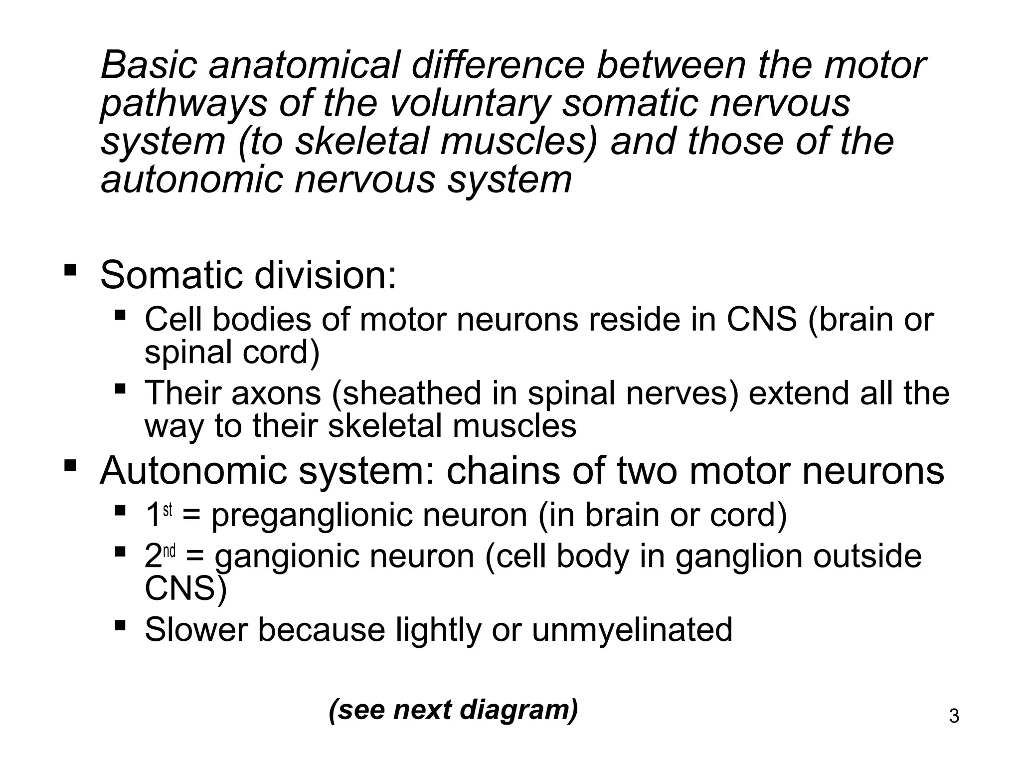
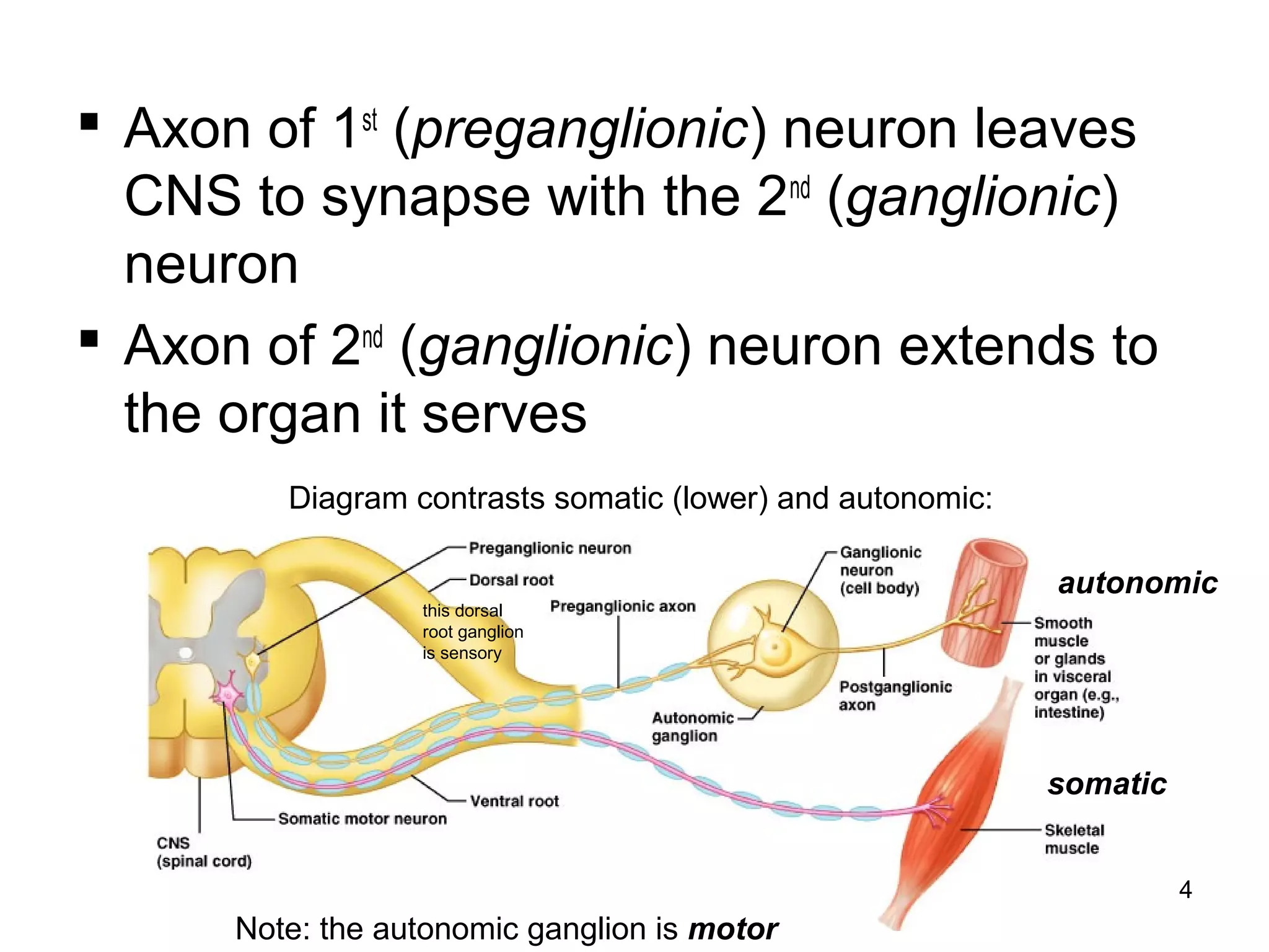
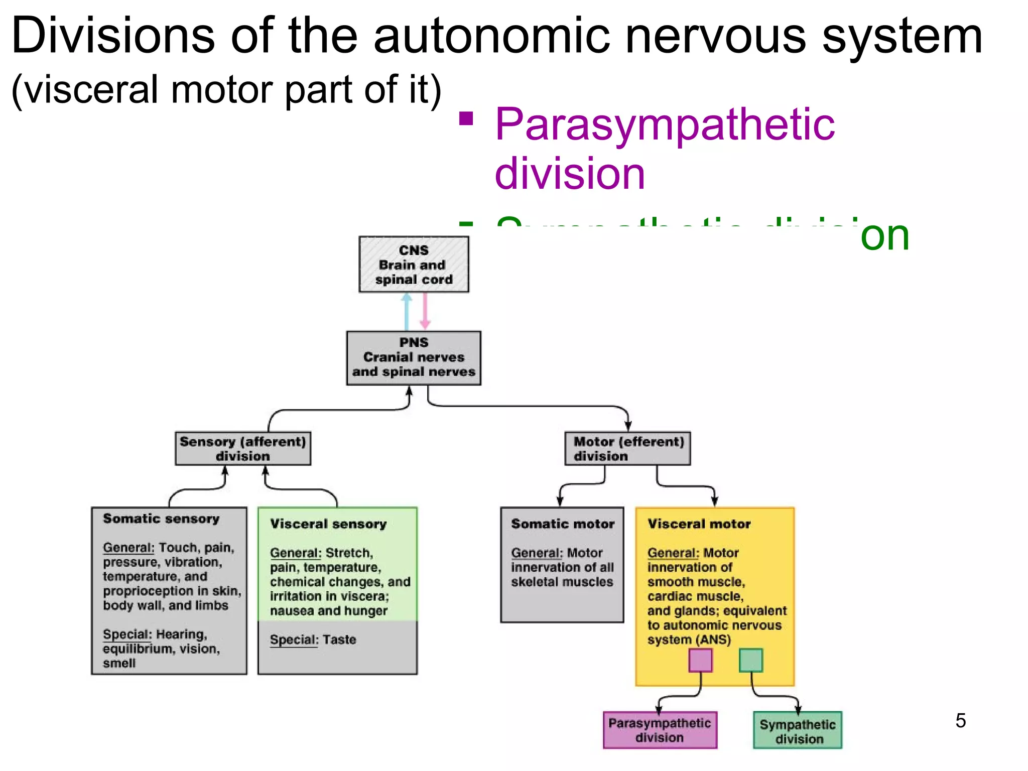
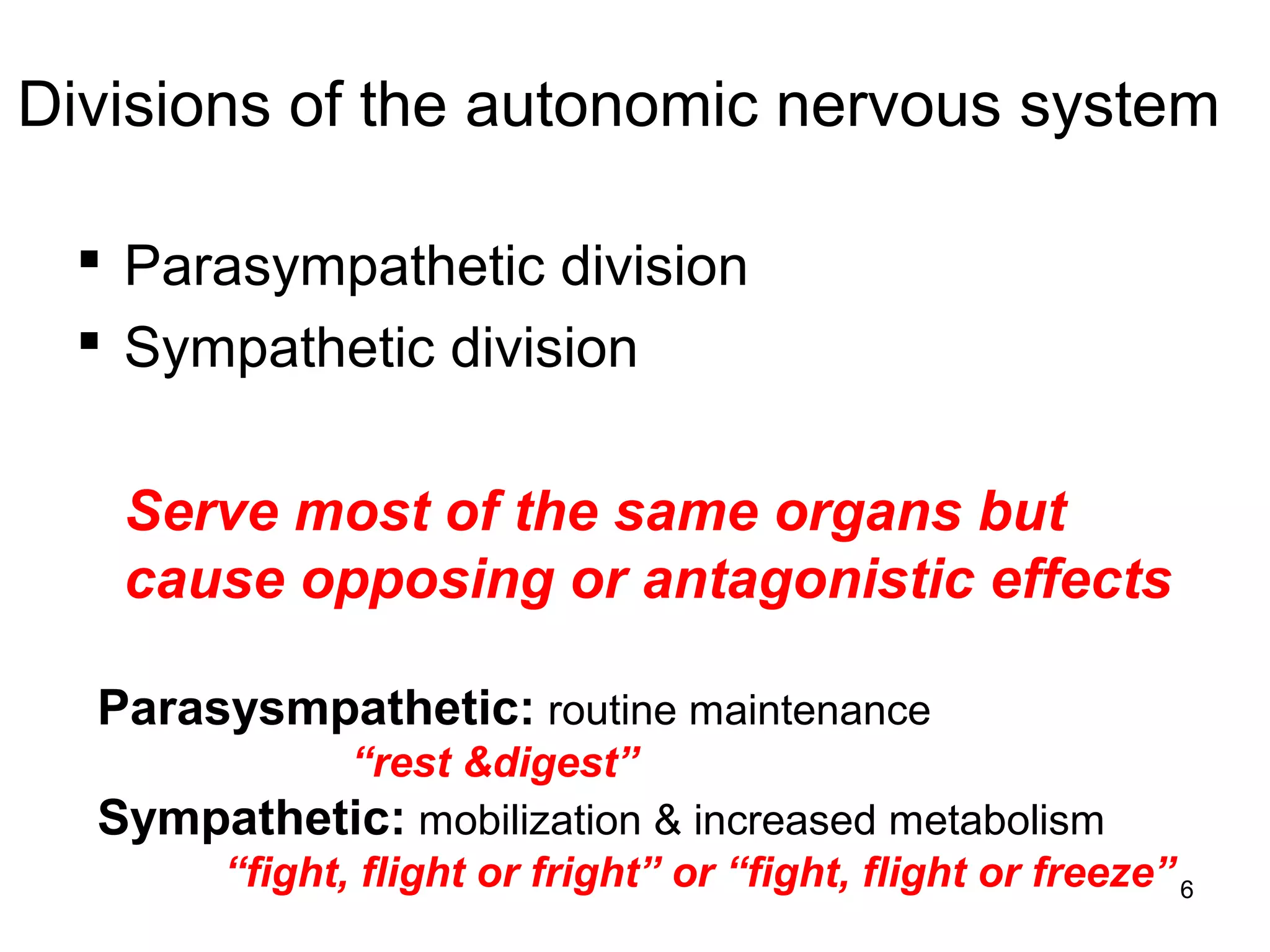
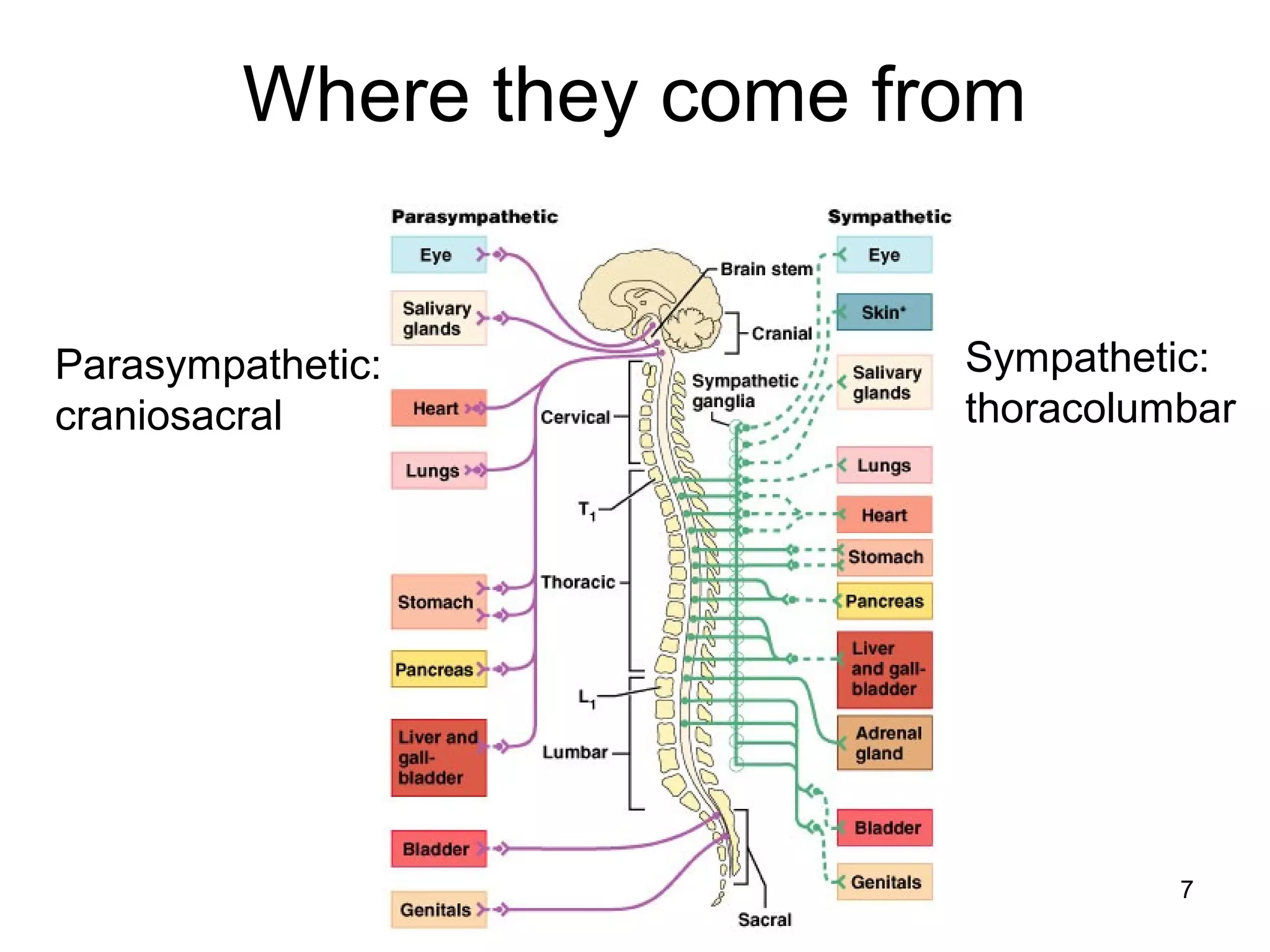
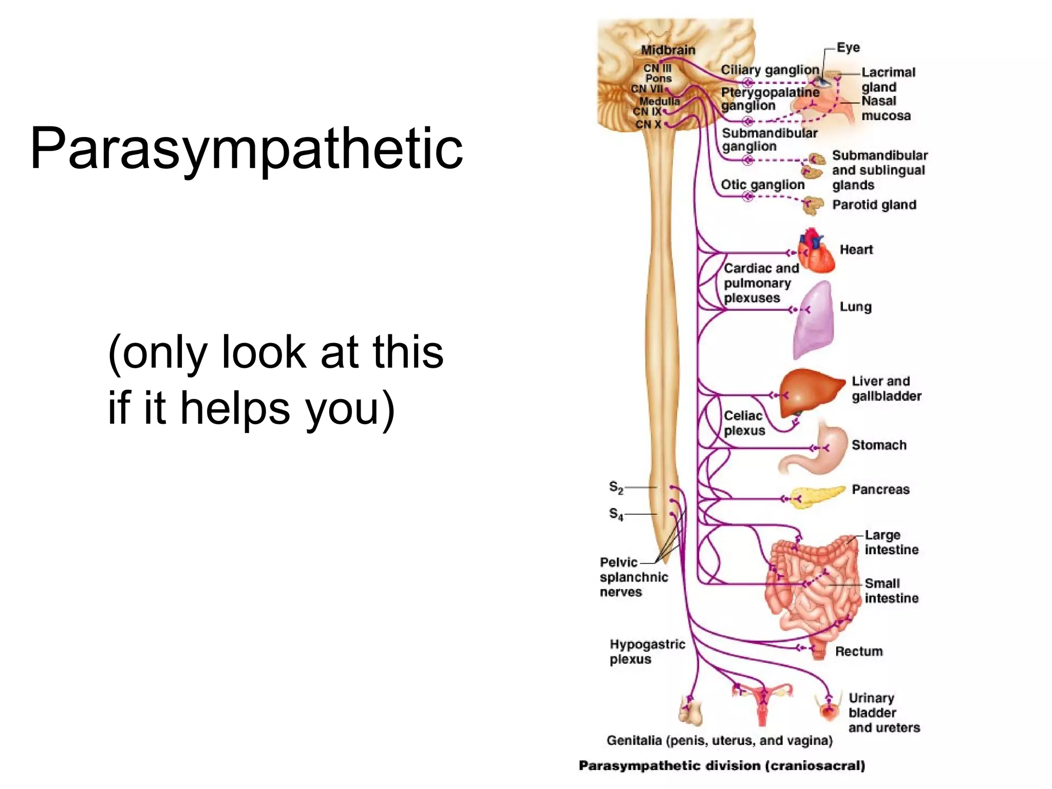
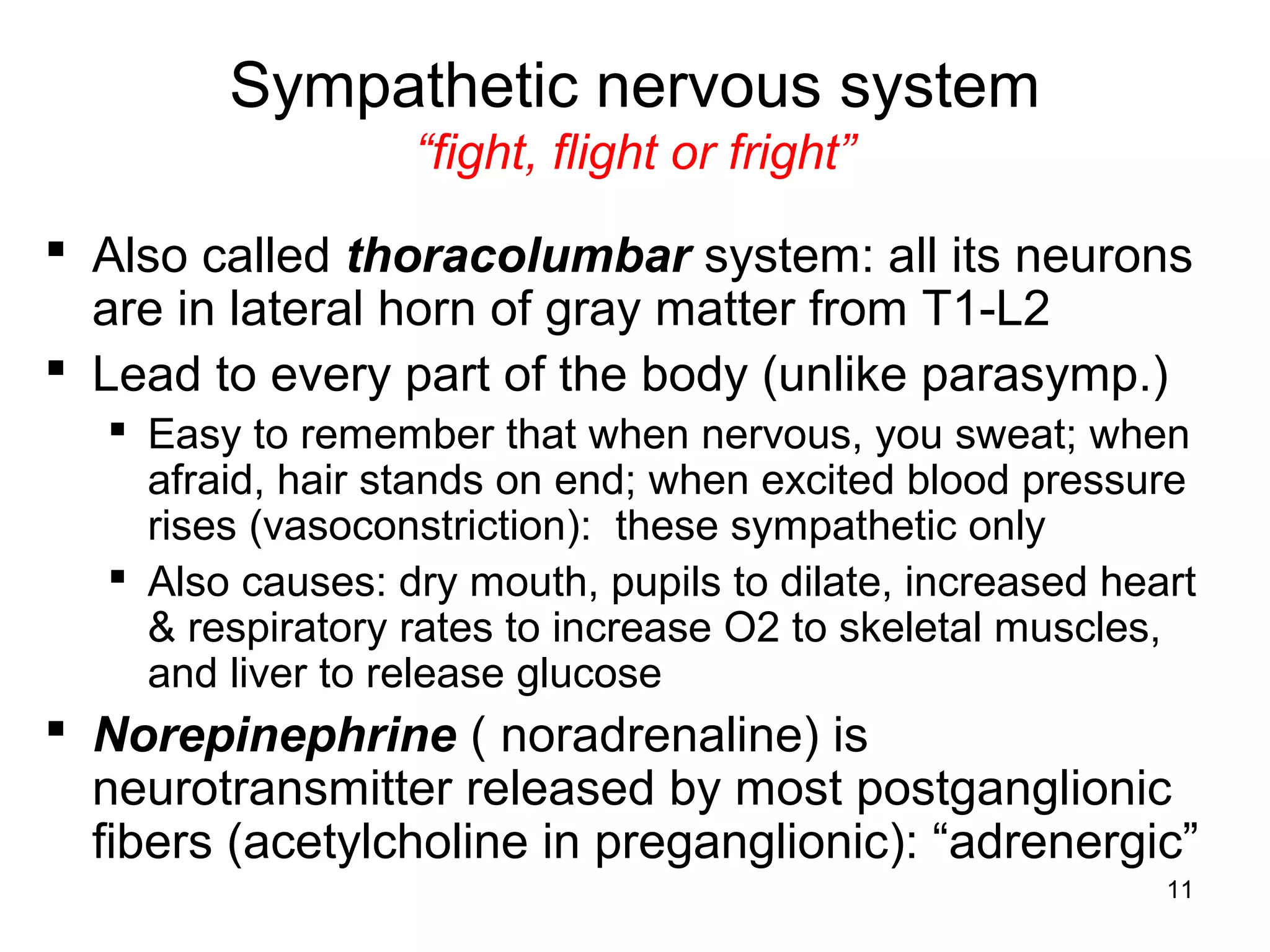
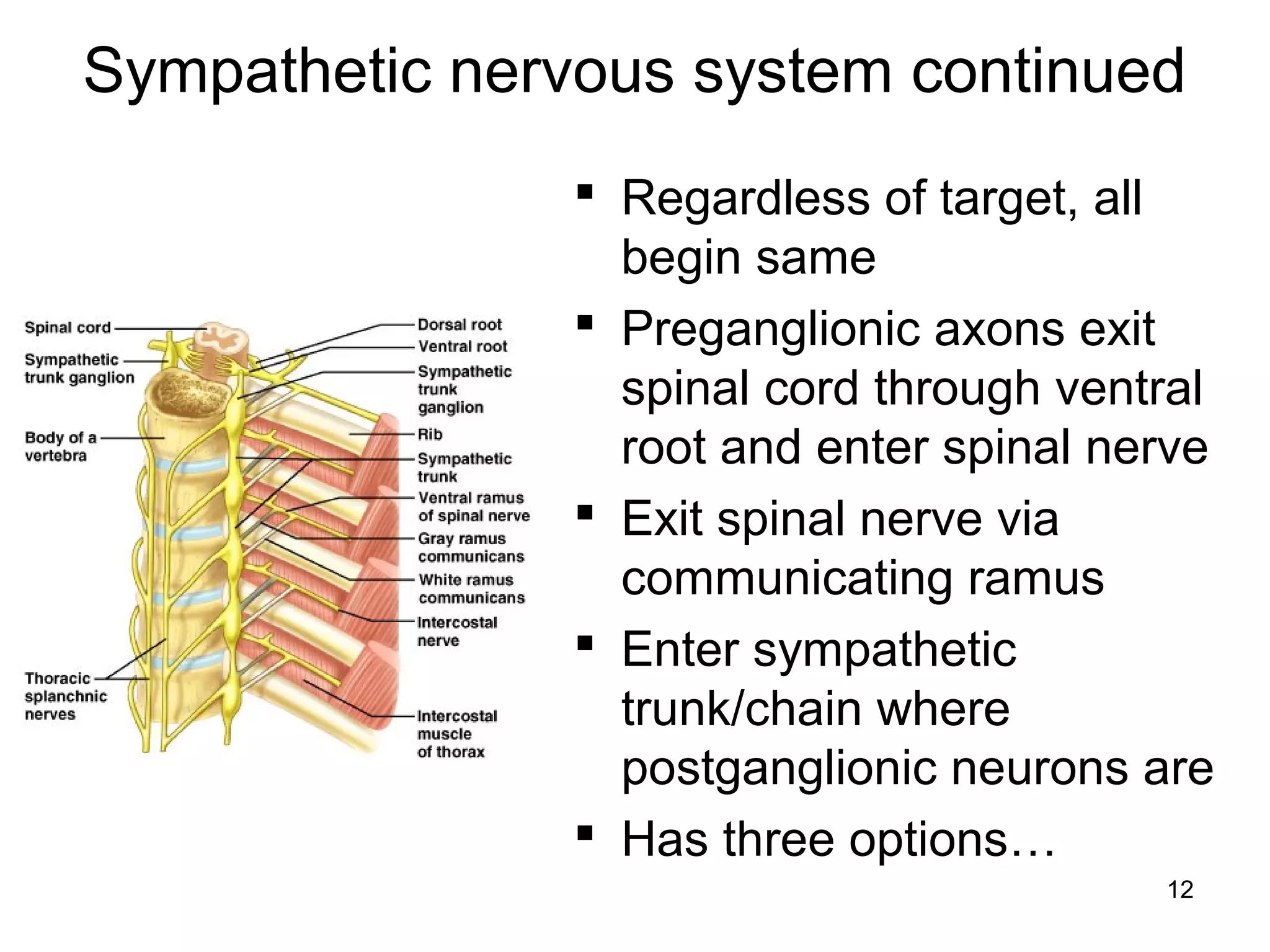
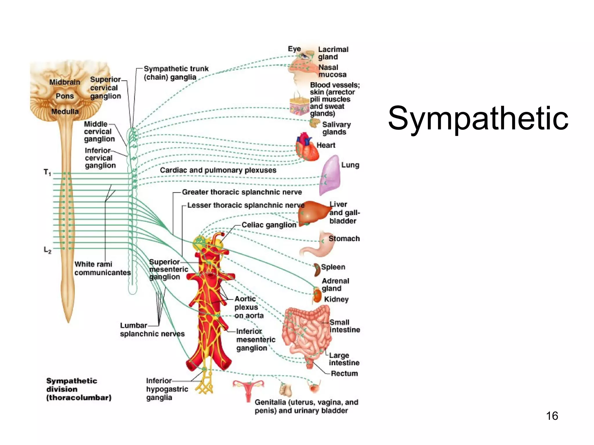
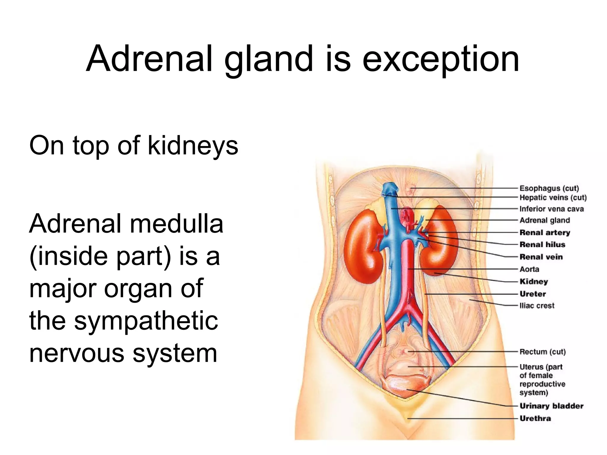
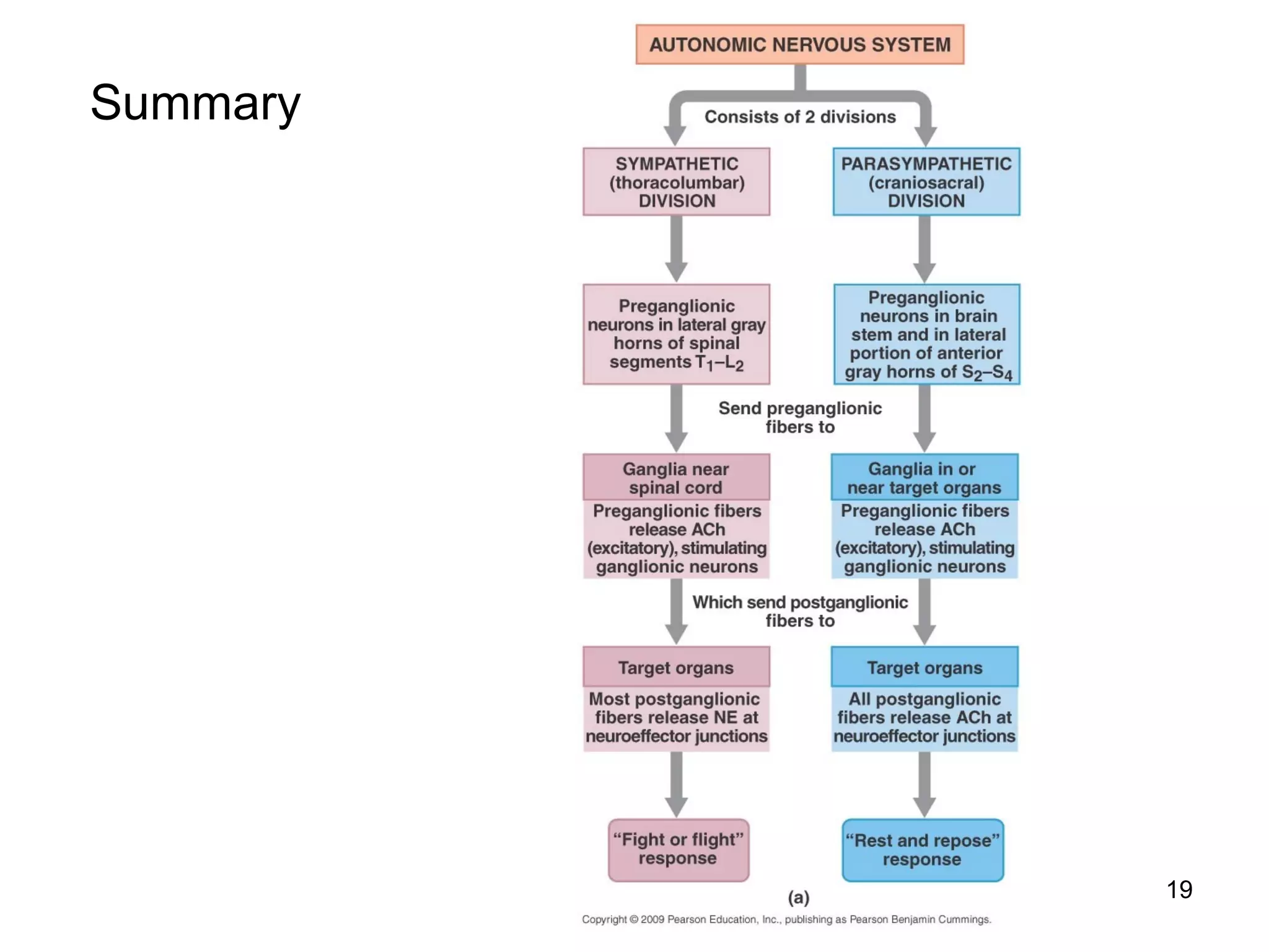
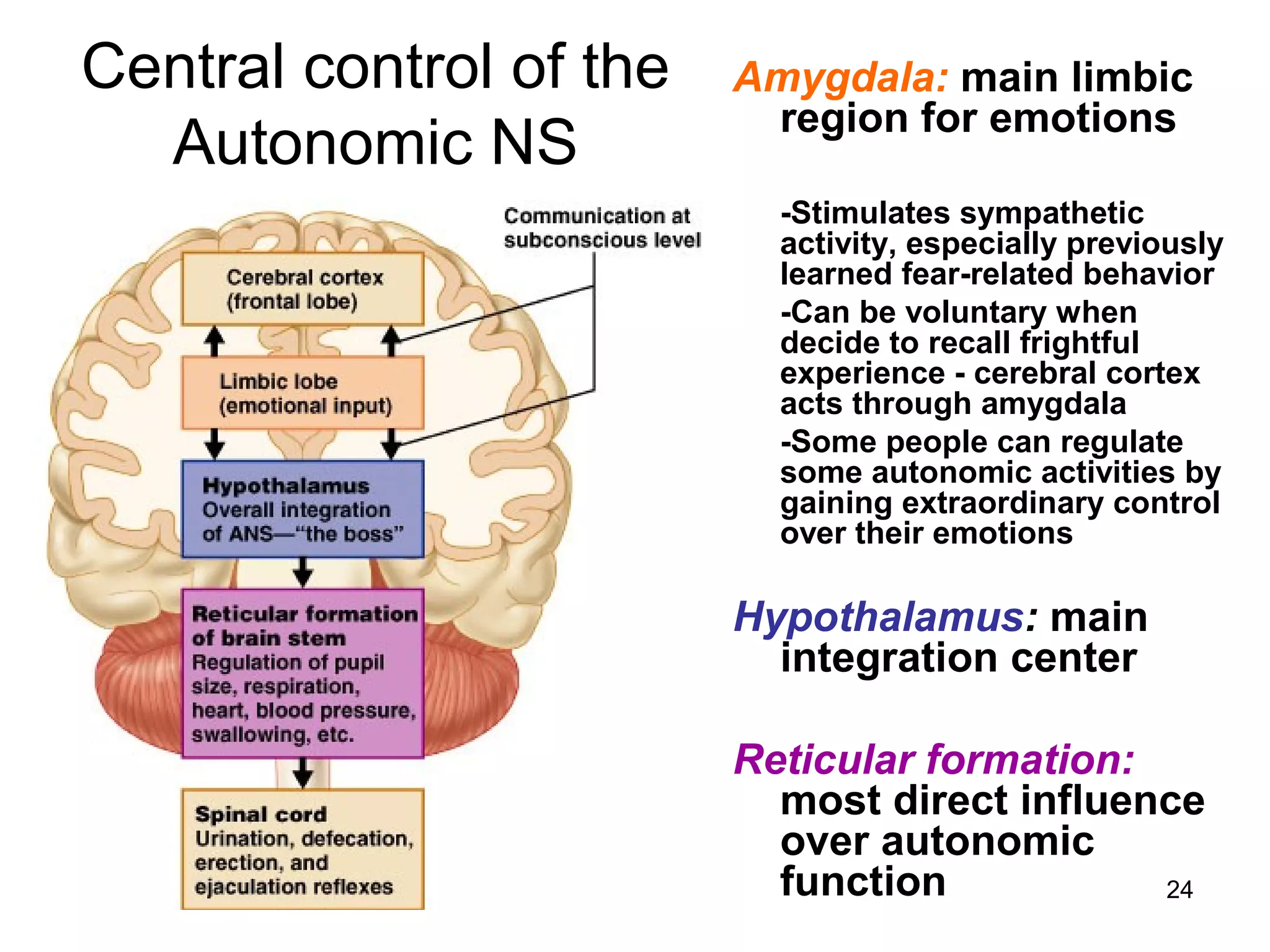

The autonomic nervous system regulates involuntary body functions and is divided into the sympathetic and parasympathetic divisions. The sympathetic division is responsible for the fight or flight response and increases heart rate and metabolism. The parasympathetic division is responsible for rest and digest functions and decreases heart rate and increases digestive functions. Both divisions contain two neurons, with the preganglionic neuron in the CNS and postganglionic neuron in peripheral ganglia. The autonomic nervous system controls functions of the heart, smooth muscles and glands.


