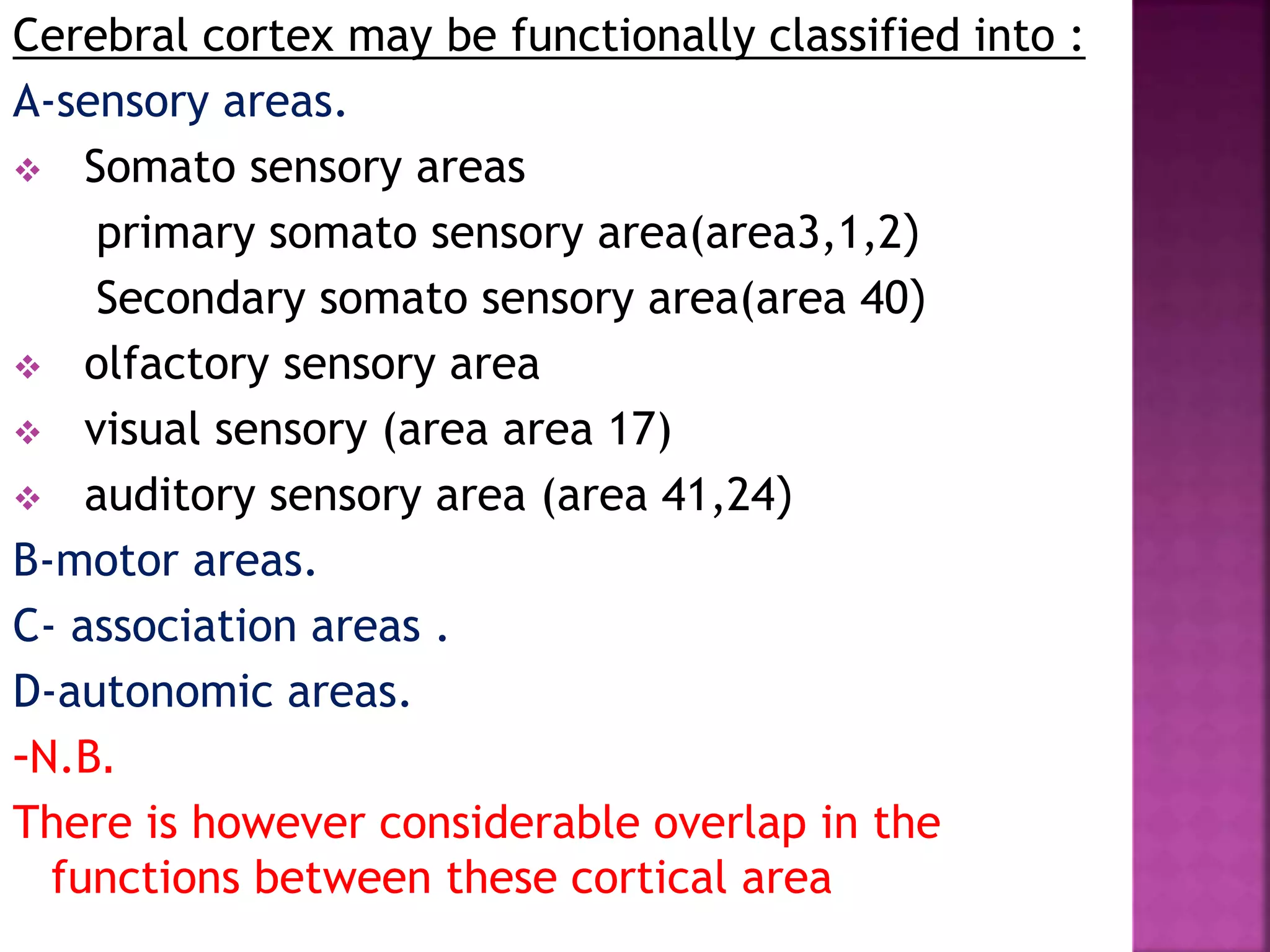
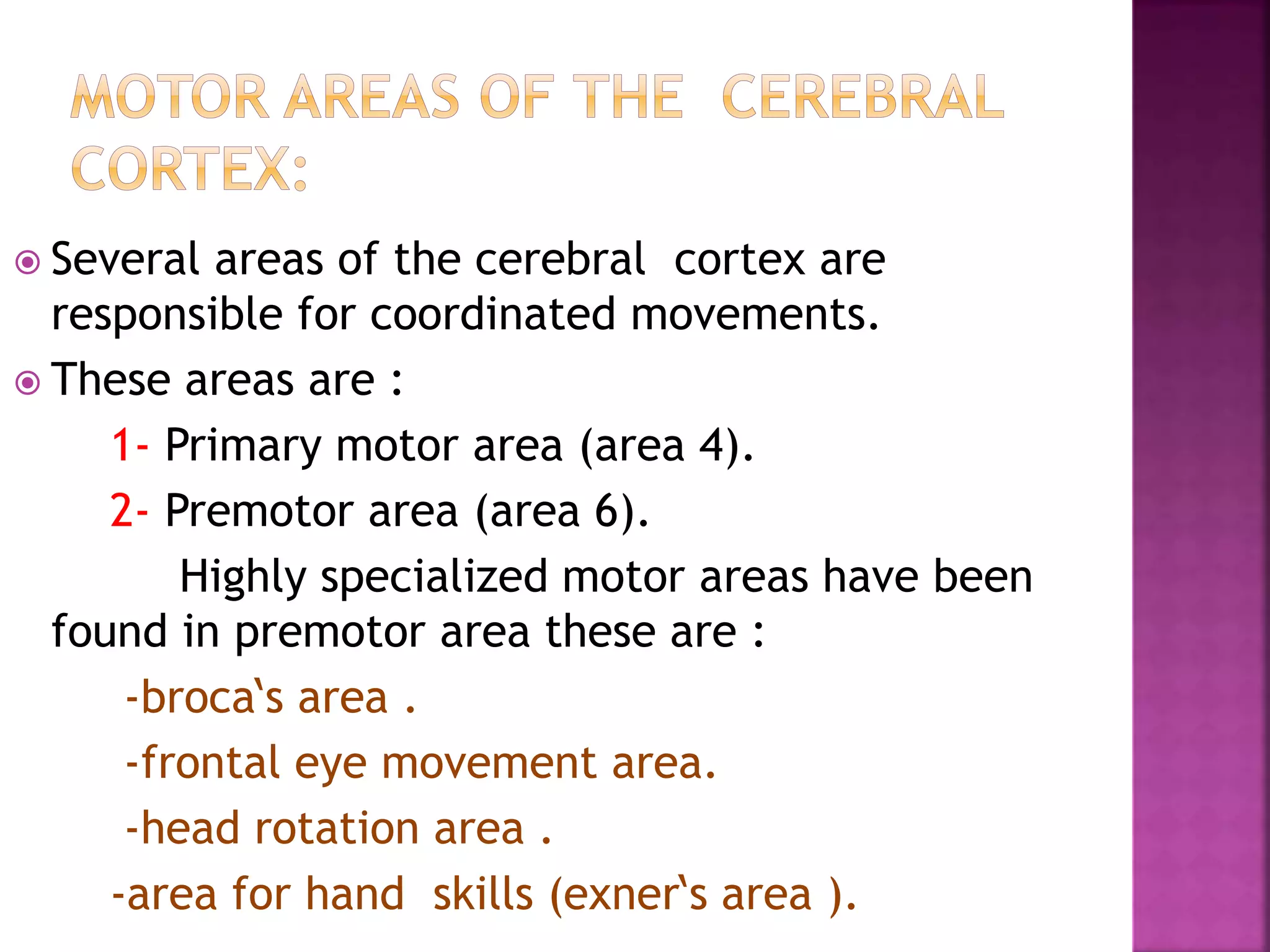
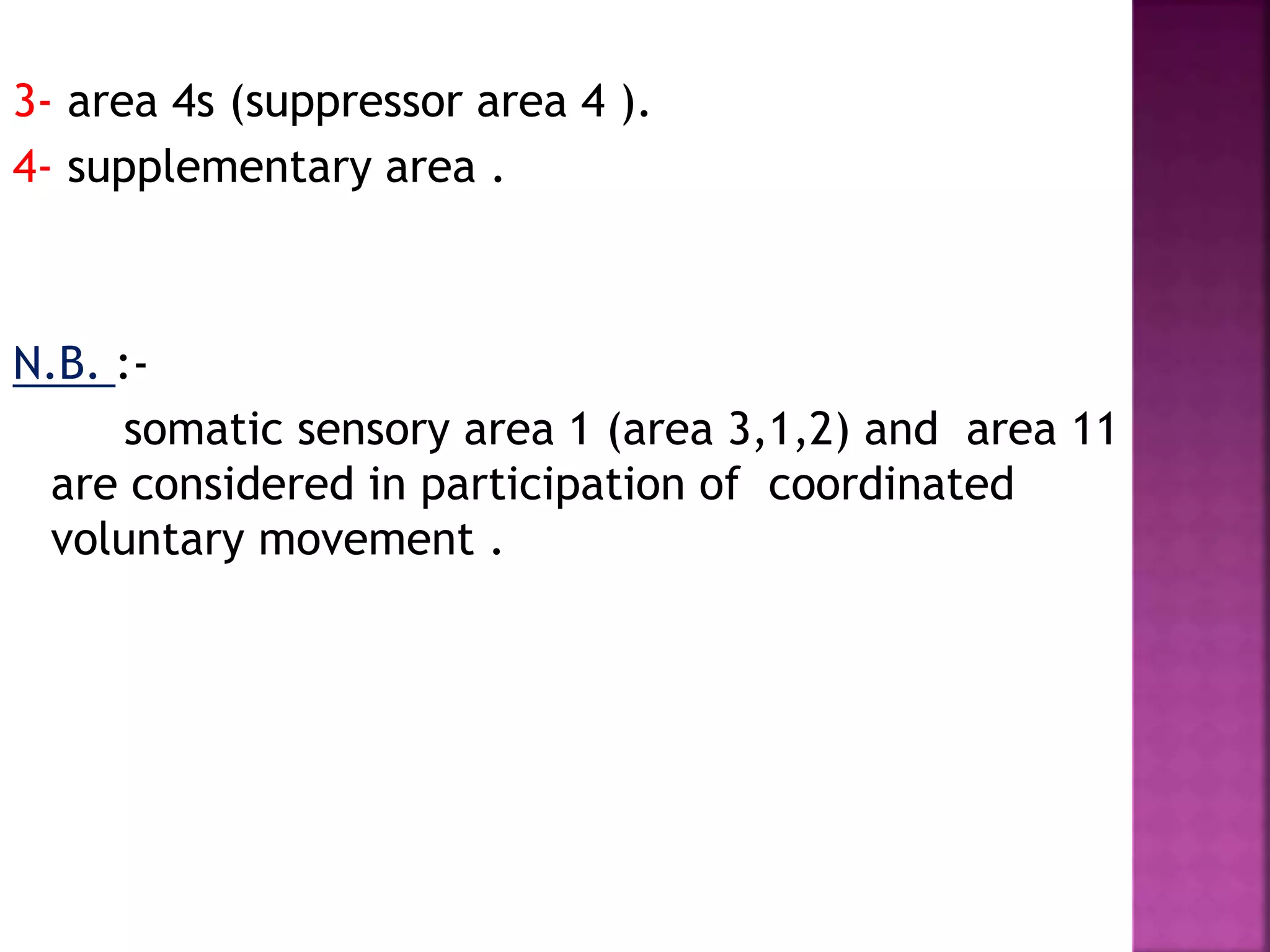
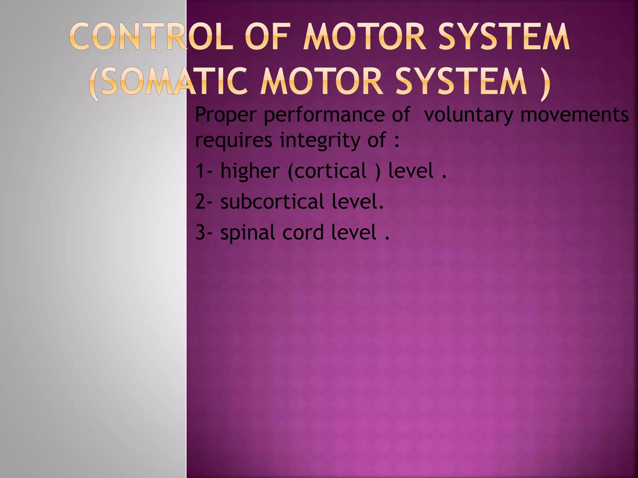
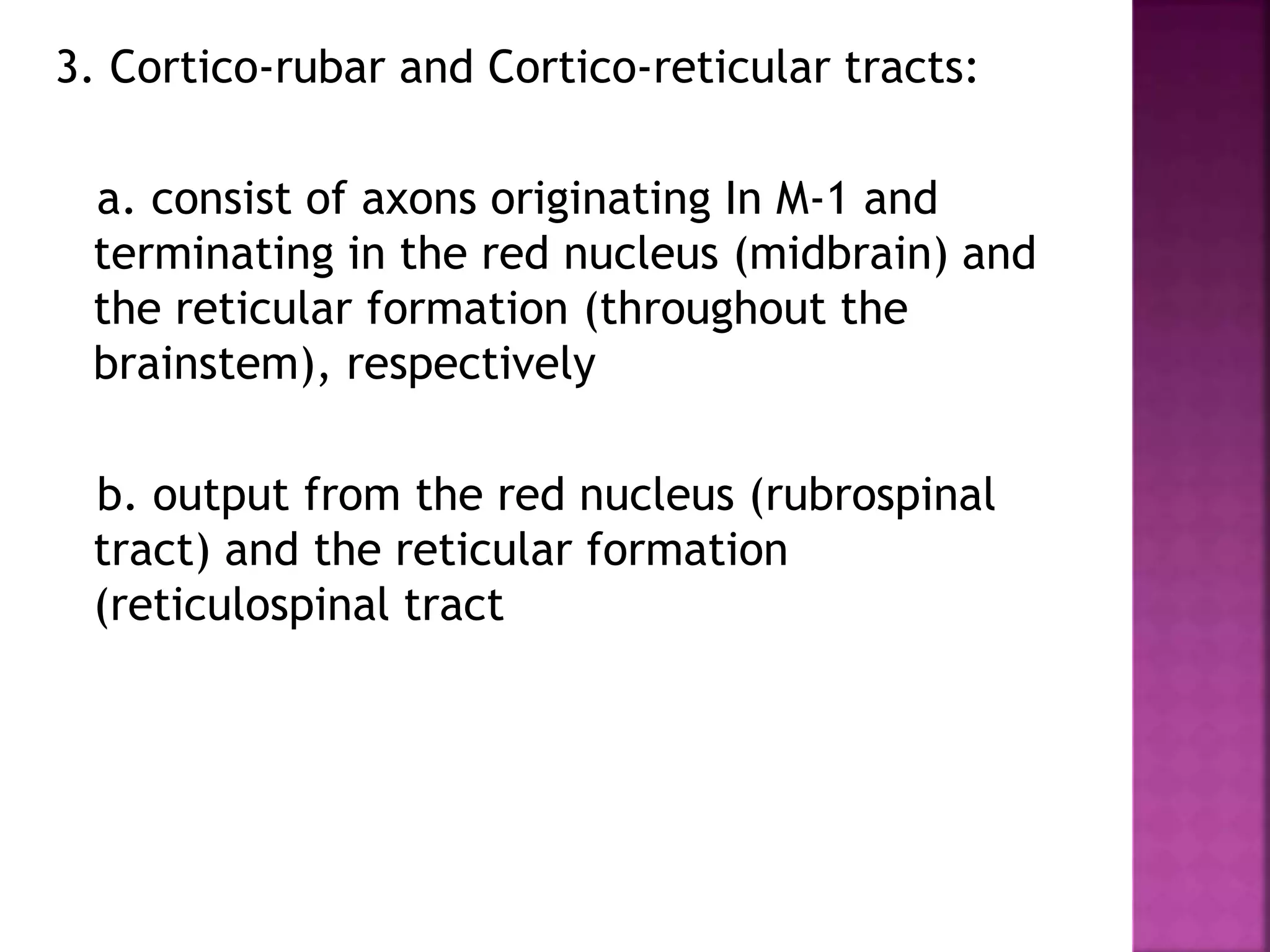
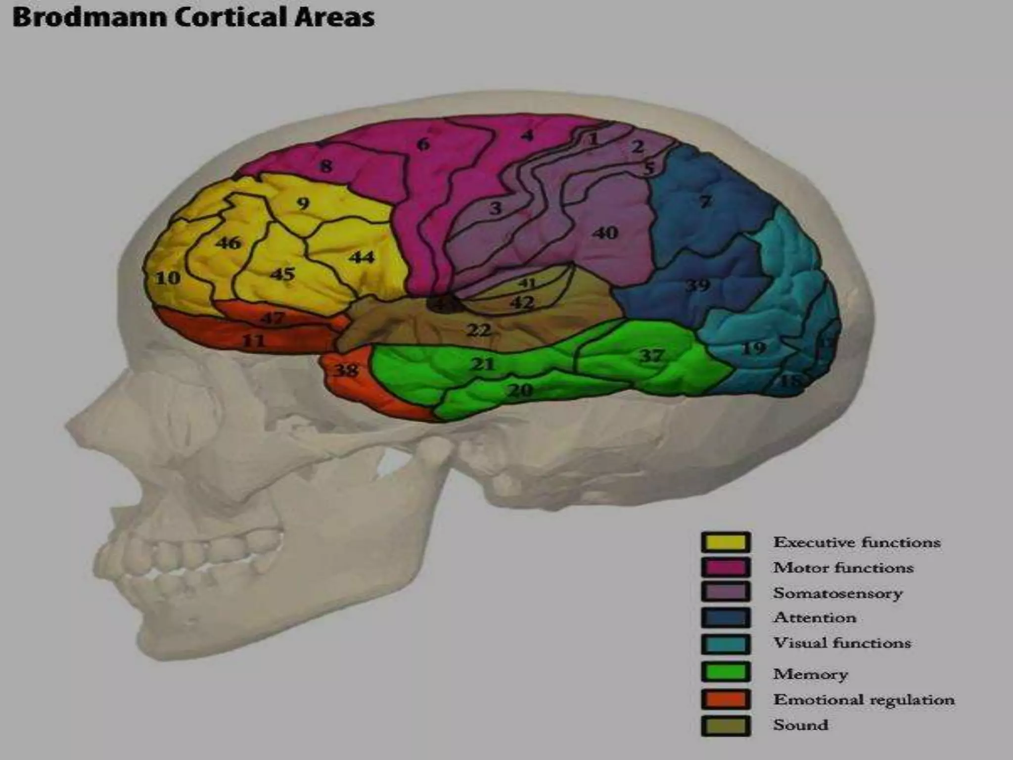
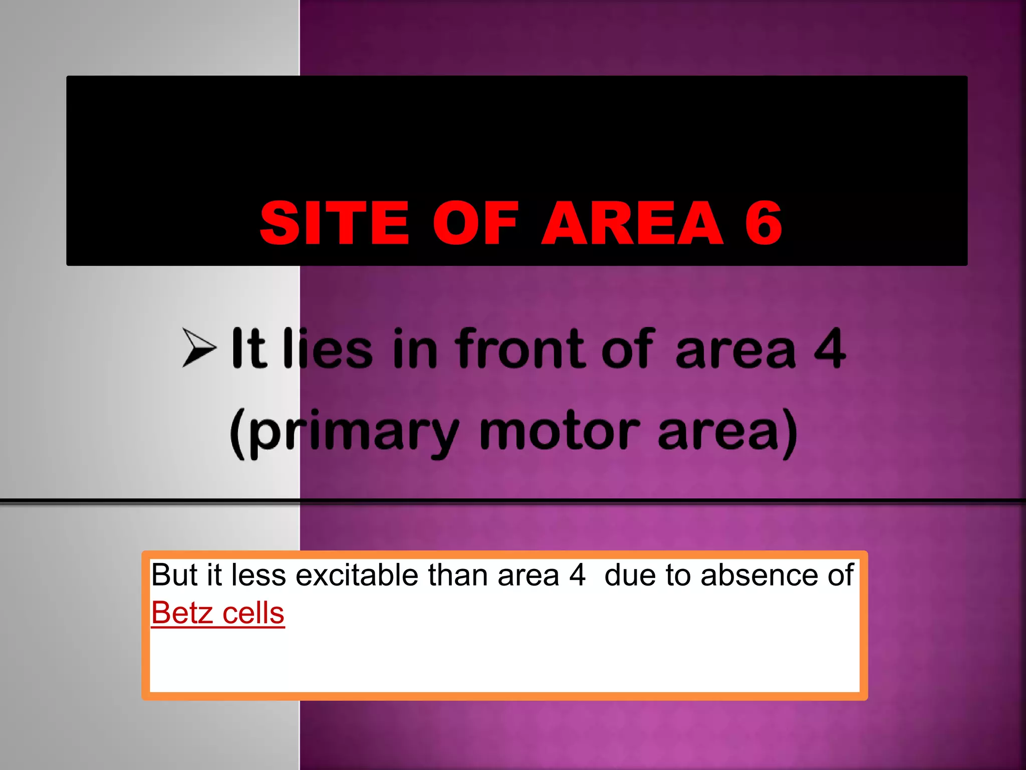
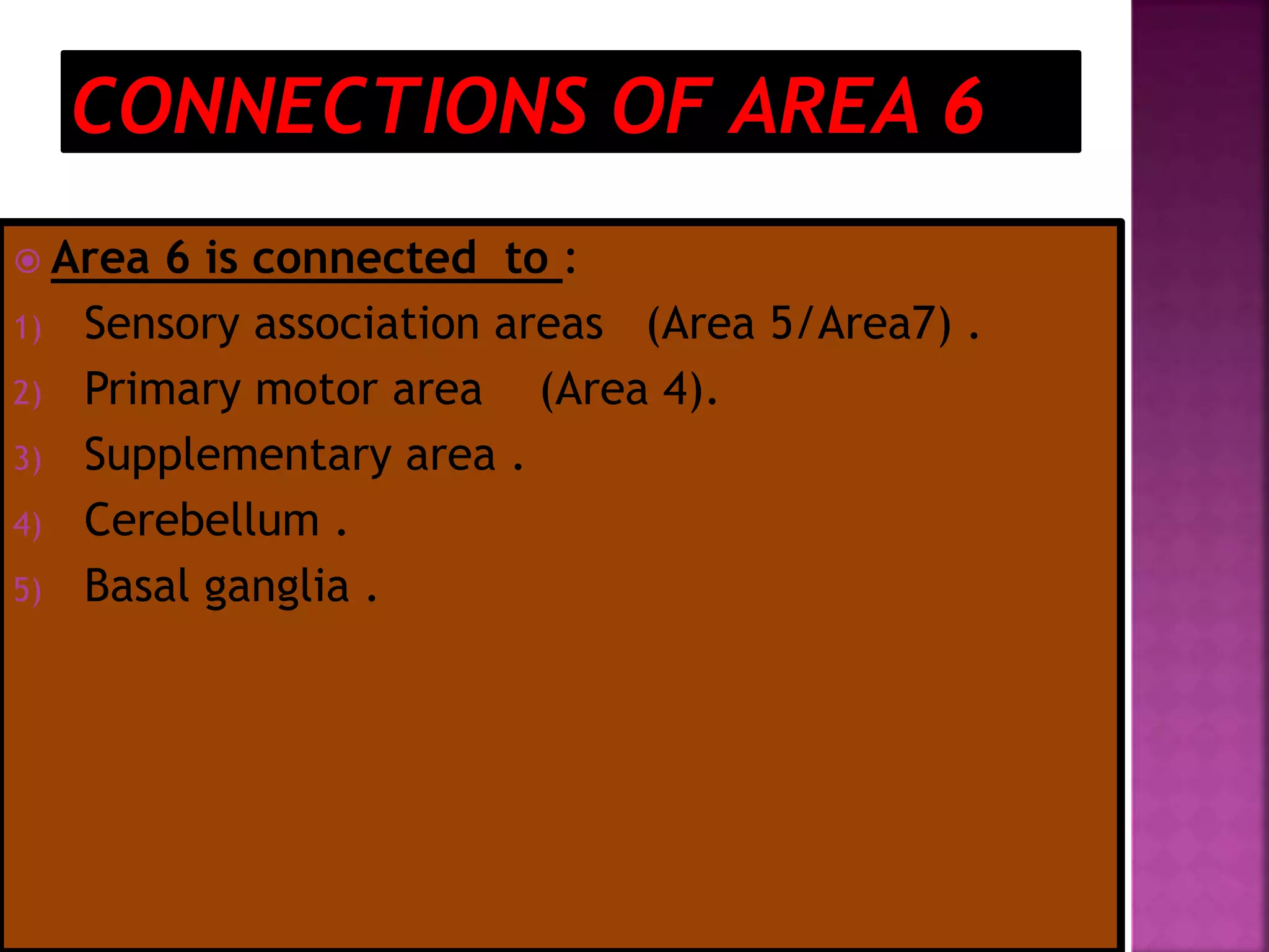
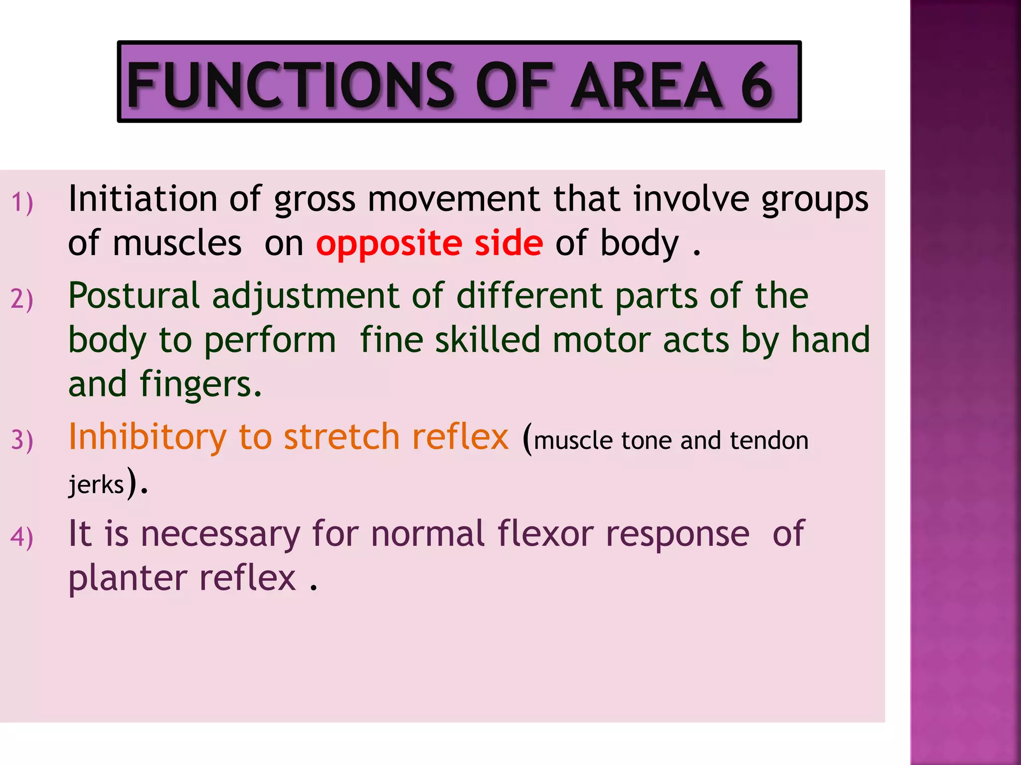
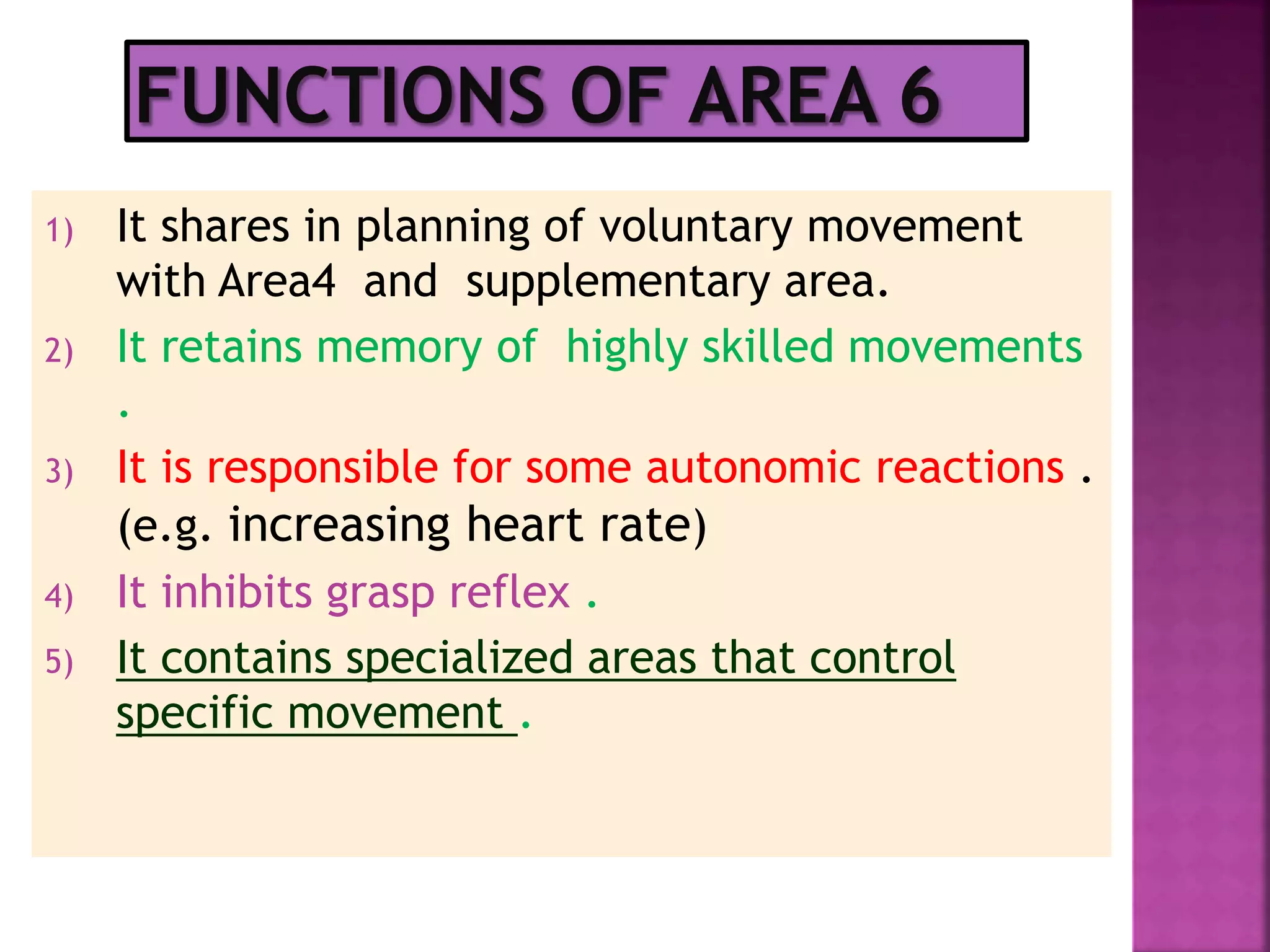
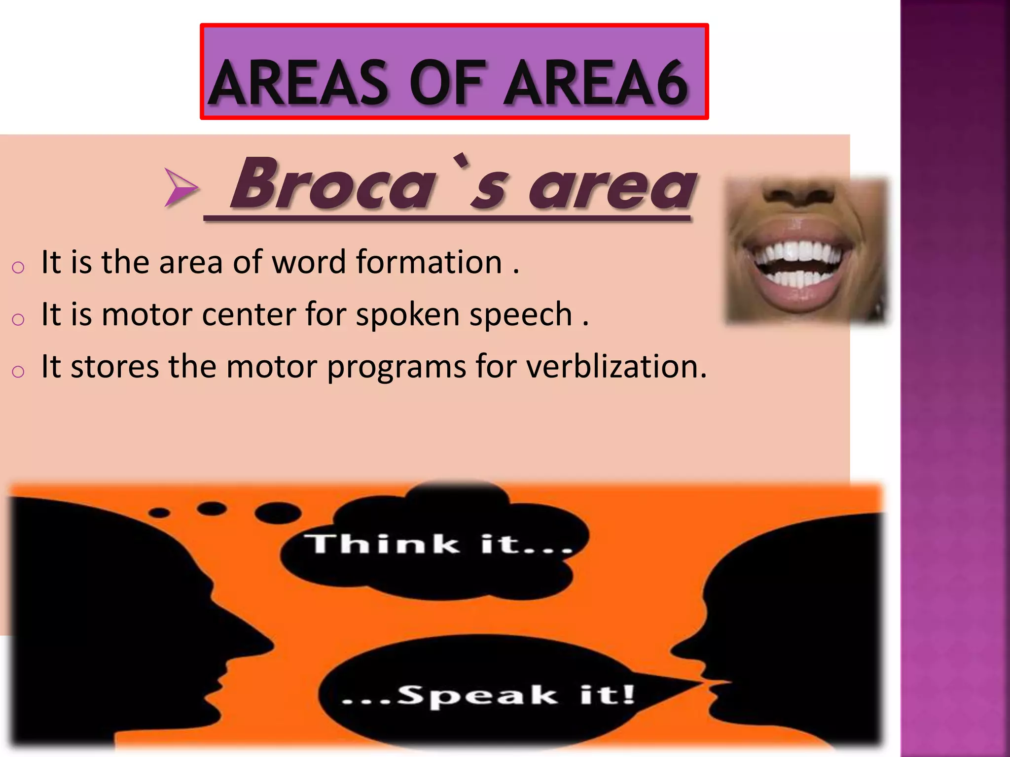
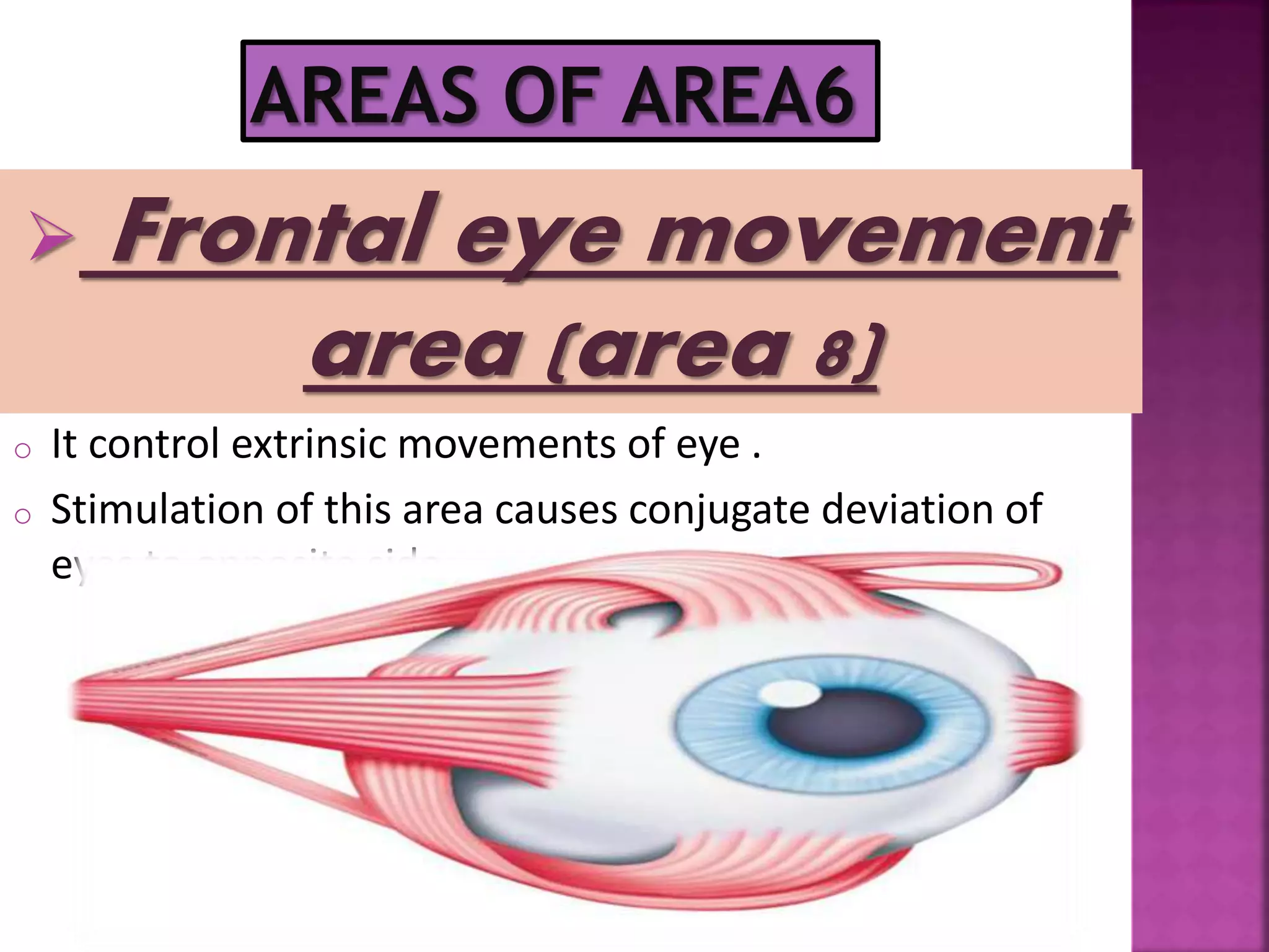
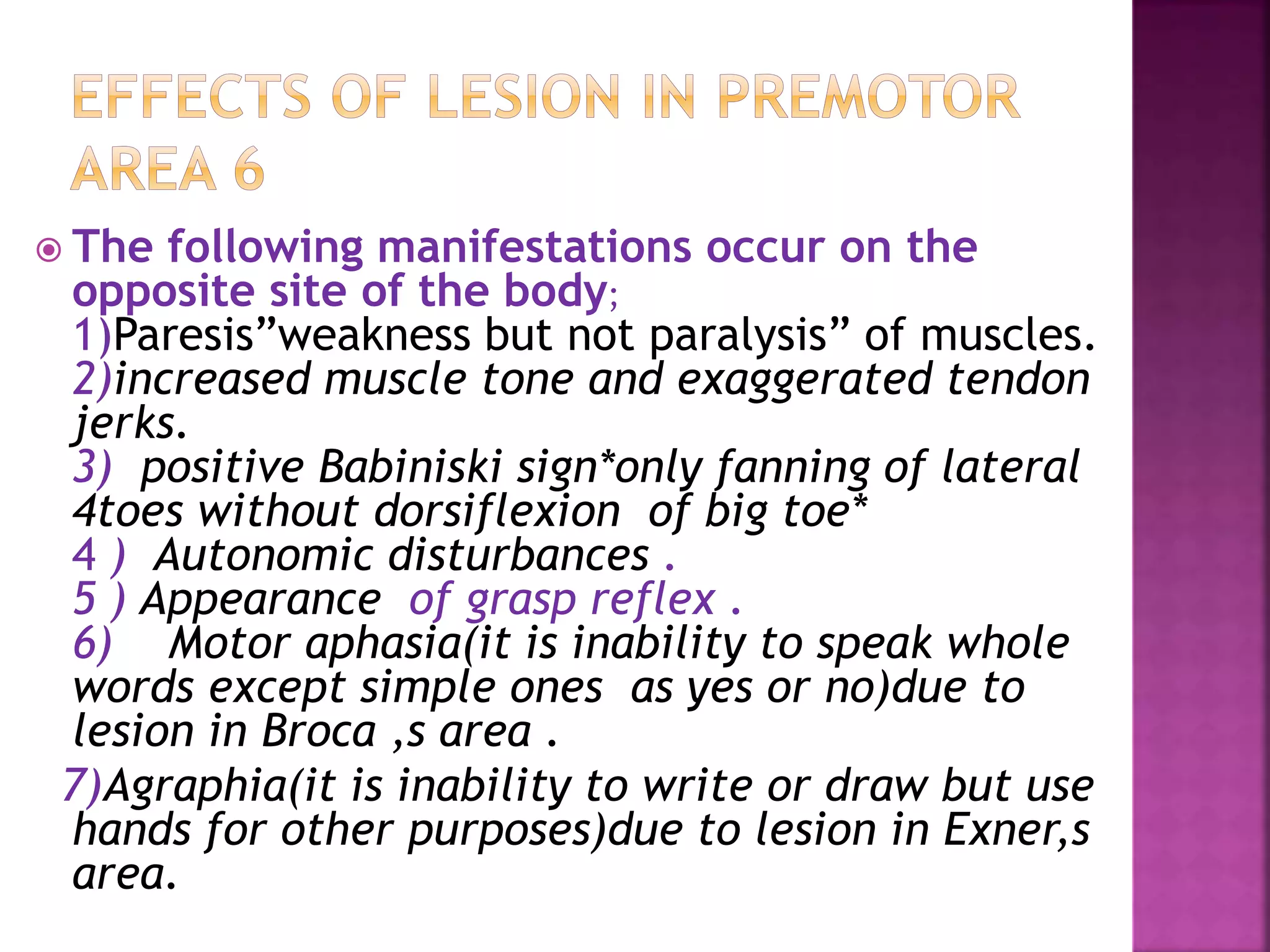
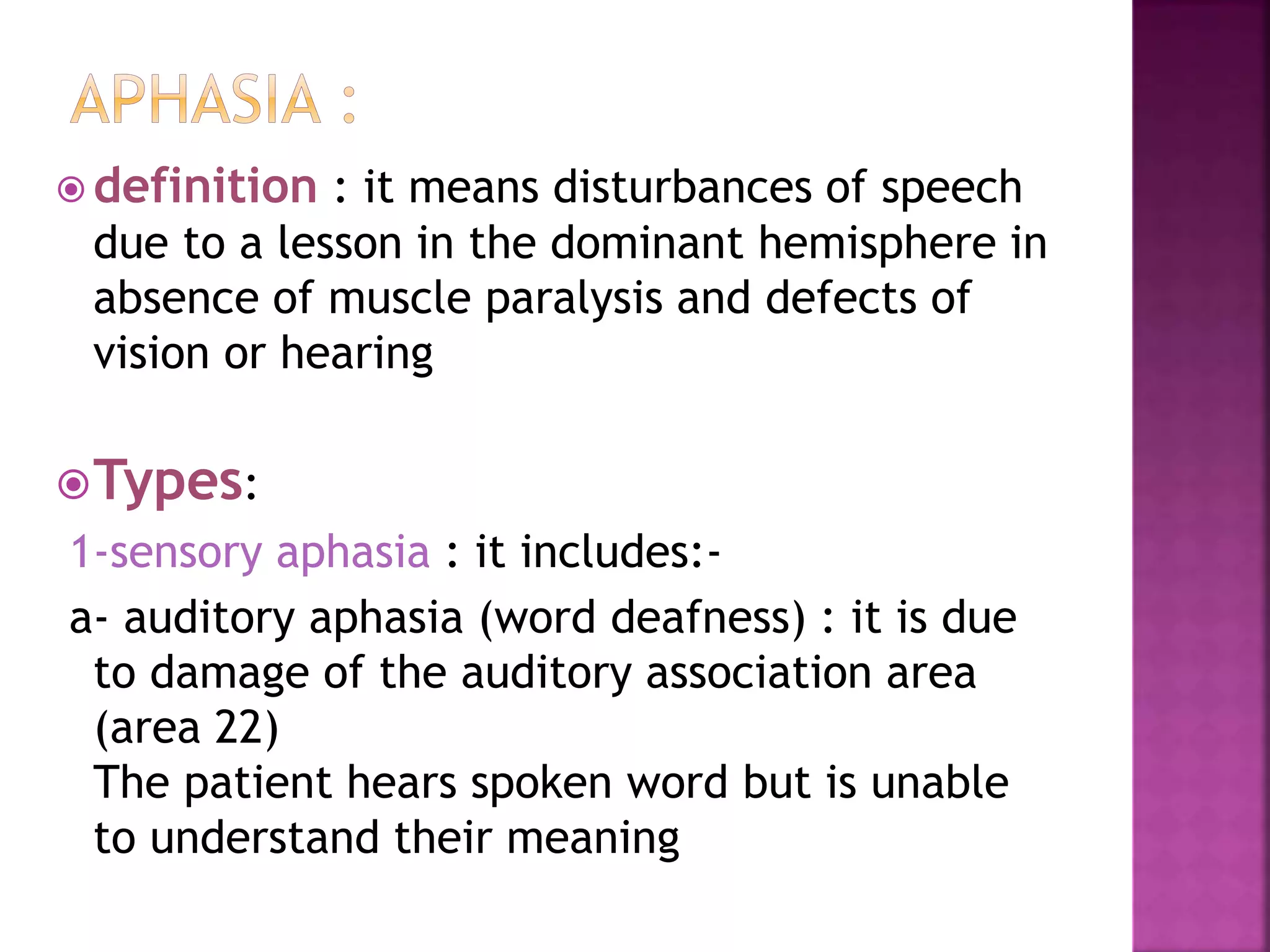
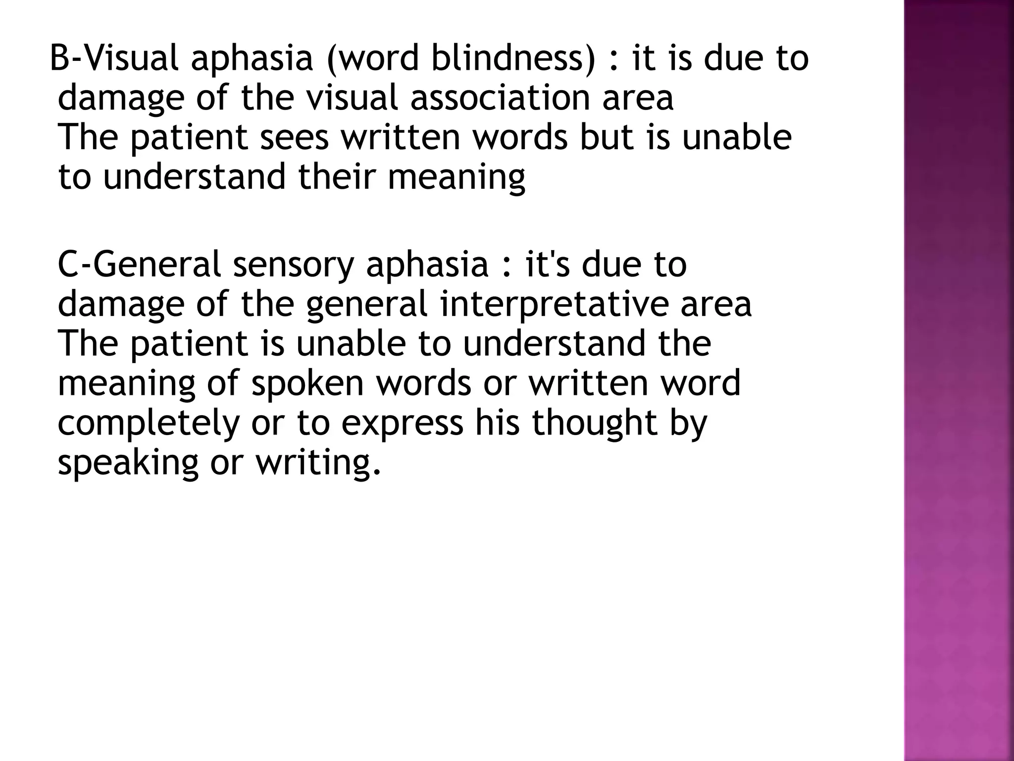
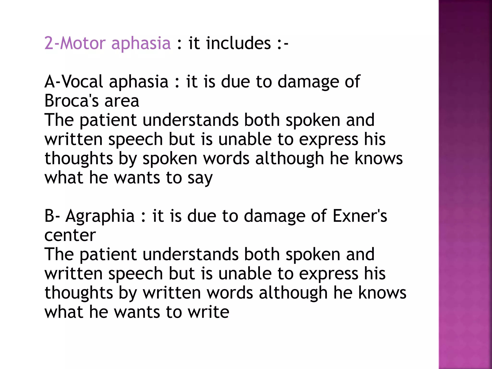
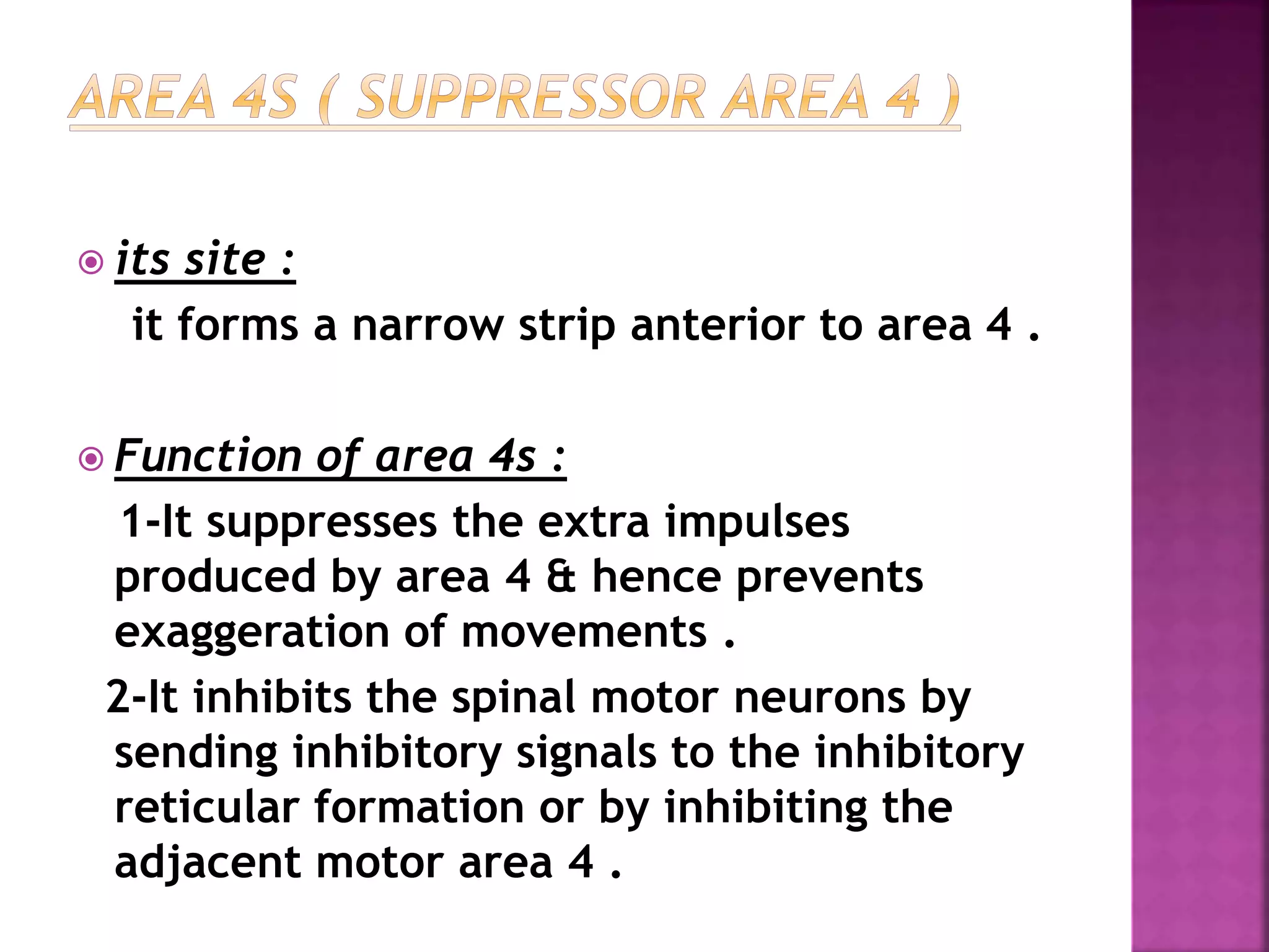
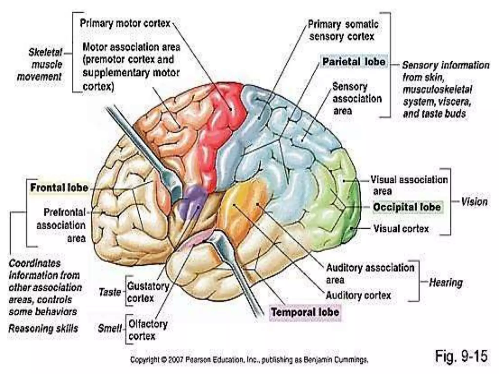
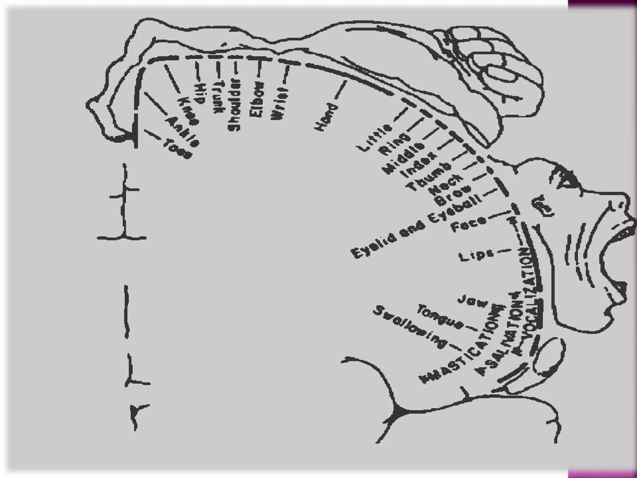
motor areas of cerebral cortex with reference to their lesion disorders . and some of the tracts connecting them to the motor nerves nuclei in the spinal cord and cranial nerves
2. Introduction.
Control of motor system.
Area 4.
Area 6.
Aphasia .
Supplementary Area.
Area 4s .
References.
3. Motor nervous system
Most of voluntary movements initiated
by cerebral cortex achieved when
the cortex activates patterns of
function stored in lower brain areas
{brainstem, basal ganglia,
cerebellum}
these centers send specific control
signals to anterior motor neurons of
spinal cord that control muscle
4. Cerebral cortex
• Cerebral cortex is the highest centre of
brain.
• It has a total surface area 0.2-0.25 m of
which about one third is exposed.
• The total cerebral cortex contain 100 billion
neurons.
It is divided by mean of primary fissures into
[frontal, parietal ,temporal,occipital,limbic
lobe]
It is divided by mean of secondary fissures
into gyri [eg. post central gyrus]
5. Cerebral cortex may be functionally classified into :
A-sensory areas.
Somato sensory areas
primary somato sensory area(area3,1,2(
Secondary somato sensory area(area 40(
olfactory sensory area
visual sensory (area area 17)
auditory sensory area (area 41,24(
B-motor areas.
C- association areas .
D-autonomic areas.
-N.B.
There is however considerable overlap in the
functions between these cortical area
6. Several areas of the cerebral cortex are
responsible for coordinated movements.
These areas are :
1- Primary motor area (area 4).
2- Premotor area (area 6).
Highly specialized motor areas have been
found in premotor area these are :
-broca‘s area .
-frontal eye movement area.
-head rotation area .
-area for hand skills (exner‘s area ).
7. 3- area 4s (suppressor area 4 ).
4- supplementary area .
N.B. :-
somatic sensory area 1 (area 3,1,2) and area 11
are considered in participation of coordinated
voluntary movement .
9. It is carried out in 2 stages :
(1) planning : this occurs as follows:
Ideas and purpose of movements originate in
cortical association areas , which discharge signals
to both basal ganglia as well as lateral cerebellum
(cerebrocerebellum).
Basal ganglia converts thoughts into a motor plan
and put program of performance (especially for
slow movements) then discharge to cortical motor
area (which also , shares in planning and
programming processes ,( especially premotor and
supplementary areas )
Cerebrocerbellum shares in planning and
programming (especially for rapid movements)
10. (2) Execution : this occurs as follows :
Motor cortex discharges signals to spinal and cranial
motor neurons as well as to intermediate
cerebellum (spinocerebellum ).
Motor neurons discharge final signals to skeletal
muscles .
Skeletal muscles contract according to the plan
Producing required movement coordinately .
12. PRIMARY MOTOR CORTEX (also known as M-1
or Brodmann’s Area 4; part of the neocortex,
“gray matter”)
13. Position:-
-Located in precentral gyrus area of the frontal
lobe of the cerebral cortex
body representation :-
-body representation is mainly contralateral;
exception muscles of respiration , muscles of the
upper part of the face and abdominal muscles
which are bilateraly respresented
-it is inverted ;the body is represented upside
down with the exception of the head which is not
inverted
14. -small, distorted, discontinuous map of the body ,
larger areas devoted to body regions characterized
by fine or complex movements "delicate highly
skilled movements “ and smaller areas to body
regions characterized by gross movements involving
few muscles
-Hand, face, intraoral and, to some extent, foot
muscles are particularly well represented while
muscles of the thigh are represented by small areas
“gross movement”
16. 1-the main function is to initiate and control fine descrete
movements of the distal parts of the body on the opposite
side
2-shares area 6 in its function in controlling gross movements
3-facilitates stretch reflex
4-necessary for superficial somatic reflexes
5-electrical stimulation of a particular area of area 4 leads to
contraction of a particular skeletal muscle or group of
muscles that is/are responsible for fine discrete
movements
6-histologically the cerebral cortex is formed of many layers
of cell arranged in columns. Stimulation of a given motor
column of area 4 may activate a single muscle
7- It shares information of the pyramidal tracts by 30% of
fibers of the pyramidal tract
17. unilateral lesion leads to the following
manifestations on the opposite side of the
body :
a)Monoplegia :- paralysis of muscles of one
limb
*permanent loss of fine movements and
temporary loss of gross movements
*later ,there is recovery of gross movements.
18. b)Hypotonia and weak tendon jerk:
due to loss of facilitatory impulses to stretch
reflex.
c)positive Babniski sign:
scratch of lateral side of sole of foot leading to
dorsiflexion of big toe (one component of Babniski
sign ) without fanning of the other toes (the
second component of this sign)
19. d) loss of the superficial reflexes:
e.g(flexion withdrawal reflex , planter reflex
,Grasp reflex)
20. descending tracts :-
1. Corticospinal tract:
a. axons pass through the internal capsule (a frequent site of stroke
injury), then through the ipsilateral diencephalon, midbrain, pons,
and into the medulla, where they form the medullary pyamids
Note: the corticospinal tract is also termed the “pyramidal tract”
(named for the medullary pyramids, not for the cortical pyramidal
cells)
b. as the axons pass from the medulla into the spinal cord, most (80-
90%) decussate (cross to the opposite side) and descend in the white
matter of the contralateral cord, forming the lateral corticospinal
tract, until they reach the region containing the alpha motoneurons
of the muscles they represent; the majority of these motoneurons
innervate flexors
c. the non-decussating axons descend in the ipsilateral cord, forming
the ventral corticospinal tract, until they reach the level of region of
the body they represent, and then they decussate also; the majority
of these motoneurons control extensors
21. 2. Corticobulbar tract:
a. axons pass through the internal capsule and
terminate either directly on alpha motorneurons or on
interneurons innervating alpha motor neurons in the
brainstem controlling somatic motor activity in the
head (e.g. muscles controlling eye movement, muscles
of mastication, muscles of facial expression)
b. axons innervating some somatic motor nerve cranial
nuclei decussate before their termination and so
control contralateral muscles; corticobulbar tracts
controlling other motor nuclei split, some axons
decussating and others descending ipsilateral, resulting
in bilateral innervation
c. cranial nerve innervation is either direct or through
interneurons, as in the corticospinal tract
22. 3. Cortico-rubar and Cortico-reticular tracts:
a. consist of axons originating In M-1 and
terminating in the red nucleus (midbrain) and
the reticular formation (throughout the
brainstem), respectively
b. output from the red nucleus (rubrospinal
tract) and the reticular formation
(reticulospinal tract
23.
24.
25. But it less excitable than area 4 due to absence of
Betz cells
26.
27. It control movement of opposite side of body.
But:
There is Bilateral representation of
muscles that moves together as:
1. Muscles of eyes.
2. Respiratory muscles.
3. Abdominal muscles.
28. The body is represented upside down.
But
Representation of head is not inverted
(eye brows upward /lips are downward)
29. 1) Muscles that are responsible for delicate
highly skilled movement are
represented by wide areas.
(e.g. muscles of fingers)
2) Muscles that are responsible for gross
movement are represented by small
areas .
(e.g. muscles of thigh)
30. Area 6 is connected to :
1) Sensory association areas (Area 5/Area7) .
2) Primary motor area (Area 4).
3) Supplementary area .
4) Cerebellum .
5) Basal ganglia .
31. 1) Initiation of gross movement that involve groups
of muscles on opposite side of body .
2) Postural adjustment of different parts of the
body to perform fine skilled motor acts by hand
and fingers.
3) Inhibitory to stretch reflex (muscle tone and tendon
jerks).
4) It is necessary for normal flexor response of
planter reflex .
32. 1) It shares in planning of voluntary movement
with Area4 and supplementary area.
2) It retains memory of highly skilled movements
.
3) It is responsible for some autonomic reactions .
(e.g. increasing heart rate)
4) It inhibits grasp reflex .
5) It contains specialized areas that control
specific movement .
33. Broca`s area
o It is the area of word formation .
o It is motor center for spoken speech .
o It stores the motor programs for verblization.
34. Frontal eye movement
area (area 8)
o It control extrinsic movements of eye .
o Stimulation of this area causes conjugate deviation of
eyes to opposite side.
35. Head rotation area
o It directs head towards different visual objects.
36. Exner`s area
o It is the area of hand skills .
o It stores the motor programs for skilled movements
of hand and fingers .
37. The following manifestations occur on the
opposite site of the body;
1)Paresis”weakness but not paralysis” of muscles.
2)increased muscle tone and exaggerated tendon
jerks.
3) positive Babiniski sign*only fanning of lateral
4toes without dorsiflexion of big toe*
4 ) Autonomic disturbances .
5 ) Appearance of grasp reflex .
6) Motor aphasia(it is inability to speak whole
words except simple ones as yes or no)due to
lesion in Broca ,s area .
7)Agraphia(it is inability to write or draw but use
hands for other purposes)due to lesion in Exner,s
area.
38. definition : it means disturbances of speech
due to a lesson in the dominant hemisphere in
absence of muscle paralysis and defects of
vision or hearing
Types:
1-sensory aphasia : it includes:-
a- auditory aphasia (word deafness) : it is due
to damage of the auditory association area
(area 22)
The patient hears spoken word but is unable
to understand their meaning
39. B-Visual aphasia (word blindness) : it is due to
damage of the visual association area
The patient sees written words but is unable
to understand their meaning
C-General sensory aphasia : it's due to
damage of the general interpretative area
The patient is unable to understand the
meaning of spoken words or written word
completely or to express his thought by
speaking or writing.
40. 2-Motor aphasia : it includes :-
A-Vocal aphasia : it is due to damage of
Broca's area
The patient understands both spoken and
written speech but is unable to express his
thoughts by spoken words although he knows
what he wants to say
B- Agraphia : it is due to damage of Exner's
center
The patient understands both spoken and
written speech but is unable to express his
thoughts by written words although he knows
what he wants to write
41. its site :
it forms a narrow strip anterior to area 4 .
Function of area 4s :
1-It suppresses the extra impulses
produced by area 4 & hence prevents
exaggeration of movements .
2-It inhibits the spinal motor neurons by
sending inhibitory signals to the inhibitory
reticular formation or by inhibiting the
adjacent motor area 4 .
42.
43.
44. It lies anterior and superior to
premotor Area (Area 6)
49. i. Postural adjustment of different parts of body to
perform skilled motor acts by the hands and fingers .
ii. It is involved in planning and programming of specific
sequence of motor commands .
iii. It evokes complex movements involving both sides of
the body .
50. 1-central nervous system “faculty of
medicine tanta university”.
2-www.acbrown.com
3-www.en.wikipedia.org