Downloaded 2,151 times
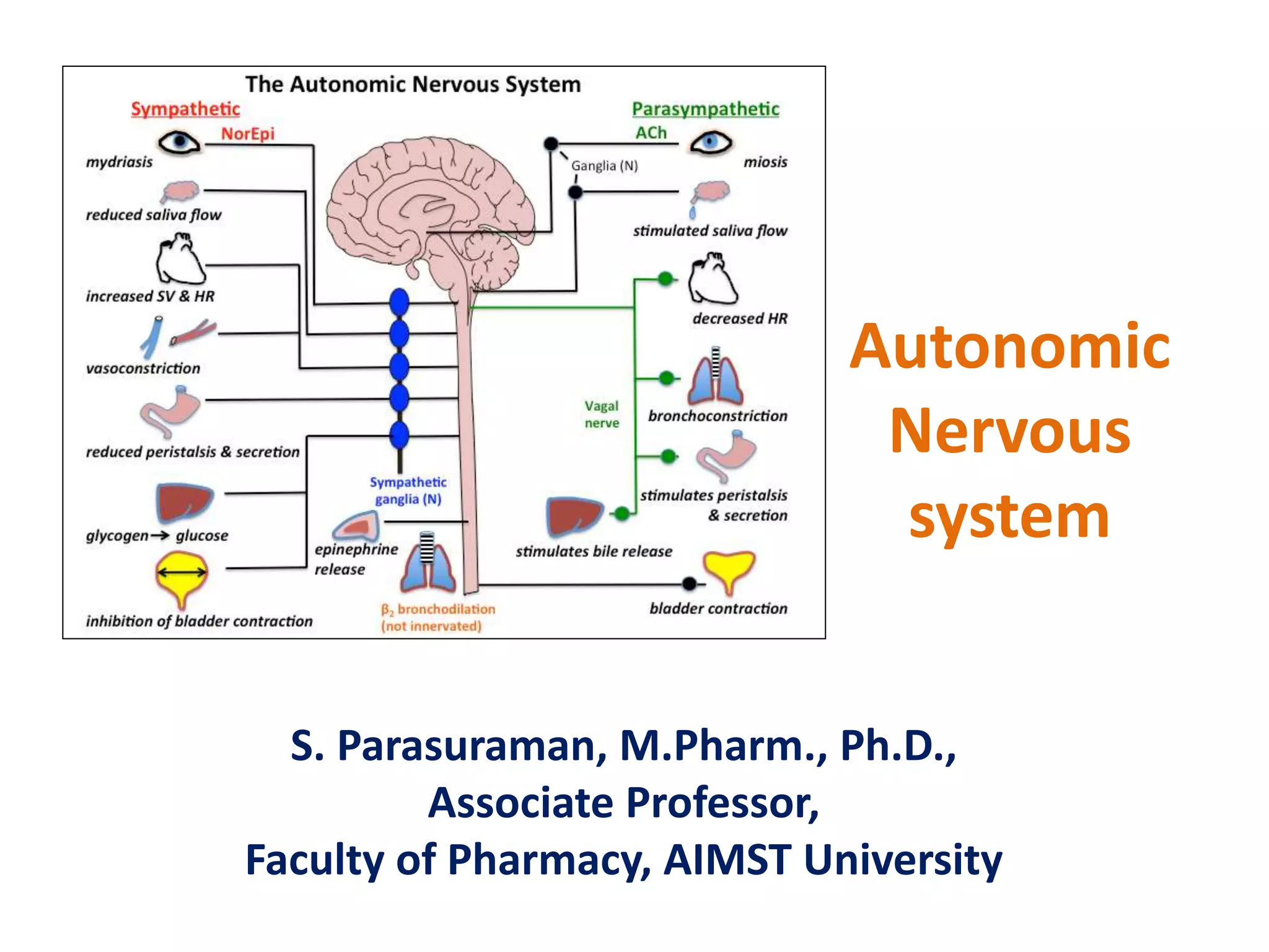


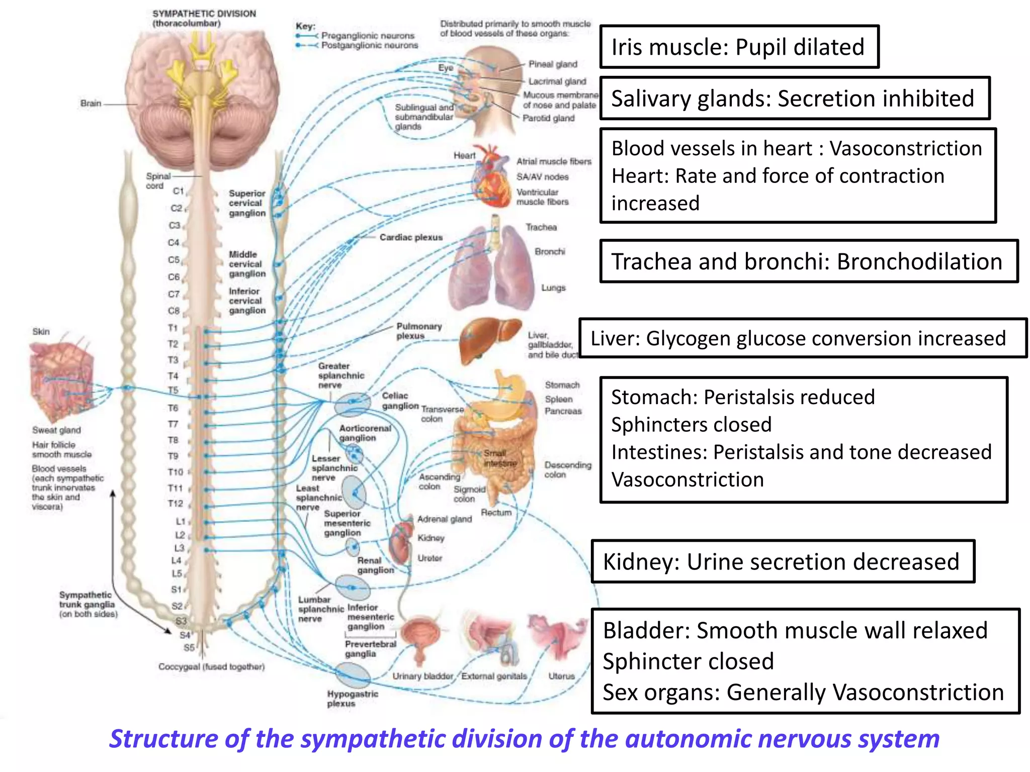


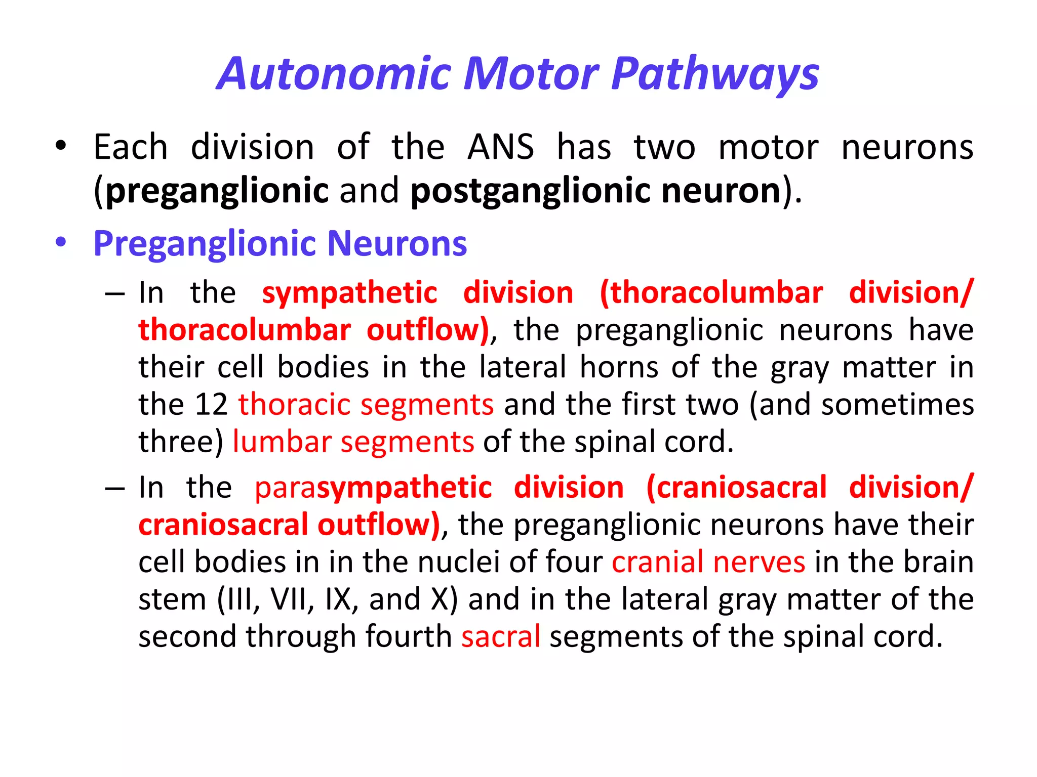


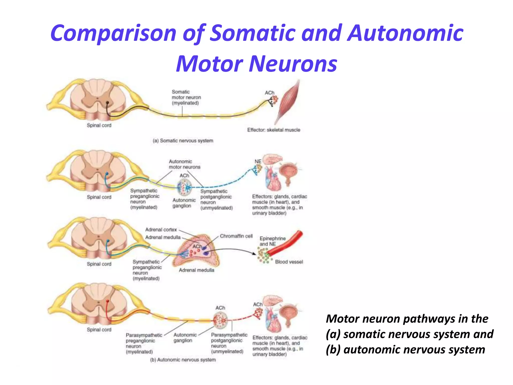


The document provides information about the autonomic nervous system (ANS). It describes the ANS as having two main divisions - the sympathetic and parasympathetic nervous systems. The sympathetic system prepares the body for "fight or flight" responses, while the parasympathetic system allows for "rest and digest" functions. Key differences between the two divisions are described, including their origins in the spinal cord/brain and targets in the body. The pathways of preganglionic and postganglionic neurons, as well as autonomic ganglia, are outlined. Neurotransmitters and receptors of each division are also detailed.


