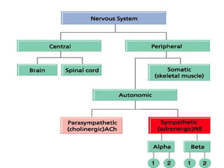
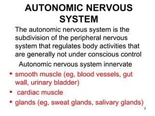
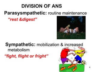
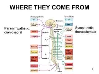
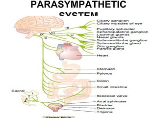
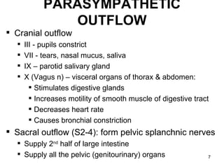
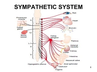
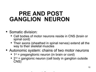
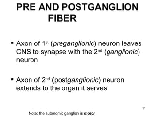
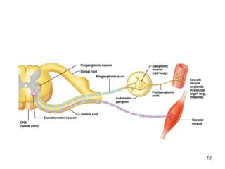
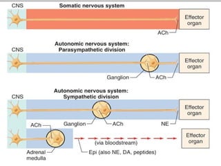
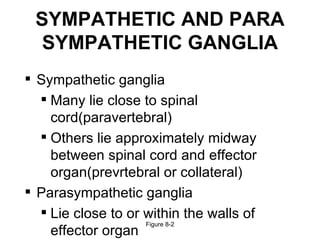
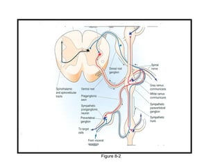
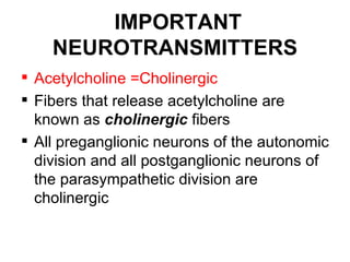
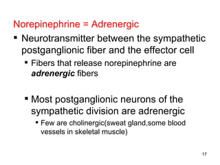
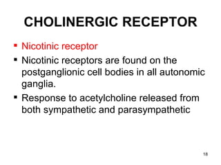
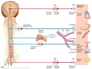
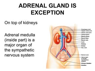
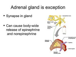
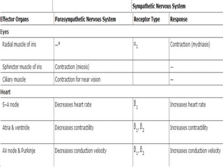
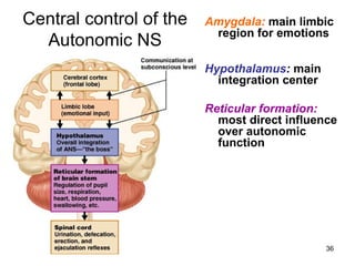
FUNCTIONAL ASPECTS OF THE
AUTONOMIC NERVOUS SYSTEM
AUTONOMIC NERVOUS
          SYSTEM
  The autonomic nervous system is the
  subdivision of the peripheral nervous
  system that regulates body activities that
  are generally not under conscious control
   Autonomic nervous system innervate
 smooth muscle (eg, blood vessels, gut
  wall, urinary bladder)
 cardiac muscle
 glands (eg, sweat glands, salivary glands)
                                               3
DIVISION OF ANS
Parasysmpathetic: routine maintenance
  “rest &digest”


Sympathetic: mobilization & increased
  metabolism
“fight, flight or fright”

                                        4
WHERE THEY COME FROM


Parasympathetic:   Sympathetic:
craniosacral       thoracolumbar


                             5
PARASYMPATHETIC
    SYSTEM


                  6
PARASYMPATHETIC
                OUTFLOW
 Cranial outflow
     III - pupils constrict
     VII - tears, nasal mucus, saliva
     IX – parotid salivary gland
     X (Vagus n) – visceral organs of thorax & abdomen:
         Stimulates digestive glands
         Increases motility of smooth muscle of digestive tract
         Decreases heart rate
         Causes bronchial constriction
 Sacral outflow (S2-4): form pelvic splanchnic nerves
   Supply 2nd half of large intestine
   Supply all the pelvic (genitourinary) organs               7
SYMPATHETIC SYSTEM


                     8
SYMPATHETIC OUTFLOW
                  “fight, flight or fright”
 Also called thoracolumbar system: all its neurons
  are in lateral horn of gray matter from T1-L2

 Lead to every part of the body (unlike parasymp.)
   Easy to remember that when nervous, you sweat; when
    afraid, hair stands on end; when excited blood pressure
    rises (vasoconstriction): these sympathetic only
   Also causes: dry mouth, pupils to dilate, increased heart
    & respiratory rates to increase O2 to skeletal muscles,
    and liver to release glucose


                                                            9
PRE AND POST
  GANGLION NEURON

 Somatic division:
   Cell bodies of motor neurons reside in CNS (brain or
    spinal cord)
   Their axons (sheathed in spinal nerves) extend all the
    way to their skeletal muscles
 Autonomic system: chains of two motor neurons
   1st = preganglionic neuron (in brain or cord)
   2nd = gangionic neuron (cell body in ganglion outside
    CNS)
                                                            10
PRE AND POSTGANGLION
      FIBER

 Axon of 1st (preganglionic) neuron leaves
  CNS to synapse with the 2nd (ganglionic)
  neuron

 Axon of 2nd (postganglionic) neuron
  extends to the organ it serves

                                              11
     Note: the autonomic ganglion is motor
12
13
SYMPATHETIC AND PARA
  SYMPATHETIC GANGLIA
 Sympathetic ganglia
   Many lie close to spinal
    cord(paravertebral)
   Others lie approximately midway
    between spinal cord and effector
    organ(prevrtebral or collateral)
 Parasympathetic ganglia
   Lie close to or within the walls of
                    Figure 8-2
    effector organ
Figure 8-2
IMPORTANT
    NEUROTRANSMITTERS
 Acetylcholine =Cholinergic
 Fibers that release acetylcholine are
  known as cholinergic fibers
 All preganglionic neurons of the autonomic
  division and all postganglionic neurons of
  the parasympathetic division are
  cholinergic
Norepinephrine = Adrenergic
 Neurotransmitter between the sympathetic
  postganglionic fiber and the effector cell
   Fibers that release norepinephrine are
    adrenergic fibers


   Most postganglionic neurons of the
    sympathetic division are adrenergic
      Few are cholinergic(sweat gland,some blood
       vessels in skeletal muscle)

                                                    17
CHOLINERGIC RECEPTOR
 Nicotinic receptor
 Nicotinic receptors are found on the
  postganglionic cell bodies in all autonomic
  ganglia.
 Response to acetylcholine released from
  both sympathetic and parasympathetic


                                            18
 Muscuranic receptor
 found on cell membranes of effectors
  organ (smooth muscle, cardiac muscle,
  and glands)

 they bind with acetylcholine released from
  parasympathetic postganglionic nerve
  fiber


                                           19
ADRENERGIC RECEPTOR
 Alpha receptors are located at
  sympathetic neuroeffector junctions of
  many organs.
 In general, alpha receptors mediate
  excitation or increased activity of the
  effector cells.
 alpha receptor have greater affinity for
  norepinephrine
                                             20
TYPES OF α-ADRENERGIC
                RECEPTOR
 α-adrenergic receptors are adrenergic receptors
  that respond to norepinephrine
 They are subdivided into two types:
 α1, found in smooth muscle, heart, and liver, with
  effects including vasoconstriction, intestinal
  relaxation, uterine contraction and pupillary dilation,
 α2, found in platelets, vascular smooth muscle,
  nerve termini, and pancreatic islets, with effects
  including platelet aggregation, vasoconstriction, and
  inhibition of norepinephrine release and of insulin
  secretion.
Beta receptors are also located
postsynaptically at sympathetic
neuroeffector junctions of many organs.

 beta receptors mediate relaxation or
decreased activity of the effector cells.

Heart muscle is an important exception to
this rule. Activation of beta adrenoceptors
in heart increases the automaticity and
contractility of all parts of the heart.
β-RECEPTOR TYPES
 β-adrenergic receptors respond particularly
  to epinephrine and nor epinephrine
 There are three known types of beta receptor,
  designated β1, β2 and β3.
 β1-Adrenergic receptors are located mainly in
  the heart. Sensitive to both EN and NOR EN
 β2-Adrenergic receptors are located mainly in
  the lungs, gastrointestinal tract, liver, uterus,
  vascular smooth muscle, and skeletal muscle
   sensitive to EN only.
 β3-receptors are located in fat cells.
24
ADRENAL GLAND IS
           EXCEPTION
On top of kidneys

Adrenal medulla
(inside part) is a
major organ of
the sympathetic
nervous system

                           25
Adrenal gland is exception
 Synapse in gland

 Can cause body-wide
  release of epinephrine
   and norepinephrine


                                 26
27
28
29
30
31
32
 SYMPATHETIC PARASYMPATHETIC
  TONE
 DUAL INNERVATION
 MASS DISCHARGE(ALARM OR STRESS
  RESPONSE)
 LOCAL RESPONSE
 SOME EXCEPTIONS: BLOOD VESSELS
  SWEAT GLAND, SALIVARY GLAND
                               33
VISCERAL REFLEXES
 These are the simplest functions of the ANS.
 Each visceral reflex arc consists of a receptor, a sensory
  neuron, an interneuron, and two visceral motor neurons.
 All visceral reflexes are polysynaptic.
 PS reflexes include : gastric and intestinal reflexes,
  defecation, urination, direct light reflexes, swallowing
  reflex, coughing reflex, baroreceptor reflex and sexual
  arousal.
 S reflexes:         cardioaccelaratory reflex, vasomotor
  reflex, pupillary reflex and ejaculation (in males).
VISCERAL REFLEX ARCS


                                                                                   35
*e.g. “enteric” nervous system: 3 neuron reflex arcs entirely within the wall of the gut
Central control of the   Amygdala: main limbic
                          region for emotions
  Autonomic NS
                         Hypothalamus: main
                           integration center

                         Reticular formation:
                           most direct influence
                           over autonomic
                           function


                                                36

autonomic nervous system by DR.IFRA

  • 1.
    FUNCTIONAL ASPECTS OFTHE AUTONOMIC NERVOUS SYSTEM
  • 3.
    AUTONOMIC NERVOUS SYSTEM The autonomic nervous system is the subdivision of the peripheral nervous system that regulates body activities that are generally not under conscious control Autonomic nervous system innervate  smooth muscle (eg, blood vessels, gut wall, urinary bladder)  cardiac muscle  glands (eg, sweat glands, salivary glands) 3
  • 4.
    DIVISION OF ANS Parasysmpathetic:routine maintenance “rest &digest” Sympathetic: mobilization & increased metabolism “fight, flight or fright” 4
  • 5.
    WHERE THEY COMEFROM Parasympathetic: Sympathetic: craniosacral thoracolumbar 5
  • 6.
  • 7.
    PARASYMPATHETIC OUTFLOW  Cranial outflow  III - pupils constrict  VII - tears, nasal mucus, saliva  IX – parotid salivary gland  X (Vagus n) – visceral organs of thorax & abdomen:  Stimulates digestive glands  Increases motility of smooth muscle of digestive tract  Decreases heart rate  Causes bronchial constriction  Sacral outflow (S2-4): form pelvic splanchnic nerves  Supply 2nd half of large intestine  Supply all the pelvic (genitourinary) organs 7
  • 8.
  • 9.
    SYMPATHETIC OUTFLOW “fight, flight or fright”  Also called thoracolumbar system: all its neurons are in lateral horn of gray matter from T1-L2  Lead to every part of the body (unlike parasymp.)  Easy to remember that when nervous, you sweat; when afraid, hair stands on end; when excited blood pressure rises (vasoconstriction): these sympathetic only  Also causes: dry mouth, pupils to dilate, increased heart & respiratory rates to increase O2 to skeletal muscles, and liver to release glucose 9
  • 10.
    PRE AND POST GANGLION NEURON  Somatic division:  Cell bodies of motor neurons reside in CNS (brain or spinal cord)  Their axons (sheathed in spinal nerves) extend all the way to their skeletal muscles  Autonomic system: chains of two motor neurons  1st = preganglionic neuron (in brain or cord)  2nd = gangionic neuron (cell body in ganglion outside CNS) 10
  • 11.
    PRE AND POSTGANGLION FIBER  Axon of 1st (preganglionic) neuron leaves CNS to synapse with the 2nd (ganglionic) neuron  Axon of 2nd (postganglionic) neuron extends to the organ it serves 11 Note: the autonomic ganglion is motor
  • 12.
  • 13.
  • 14.
    SYMPATHETIC AND PARA SYMPATHETIC GANGLIA  Sympathetic ganglia  Many lie close to spinal cord(paravertebral)  Others lie approximately midway between spinal cord and effector organ(prevrtebral or collateral)  Parasympathetic ganglia  Lie close to or within the walls of Figure 8-2 effector organ
  • 15.
  • 16.
    IMPORTANT NEUROTRANSMITTERS  Acetylcholine =Cholinergic  Fibers that release acetylcholine are known as cholinergic fibers  All preganglionic neurons of the autonomic division and all postganglionic neurons of the parasympathetic division are cholinergic
  • 17.
    Norepinephrine = Adrenergic Neurotransmitter between the sympathetic postganglionic fiber and the effector cell  Fibers that release norepinephrine are adrenergic fibers  Most postganglionic neurons of the sympathetic division are adrenergic  Few are cholinergic(sweat gland,some blood vessels in skeletal muscle) 17
  • 18.
    CHOLINERGIC RECEPTOR  Nicotinicreceptor  Nicotinic receptors are found on the postganglionic cell bodies in all autonomic ganglia.  Response to acetylcholine released from both sympathetic and parasympathetic 18
  • 19.
     Muscuranic receptor found on cell membranes of effectors organ (smooth muscle, cardiac muscle, and glands)  they bind with acetylcholine released from parasympathetic postganglionic nerve fiber 19
  • 20.
    ADRENERGIC RECEPTOR  Alphareceptors are located at sympathetic neuroeffector junctions of many organs.  In general, alpha receptors mediate excitation or increased activity of the effector cells.  alpha receptor have greater affinity for norepinephrine 20
  • 21.
    TYPES OF α-ADRENERGIC RECEPTOR  α-adrenergic receptors are adrenergic receptors that respond to norepinephrine  They are subdivided into two types:  α1, found in smooth muscle, heart, and liver, with effects including vasoconstriction, intestinal relaxation, uterine contraction and pupillary dilation,  α2, found in platelets, vascular smooth muscle, nerve termini, and pancreatic islets, with effects including platelet aggregation, vasoconstriction, and inhibition of norepinephrine release and of insulin secretion.
  • 22.
    Beta receptors arealso located postsynaptically at sympathetic neuroeffector junctions of many organs.  beta receptors mediate relaxation or decreased activity of the effector cells. Heart muscle is an important exception to this rule. Activation of beta adrenoceptors in heart increases the automaticity and contractility of all parts of the heart.
  • 23.
    β-RECEPTOR TYPES  β-adrenergicreceptors respond particularly to epinephrine and nor epinephrine  There are three known types of beta receptor, designated β1, β2 and β3.  β1-Adrenergic receptors are located mainly in the heart. Sensitive to both EN and NOR EN  β2-Adrenergic receptors are located mainly in the lungs, gastrointestinal tract, liver, uterus, vascular smooth muscle, and skeletal muscle sensitive to EN only.  β3-receptors are located in fat cells.
  • 24.
  • 25.
    ADRENAL GLAND IS EXCEPTION On top of kidneys Adrenal medulla (inside part) is a major organ of the sympathetic nervous system 25
  • 26.
    Adrenal gland isexception  Synapse in gland  Can cause body-wide release of epinephrine and norepinephrine 26
  • 27.
  • 28.
  • 29.
  • 30.
  • 31.
  • 32.
  • 33.
     SYMPATHETIC PARASYMPATHETIC TONE  DUAL INNERVATION  MASS DISCHARGE(ALARM OR STRESS RESPONSE)  LOCAL RESPONSE  SOME EXCEPTIONS: BLOOD VESSELS SWEAT GLAND, SALIVARY GLAND 33
  • 34.
    VISCERAL REFLEXES  Theseare the simplest functions of the ANS.  Each visceral reflex arc consists of a receptor, a sensory neuron, an interneuron, and two visceral motor neurons.  All visceral reflexes are polysynaptic.  PS reflexes include : gastric and intestinal reflexes, defecation, urination, direct light reflexes, swallowing reflex, coughing reflex, baroreceptor reflex and sexual arousal.  S reflexes: cardioaccelaratory reflex, vasomotor reflex, pupillary reflex and ejaculation (in males).
  • 35.
    VISCERAL REFLEX ARCS 35 *e.g. “enteric” nervous system: 3 neuron reflex arcs entirely within the wall of the gut
  • 36.
    Central control ofthe Amygdala: main limbic region for emotions Autonomic NS Hypothalamus: main integration center Reticular formation: most direct influence over autonomic function 36

Editor's Notes