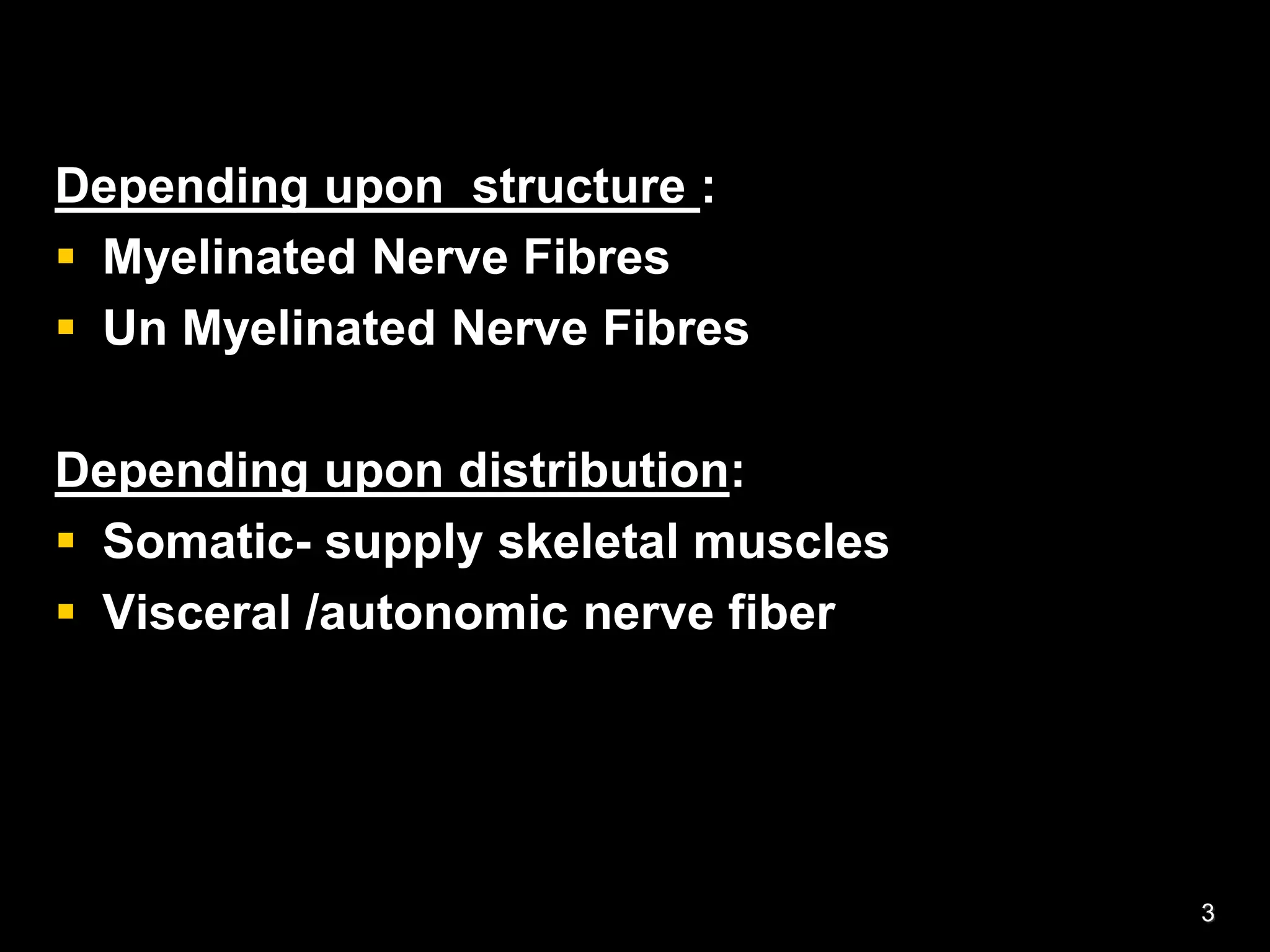
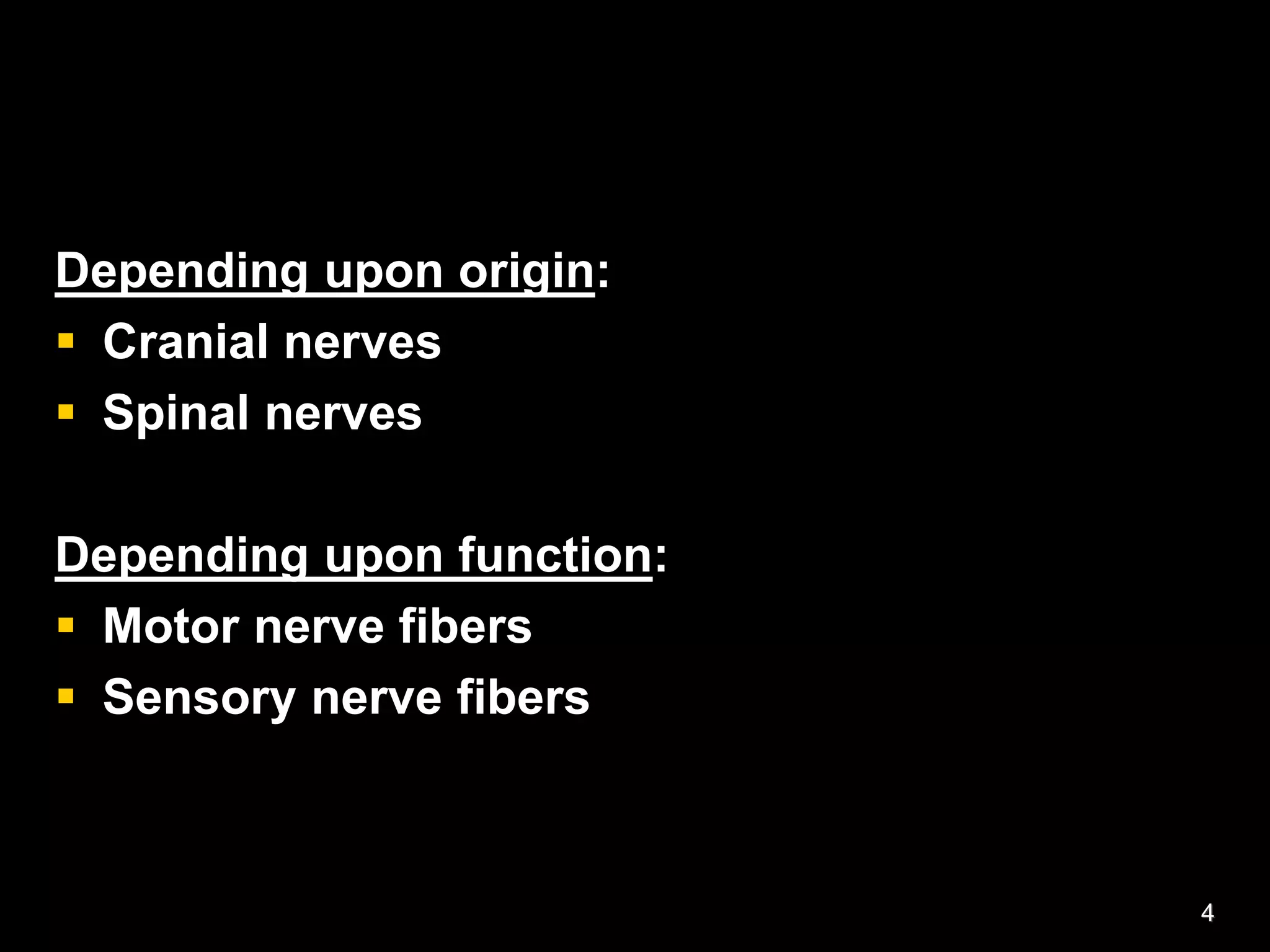
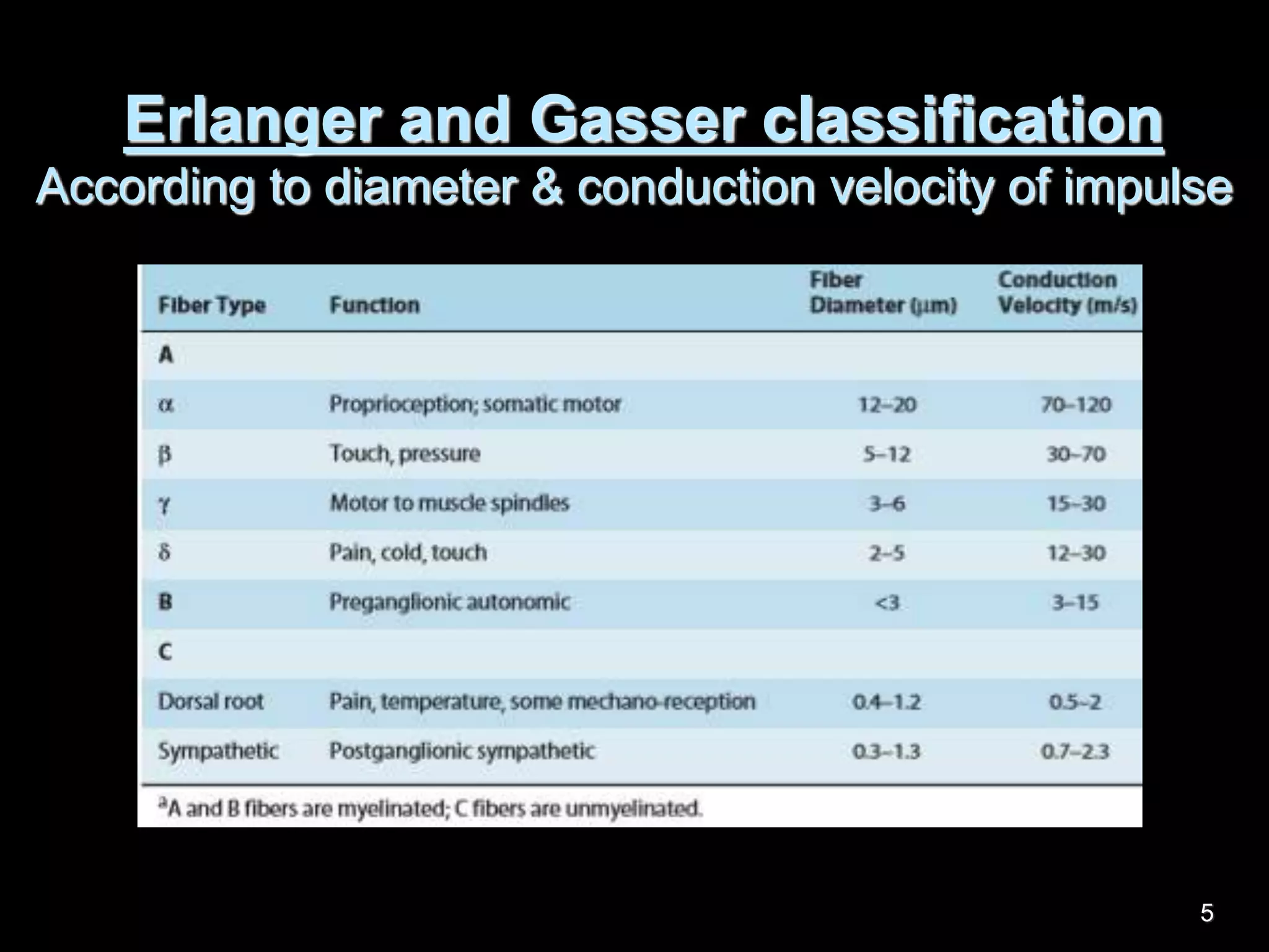
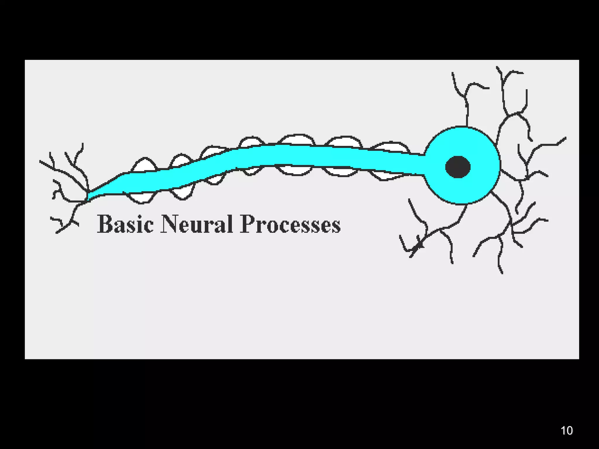
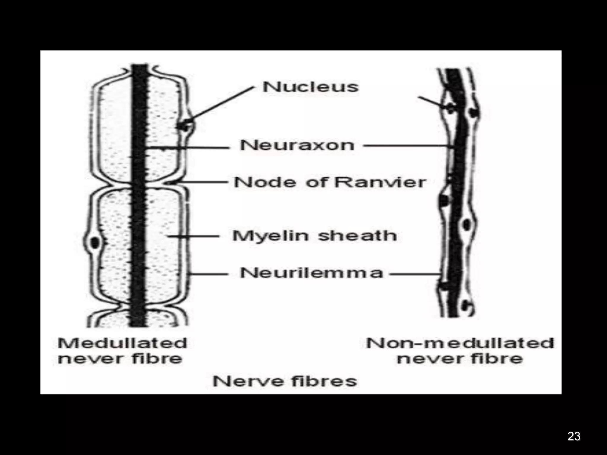
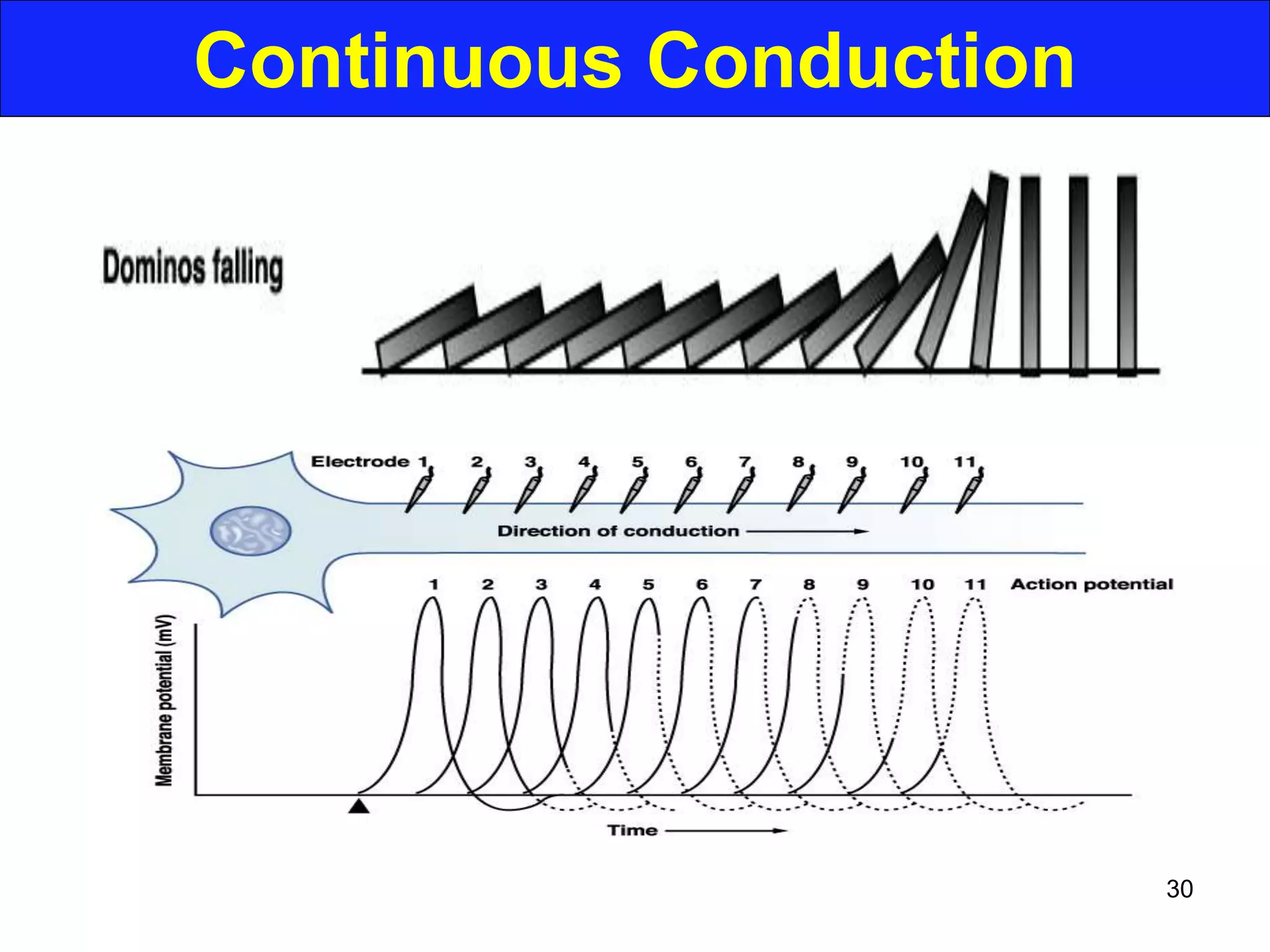
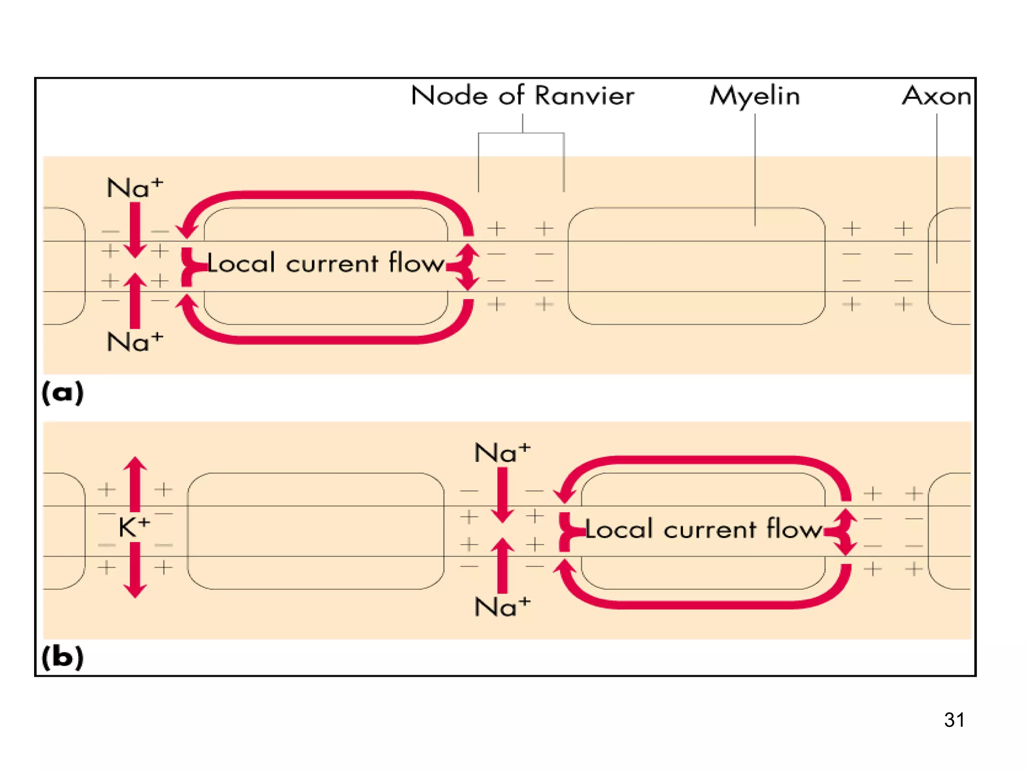
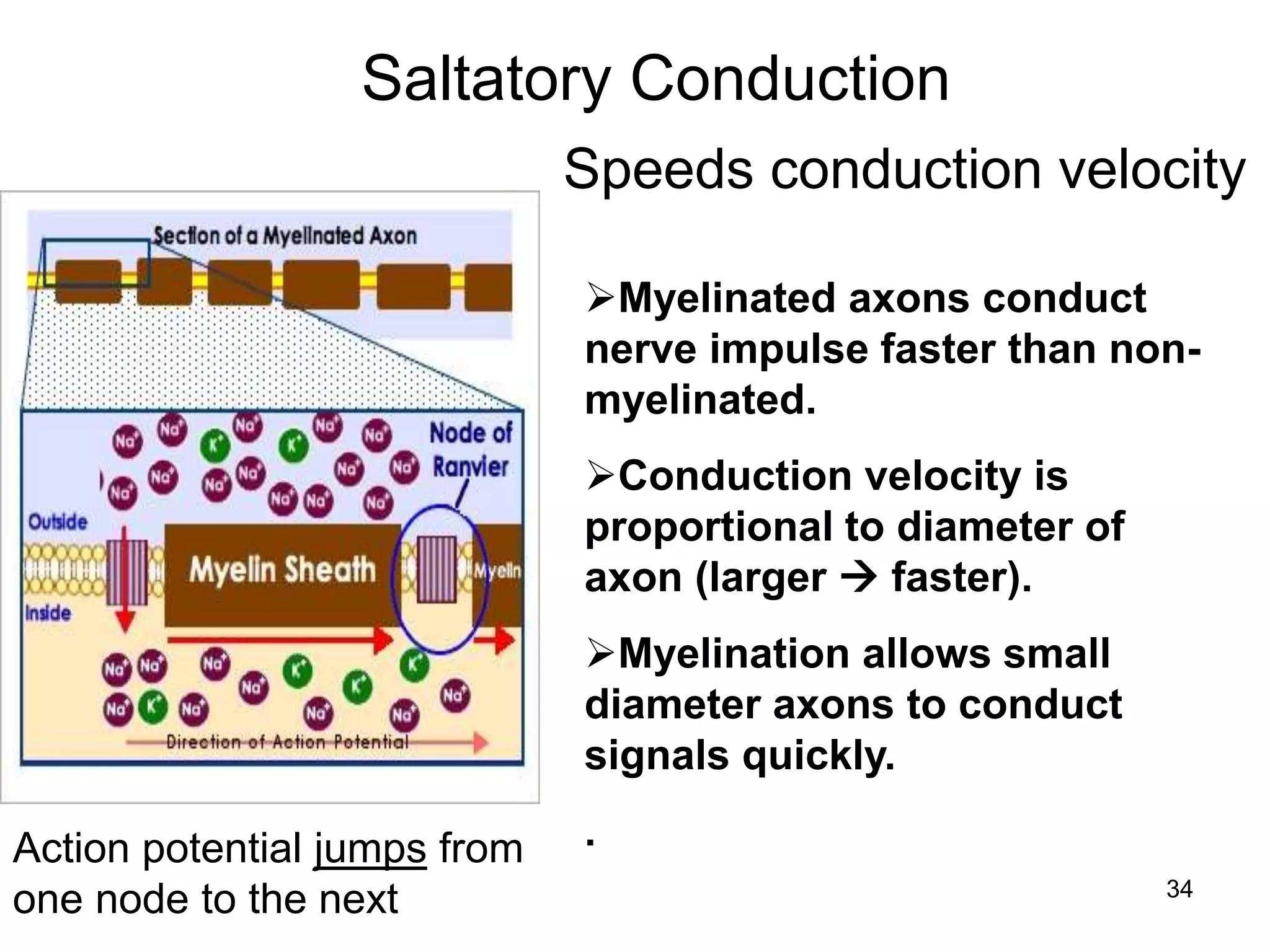
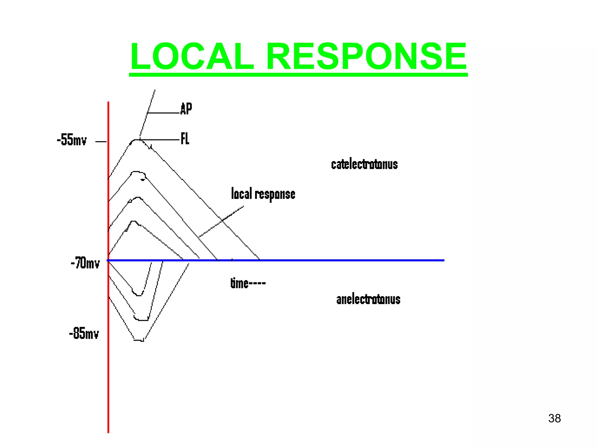
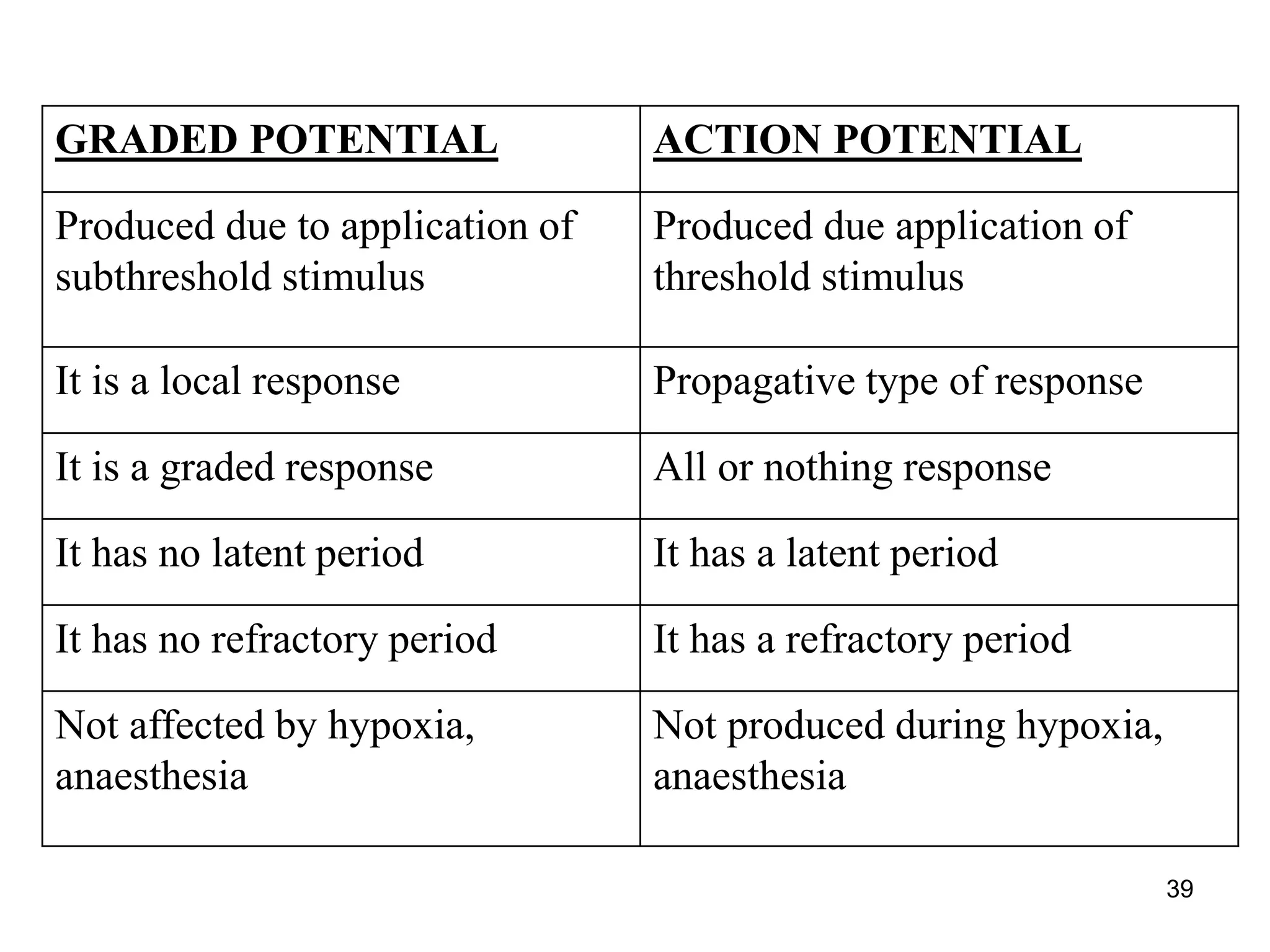
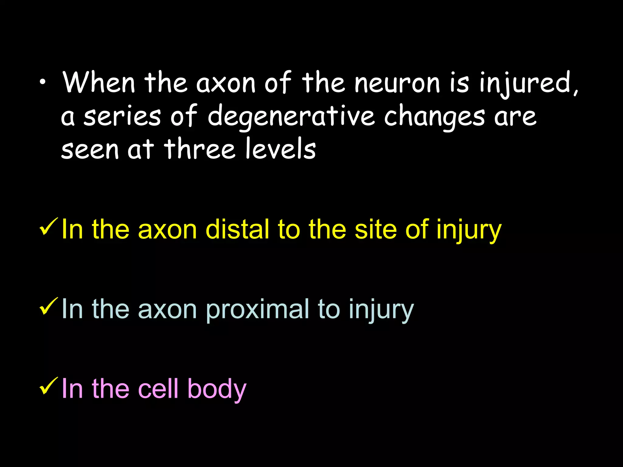
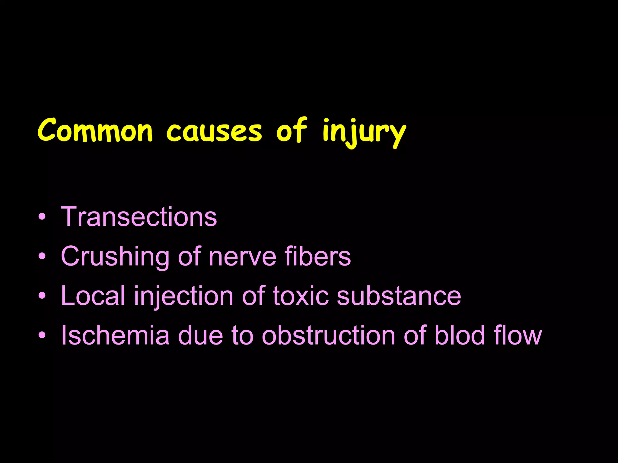
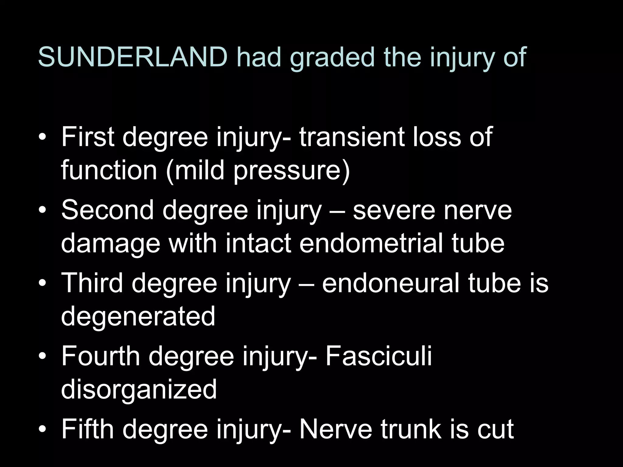
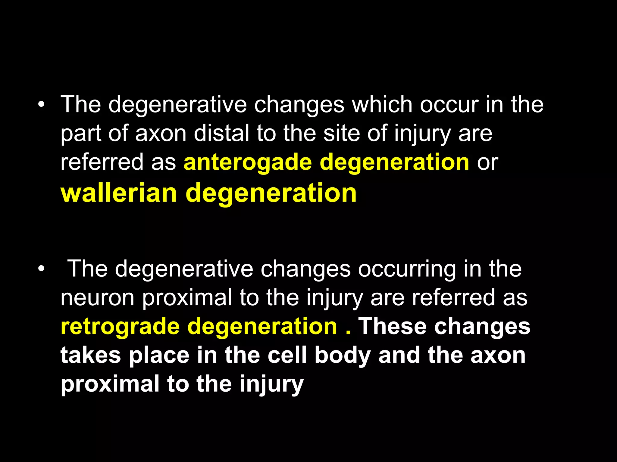
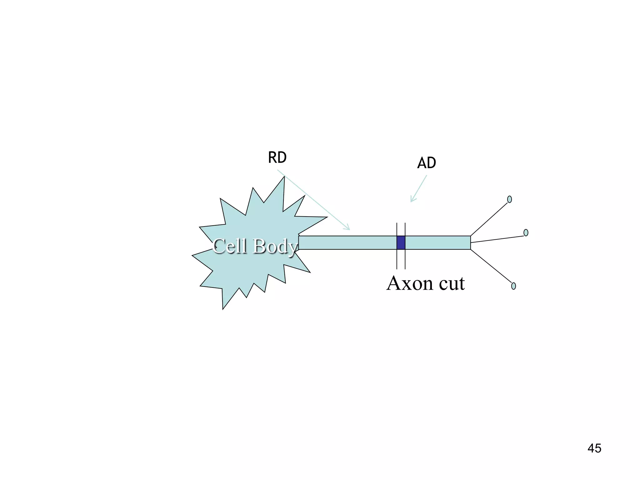
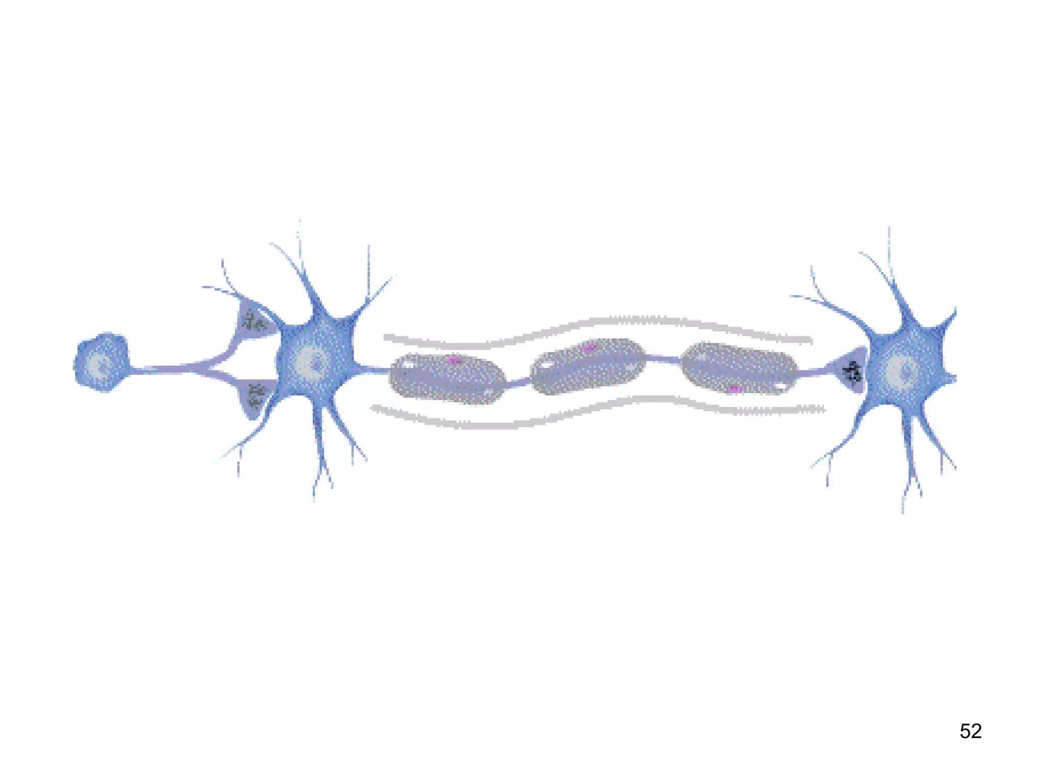
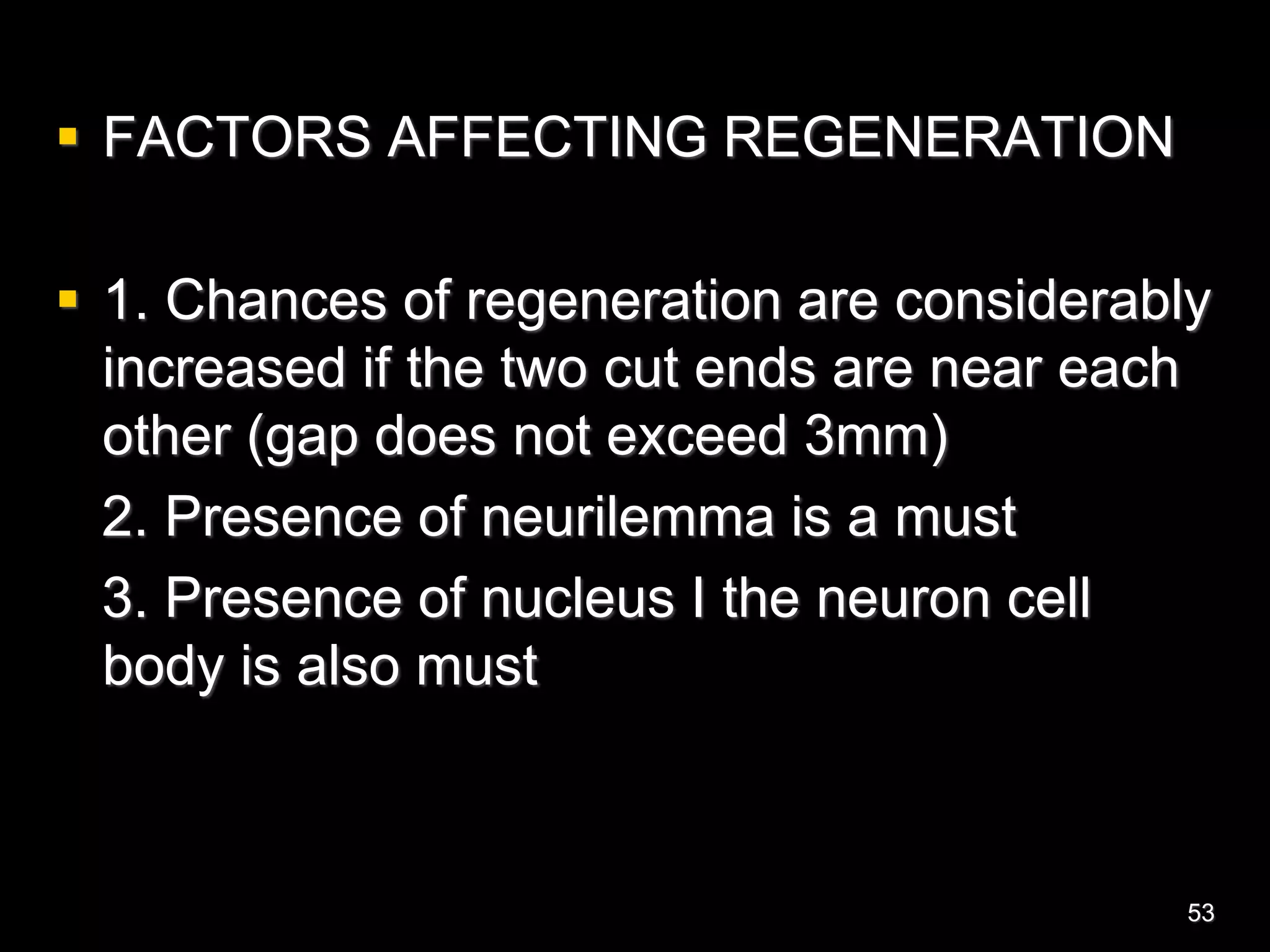
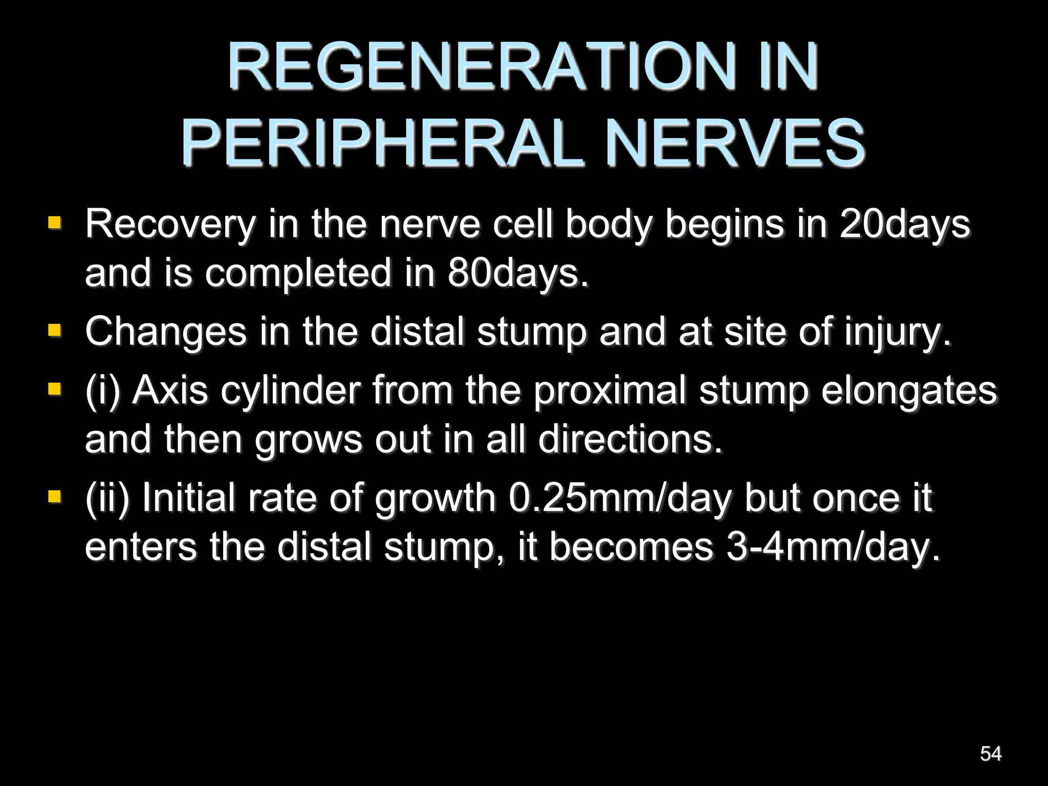
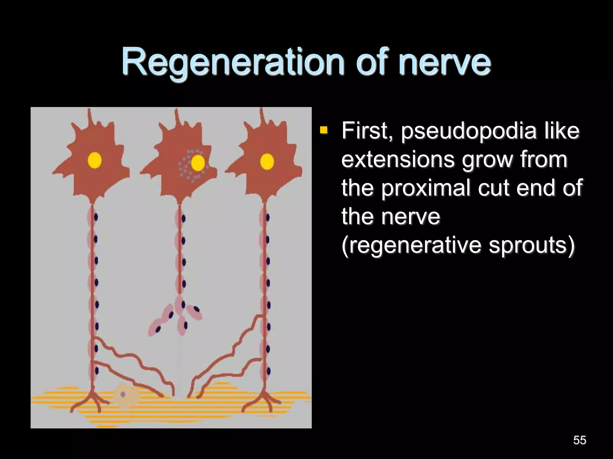
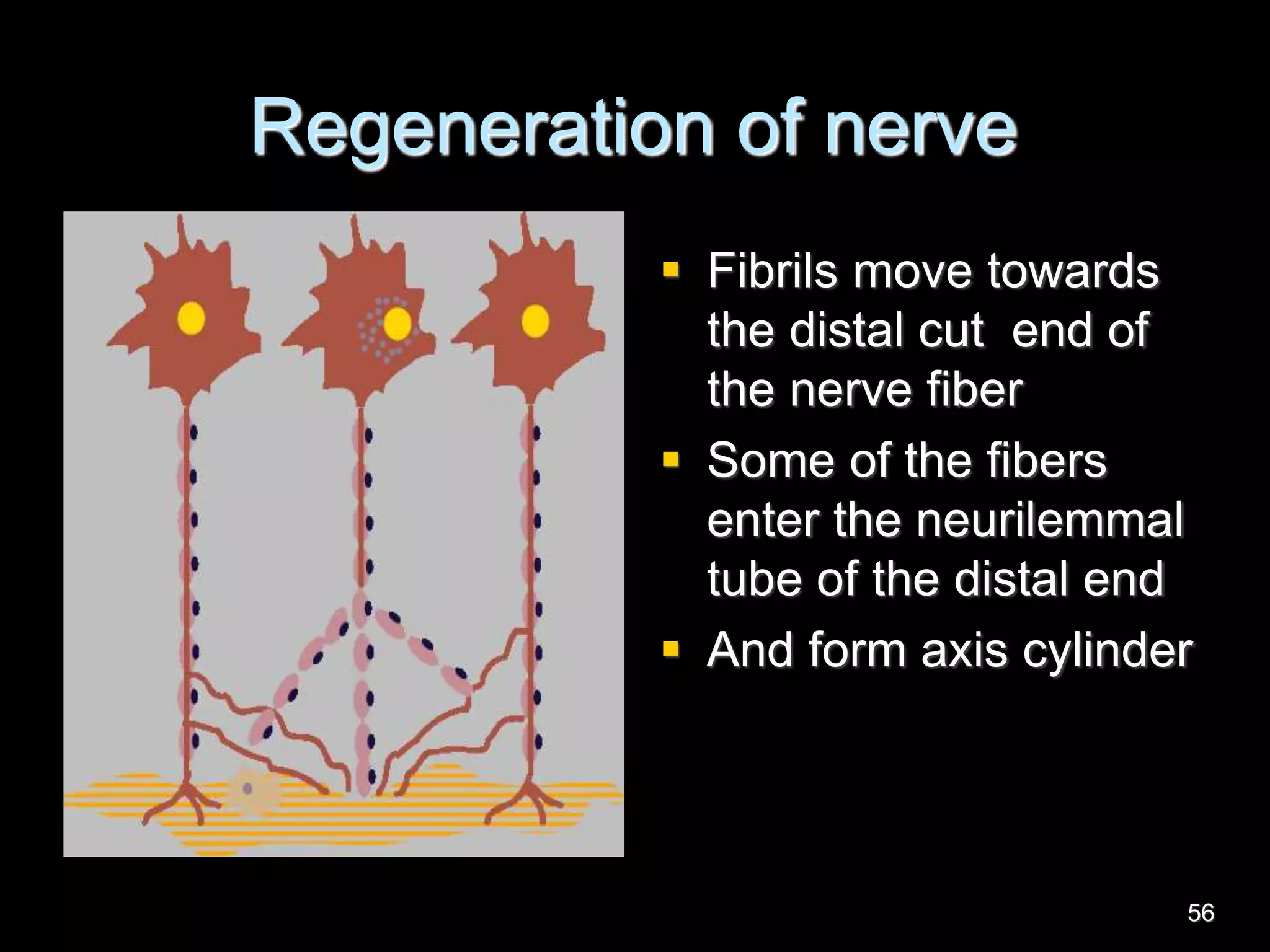
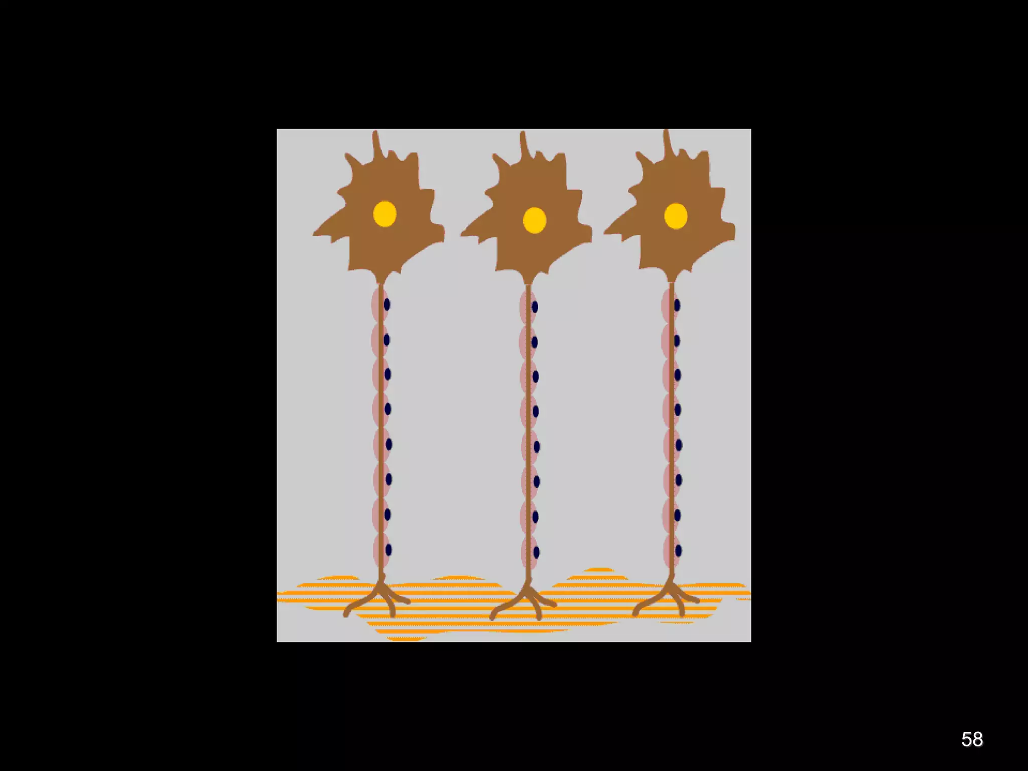
Nerve fibers are classified based on their structure, distribution, origin and function. They can be myelinated or unmyelinated. When injured, the distal portion undergoes Wallerian degeneration over 3 months as the axon and myelin sheath break down. The cell body shows chromatolysis. Regeneration is possible if the nerve ends are close together. New axonal growth occurs rapidly, entering the distal stump at 3-4mm/day. Myelination resumes over a year. Though anatomy recovers, full function returns slowly.
17. RHEOBASE- minimum strength (voltage)
of stimulus which can excite the tissue,
whatever may be the duration of the
stimulus
UTILIZATION TIME- Minimum time
required for a rheobasic current (threshold
strength) to excite the tissue.
CHRONAXIE- Minimum time at which the
stimulus with double the rheobasic
strength (Voltage) can excite the tissue.
17
18. Chronaxie
Is an index of excitability of a tissue
Can be used to compare the excitability
of the various tissues
Longer the chronaxie lesser is the
excitability
18
19. Factors affecting Excitability
1) Temperature
2) Mechanical pressure
3) Blood supply
4) Chemicals- CO2 & narcotics
5) pH- increased excitability in alkaline and
reduced excitability in acidic media.
6) Ions- Na+, Mg++ and K+ are neuro-
excitatory and Ca++ is neurosedative
19
20. Nerve and muscle are excitable
Capable of self-generation of
electrochemical impulses at
their membrane
Transmit signals along the
membrane
20
21. CONDUCTIVITY
Ability to conduct an impulse is called
Conductivity.
TWO TYPES:
Orthodromic conduction
Antidromic conduction
21
22. Factors affecting Conductivity
Temperature
Mechanical pressure
Blood supply
Chemicals
pH
Ions
Size of the nerve
Myelination
22
24. ALL OR NONE RESPONSE
A single nerve fiber obeys “all or none law”
When stimulus of sub-threshold intensity is
applied to the axon , then no action potential is
produced (NONE RESPONSE)
A response in the spike of action potential is
observed when the stimulus if of threshold intensity
There occurs no increase in the magnitude of
action potential when the strength of stimulus is
more than the threshold level (ALL RESPONSE) 24
25. REFRACTORY PERIOD
The period of time during
which an excitable cell
cannot generate another
action potential is called the
refractory period.
25
26. 1) Absolute refractory period-
it is the period during an action potential,
during which a second stimulus can’t
produce a second response.
period of action potential from firing level
until repolarization is almost 1/3rd
complete (spike potential)
26
27. 2) Relative refractory period-
it is the period during an action
potential, during which a stimulus of
higher intensity can produce a second
response
It extends FROM the end of absolute
refractory period to the start of after
depolarization phase of action potential
27
29. CONDUCTION OF IMPULSES IN THE
NERVE FIBRE
• Unmyelinated neurons undergo the
Continuous conduction of an AP.
• Myelinated neurons undergo Saltatory
conduction of an AP.
29
32. • There are two advantages of saltatory
conduction.
1.Energy is saved as sodium-potassium
pumps are only required at specific points
along the axon.
2. Conduction of an action potential is much
faster within a myelinated axon (around
120 m/sec as opposed to around 35 m/sec)
in unmyelinated ones.
32
34. Myelinated axons conduct
nerve impulse faster than non-
myelinated.
Conduction velocity is
proportional to diameter of
axon (larger faster).
Myelination allows small
diameter axons to conduct
signals quickly.
.
Saltatory Conduction
Action potential jumps from
one node to the next
Speeds conduction velocity
34
35. – one node of Ranvier depolarizes the next,
so that action potentials can skip between
nodes
35
39. GRADED POTENTIAL ACTION POTENTIAL
Produced due to application of
subthreshold stimulus
Produced due application of
threshold stimulus
It is a local response Propagative type of response
It is a graded response All or nothing response
It has no latent period It has a latent period
It has no refractory period It has a refractory period
Not affected by hypoxia,
anaesthesia
Not produced during hypoxia,
anaesthesia
39
41. • When the axon of the neuron is injured,
a series of degenerative changes are
seen at three levels
In the axon distal to the site of injury
In the axon proximal to injury
In the cell body
41
42. Common causes of injury
• Transections
• Crushing of nerve fibers
• Local injection of toxic substance
• Ischemia due to obstruction of blod flow
42
43. SUNDERLAND had graded the injury of
• First degree injury- transient loss of
function (mild pressure)
• Second degree injury – severe nerve
damage with intact endometrial tube
• Third degree injury – endoneural tube is
degenerated
• Fourth degree injury- Fasciculi
disorganized
• Fifth degree injury- Nerve trunk is cut 43
44. • The degenerative changes which occur in the
part of axon distal to the site of injury are
referred as anterogade degeneration or
wallerian degeneration
• The degenerative changes occurring in the
neuron proximal to the injury are referred as
retrograde degeneration . These changes
takes place in the cell body and the axon
proximal to the injury
44
46. • The degenerative changes
start within few hours of
injury and continue for about
3 months
46
47. CHANGES IN THE PART OF THE AXON DISTAL
TO INJURY
• Axis cylinder becomes swollen and breaks
up into small pieces
• After few days, the broken pieces appear as
debris in the space occupied by the axis
cylinder
• The myelin sheath slowly disintegrates into
fat droplets (8th to 35 day)
• Neurilemmal sheath is unaffected 47
48. • Schwann cell multiply rapidly
• The macrophages remove the debris of
axis cylinder and the fat droplets of
disintegrated myelin sheath
• Neurilemmal tube becomes empty
• Changes takes place for about 2 months
from day of injury
48
49. Changes in the action
potential and the ability of
the nerve to conduct an
impulse decreases
markedly.
49
50. Changes in the cell bodyCHANGES IN THE CELL BODY OF THE
NEURON (start within 48 hours and
continue for 15-20 days)
Nissl substances undergo disintegration
and dissolution (chromatolysis)
Golgi apparatus, mitochondria and
neurofibrils are fragmented and
eventually disappear
50
51. Changes in the cell body
Cell body draws in more fluid, enlarges
and become spherical
Nucleus is displaced to the periphery ,
sometimes extruded out of the cell
51
53. FACTORS AFFECTING REGENERATION
1. Chances of regeneration are considerably
increased if the two cut ends are near each
other (gap does not exceed 3mm)
2. Presence of neurilemma is a must
3. Presence of nucleus I the neuron cell
body is also must
53
54. REGENERATION IN
PERIPHERAL NERVES
Recovery in the nerve cell body begins in 20days
and is completed in 80days.
Changes in the distal stump and at site of injury.
(i) Axis cylinder from the proximal stump elongates
and then grows out in all directions.
(ii) Initial rate of growth 0.25mm/day but once it
enters the distal stump, it becomes 3-4mm/day.
54
55. Regeneration of nerve
First, pseudopodia like
extensions grow from
the proximal cut end of
the nerve
(regenerative sprouts)
55
56. Regeneration of nerve
Fibrils move towards
the distal cut end of
the nerve fiber
Some of the fibers
enter the neurilemmal
tube of the distal end
And form axis cylinder
56
57. In about 15 days, schwann cell starts laying down
myelin sheath round the successful fibril, which is
completed within one year.
In the nerve cell body , first the nissl granules
appear followed by golgi apparatus
cell looses excess fluid .
The nucleus occupies the central portion
Though anatomical regeneration occur the
functional recovery occurs after long period
57