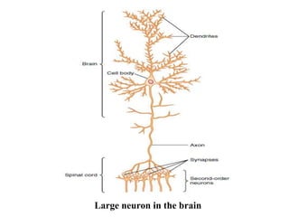
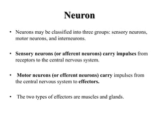
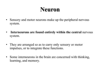
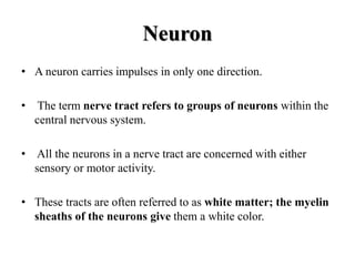
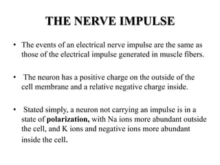
NERVOUS SYSTEM
PREPARED BY :
VANARAJ S. RABARI
M.PHARM (PHARMACOLOGY)
NERVOUS SYSTEM
• The nervous system is unique in the vast complexity of
thought processes and control actions it can perform.
• It receives each minute literally millions of bits of information
from the different sensory nerves and sensory organs and then
integrates all these to determine responses to be made by the
body.
• The nervous system is one of the regulating systems.
NERVOUS SYSTEM
• Electrochemical impulses of the nervous system make it
possible to obtain information about the external or internal
environment and do whatever is necessary to maintain
homeostasis.
• The nervous system is divided into two parts :
1. central nervous system (CNS)
2. peripheral nervous system (PNS)
NERVOUS SYSTEM
• The central nervous system (CNS) consists of the brain and
spinal cord.
• The peripheral nervous system (PNS) consists of cranial
nerves and spinal nerves.
• The PNS includes the autonomic nervous system (ANS).
• The peripheral nervous system relays information to and from
the central nervous system, and the brain is the center of
activity that integrates this information, initiates responses, and
makes us the individuals we are.
Neuron
• Nerve cells are called neurons, or nerve fibers and Neuron is
the basic functional unit of the central nervous system.
• Whatever their specific functions, all neurons have the same
physical parts.
• The cell body contains the nucleus and is essential for the
continued life of the neuron.
Neuron
• The central nervous system contains more than 100 billion
neurons.
• Dendrites are processes (extensions) that transmit impulses
toward the cell body. The one axon of a neuron transmits
impulses away from the cell body.
• In the central nervous system, the myelin sheaths are formed
by oligodendrocytes, one of the neuroglia (glial cells), the
specialized cells found only in the brain and spinal cord.
Large neuron in the brain
Neuron
• Another kind of neuroglia are microglia, which are
constantly moving, phagocytizing cellular debris, damaged
cells, and pathogens.
• The small gap or space between the axon of one neuron and
the dendrites or cell body of the next neuron is called the
synapse.
Neuron
• Neurons may be classified into three groups: sensory neurons,
motor neurons, and interneurons.
• Sensory neurons (or afferent neurons) carry impulses from
receptors to the central nervous system.
• Motor neurons (or efferent neurons) carry impulses from
the central nervous system to effectors.
• The two types of effectors are muscles and glands.
Neuron
• Sensory and motor neurons make up the peripheral nervous
system.
• Interneurons are found entirely within the central nervous
system.
• They are arranged so as to carry only sensory or motor
impulses, or to integrate these functions.
• Some interneurons in the brain are concerned with thinking,
learning, and memory.
Neuron
• A neuron carries impulses in only one direction.
• The term nerve tract refers to groups of neurons within the
central nervous system.
• All the neurons in a nerve tract are concerned with either
sensory or motor activity.
• These tracts are often referred to as white matter; the myelin
sheaths of the neurons give them a white color.
THE NERVE IMPULSE
• The events of an electrical nerve impulse are the same as
those of the electrical impulse generated in muscle fibers.
• The neuron has a positive charge on the outside of the
cell membrane and a relative negative charge inside.
• Stated simply, a neuron not carrying an impulse is in a
state of polarization, with Na ions more abundant outside
the cell, and K ions and negative ions more abundant
inside the cell.
THE NERVE IMPULSE
• A stimulus (such as a neurotransmitter) makes the membrane
very permeable to Na ions, which rush into the cell. This
brings about depolarization.
• As soon as depolarization takes place, the neuron membrane
becomes very permeable to K ions, which rush out of the cell.
This restores the positive charge outside and the negative
charge inside, and is called repolarization.
• The term action potential refers to depolarization followed by
repolarization.
THE SPINAL CORD
• The spinal cord transmits impulses to and from the brain
and is the integrating center for the spinal cord reflexes.
• Enclosed within the vertebral canal and the meninges, the
spinal cord is well protected from mechanical injury.
• In length, the spinal cord extends from the foramen magnum
of the occipital bone to the disc between the first and second
lumbar vertebrae.
THE SPINAL CORD
• Ascending tracts (such as the dorsal columns and
spinothalamic tracts) carry sensory impulses to the brain.
• Descending tracts (such as the corticospinal and rubrospinal
tracts) carry motor impulses away from the brain.
• central canal; this contains cerebrospinal fluid and is
continuous with cavities in the brain called ventricles.
SPINAL CORD
THE BRAIN
• The major parts are the medulla, pons, and midbrain
(collectively called the brain stem), the cerebellum, the
hypothalamus, the thalamus, and the cerebrum.
Parts of brain
VENTRICLES
• The ventricles are four cavities within the brain: two lateral
ventricles, the third ventricle, and the fourth ventricle.
• Each ventricle contains a capillary network called a choroid
plexus, which forms cerebrospinal fluid (CSF) from blood
plasma.
• Cerebrospinal fluid is the tissue fluid of the central nervous
system.
MEDULLA
• The medulla extends from the spinal cord to the pons and is
anterior to the cerebellum.
• The medulla contains cardiac centers that regulate heart rate,
vasomotor centers that regulate the diameter of blood vessels
and, thereby, blood pressure, and respiratory centers that
regulate breathing.
PONS
• The pons bulges anteriorly from the upper part of the
medulla
• Within the pons are two respiratory centers that work with
those in the medulla to produce a normal breathing rhythm.
• The many other neurons in the pons connect the medulla with
other parts of the brain.
MIDBRAIN
• The midbrain extends from the pons to the
hypothalamus and encloses the cerebral aqueduct, a
tunnel that connects the third and fourth ventricles.
• Several different kinds of reflexes are integrated in the
midbrain, including visual and auditory reflexes.
• If you see a wasp flying toward you, you automatically
duck or twist away; this is a visual reflex, as is the
coordinated movement of the eyeballs.
CEREBELLUM
• The cerebellum is separated from the medulla and pons by
the fourth ventricle and is inferior to the occipital lobes of the
cerebrum.
• As you already know, many of the functions of the cerebellum
are concerned with movement.
• These include coordination, regulation of muscle tone, the
appropriate trajectory and endpoint of movements, and the
maintenance of posture and equilibrium.
HYPOTHALAMUS
• Located superior to the pituitary gland and inferior to the
thalamus, the hypothalamus is a small area of the brain with
many diverse functions: Production of antidiuretic hormone
(ADH) and oxytocin;
• The hypothalamus produces growth hormone releasing
hormone (GHRH), which stimulates the anterior pituitary
gland to secrete growth hormone (GH).
HYPOTHALAMUS
• Regulation of body temperature by promoting responses such
as sweating in a warm environment or shivering in a cold
environment.
• Regulation of food intake; the hypothalamus is believed to
respond to changes in blood nutrient levels, to chemicals
secreted by fat cells, and to hormones secreted by the
gastrointestinal tract.
THALAMUS
• The thalamus is superior to the hypothalamus and
inferior to the cerebrum.
• The third ventricle is a narrow cavity that passes through
both the thalamus and hypothalamus.
• Many of the functions of the thalamus are concerned with
sensation.
• Sensory impulses to the brain (except those for the sense of
smell) follow neuron pathways that first enter the thalamus,
which groups the impulses before relaying them to the
cerebrum, where sensations are felt.
CEREBRUM
• The largest part of the human brain is the cerebrum, which
consists of two hemispheres separated by the longitudinal
fissure.
• The largest part of the human brain is the cerebrum, which
consists of two hemispheres separated by the longitudinal
fissure.
• The surface of the cerebrum is gray matter called the cerebral
cortex.
Frontal Lobes
• Within the frontal lobes are the motor areas that generate
the impulses for voluntary movement.
• The largest portions are for movement of the hands and face,
those areas with many muscles capable of very fine or precise
movements.
• It is the large size of the motor area devoted to them that gives
these muscles their precision.
• The left motor area controls movement on the right side of
the body, and the right motor area controls the left side of the
body.
Parietal Lobes
• The general sensory areas in the parietal lobes receive
impulses from receptors in the skin and feel and interpret the
cutaneous sensations.
• The left area is for the right side of the body and vice versa.
• These areas also receive impulses from stretch receptors in
muscles for conscious muscle sense.
Temporal Lobes
• The olfactory areas in the temporal lobes receive impulses
from receptors in the nasal cavities for the sense of smell.
Occipital Lobes
• Impulses from the retinas of the eyes travel along the optic
nerves to the visual areas in the occipital lobes.
Basal Ganglia
• The basal ganglia are paired masses of gray matter within
the white matter of the cerebral hemispheres.
• Their functions are certain subconscious aspects of voluntary
movement, and they work with the cerebellum.
Corpus Callosum
• the corpus callosum is a band of nerve fibers that connects
the left and right cerebral hemispheres.
• This enables each hemisphere to know of the activity of the
other.
MENINGES AND CEREBROSPINAL FLUID
(CSF)
• The connective tissue membranes that cover the brain and
spinal cord are called meninges.
• Three layers :
1. dura mater
2. Arachnoid membrane
3. Pia mater
MENINGES AND CEREBROSPINAL FLUID
(CSF)
• Between the arachnoid and the pia mater is the subarachnoid
space, which contains cerebrospinal fluid (CSF), the tissue
fluid of the central nervous system.
Structure of meninges
THE AUTONOMIC NERVOUS SYSTEM
• The autonomic nervous system (ANS) is actually part of the
peripheral nervous system in that it consists of motor portions
of some cranial and spinal nerves.
• The ANS has two divisions: sympathetic and
parasympathetic.
• The activity of both divisions is integrated by the
hypothalamus.
SYMPATHETIC DIVISION
• Another name for the sympathetic division is thoracolumbar
division, which tells us where the sympathetic preganglionic
neurons originate.
• The sympathetic division is dominant in stressful situations,
which include anger, fear, or anxiety, as well as exercise.
PARASYMPATHETIC DIVISION
• The other name for the parasympathetic division is the
craniosacral division.
• The cell bodies of parasympathetic preganglionic neurons are
in the brain stem and the sacral segments of the spinal cord.
• In the parasympathetic division, one preganglionic neuron
synapses with just a few postganglionic neurons to only one
effector.
PARASYMPATHETIC DIVISION
• The parasympathetic division dominates in relaxed (non-
stress) situations to promote normal functioning of several
organ systems.
• Digestion will be efficient, with increased secretions and
peristalsis; defecation and urination may occur; and the heart
will beat at a normal resting rate.
NEUROTRANSMITTERS
• Recall that neurotransmitters enable nerve impulses to cross
synapses. In autonomic pathways there are two synapses: one
between preganglionic and postganglionic neurons, and the
second between postganglionic neurons and visceral effectors.
• Acetylcholine is the transmitter released by all
preganglionic neurons, both sympathetic and para-
sympathetic; it is inactivated by cholinesterase in
postganglionic neurons.
• Parasympathetic postganglionic neurons all release
acetylcholine at the synapses with their visceral effectors.
NEUROTRANSMITTERS
• Most sympathetic postganglionic neurons release the
transmitter norepinephrine at the synapses with the effector
cells.
References
• Valerie C. Scanlon., Tina Sanders Essential of anatomy and
physiology; 5th Edn; F.A DAVIS Company, Philadelphia,
2007, pp 163-191.
• Guyton, Hall., Textbook of Medical Physiology;12th Edn;
Saunders Elsevier, Philadelphia, 2011, pp 543-557.
Thank You

Nervous system

  • 1.
    NERVOUS SYSTEM PREPARED BY: VANARAJ S. RABARI M.PHARM (PHARMACOLOGY)
  • 2.
    NERVOUS SYSTEM • Thenervous system is unique in the vast complexity of thought processes and control actions it can perform. • It receives each minute literally millions of bits of information from the different sensory nerves and sensory organs and then integrates all these to determine responses to be made by the body. • The nervous system is one of the regulating systems.
  • 3.
    NERVOUS SYSTEM • Electrochemicalimpulses of the nervous system make it possible to obtain information about the external or internal environment and do whatever is necessary to maintain homeostasis. • The nervous system is divided into two parts : 1. central nervous system (CNS) 2. peripheral nervous system (PNS)
  • 4.
    NERVOUS SYSTEM • Thecentral nervous system (CNS) consists of the brain and spinal cord. • The peripheral nervous system (PNS) consists of cranial nerves and spinal nerves. • The PNS includes the autonomic nervous system (ANS). • The peripheral nervous system relays information to and from the central nervous system, and the brain is the center of activity that integrates this information, initiates responses, and makes us the individuals we are.
  • 5.
    Neuron • Nerve cellsare called neurons, or nerve fibers and Neuron is the basic functional unit of the central nervous system. • Whatever their specific functions, all neurons have the same physical parts. • The cell body contains the nucleus and is essential for the continued life of the neuron.
  • 6.
    Neuron • The centralnervous system contains more than 100 billion neurons. • Dendrites are processes (extensions) that transmit impulses toward the cell body. The one axon of a neuron transmits impulses away from the cell body. • In the central nervous system, the myelin sheaths are formed by oligodendrocytes, one of the neuroglia (glial cells), the specialized cells found only in the brain and spinal cord.
  • 7.
    Large neuron inthe brain
  • 8.
    Neuron • Another kindof neuroglia are microglia, which are constantly moving, phagocytizing cellular debris, damaged cells, and pathogens. • The small gap or space between the axon of one neuron and the dendrites or cell body of the next neuron is called the synapse.
  • 9.
    Neuron • Neurons maybe classified into three groups: sensory neurons, motor neurons, and interneurons. • Sensory neurons (or afferent neurons) carry impulses from receptors to the central nervous system. • Motor neurons (or efferent neurons) carry impulses from the central nervous system to effectors. • The two types of effectors are muscles and glands.
  • 10.
    Neuron • Sensory andmotor neurons make up the peripheral nervous system. • Interneurons are found entirely within the central nervous system. • They are arranged so as to carry only sensory or motor impulses, or to integrate these functions. • Some interneurons in the brain are concerned with thinking, learning, and memory.
  • 11.
    Neuron • A neuroncarries impulses in only one direction. • The term nerve tract refers to groups of neurons within the central nervous system. • All the neurons in a nerve tract are concerned with either sensory or motor activity. • These tracts are often referred to as white matter; the myelin sheaths of the neurons give them a white color.
  • 12.
    THE NERVE IMPULSE •The events of an electrical nerve impulse are the same as those of the electrical impulse generated in muscle fibers. • The neuron has a positive charge on the outside of the cell membrane and a relative negative charge inside. • Stated simply, a neuron not carrying an impulse is in a state of polarization, with Na ions more abundant outside the cell, and K ions and negative ions more abundant inside the cell.
  • 13.
    THE NERVE IMPULSE •A stimulus (such as a neurotransmitter) makes the membrane very permeable to Na ions, which rush into the cell. This brings about depolarization. • As soon as depolarization takes place, the neuron membrane becomes very permeable to K ions, which rush out of the cell. This restores the positive charge outside and the negative charge inside, and is called repolarization. • The term action potential refers to depolarization followed by repolarization.
  • 14.
    THE SPINAL CORD •The spinal cord transmits impulses to and from the brain and is the integrating center for the spinal cord reflexes. • Enclosed within the vertebral canal and the meninges, the spinal cord is well protected from mechanical injury. • In length, the spinal cord extends from the foramen magnum of the occipital bone to the disc between the first and second lumbar vertebrae.
  • 15.
    THE SPINAL CORD •Ascending tracts (such as the dorsal columns and spinothalamic tracts) carry sensory impulses to the brain. • Descending tracts (such as the corticospinal and rubrospinal tracts) carry motor impulses away from the brain. • central canal; this contains cerebrospinal fluid and is continuous with cavities in the brain called ventricles.
  • 16.
  • 17.
    THE BRAIN • Themajor parts are the medulla, pons, and midbrain (collectively called the brain stem), the cerebellum, the hypothalamus, the thalamus, and the cerebrum.
  • 18.
  • 19.
    VENTRICLES • The ventriclesare four cavities within the brain: two lateral ventricles, the third ventricle, and the fourth ventricle. • Each ventricle contains a capillary network called a choroid plexus, which forms cerebrospinal fluid (CSF) from blood plasma. • Cerebrospinal fluid is the tissue fluid of the central nervous system.
  • 20.
    MEDULLA • The medullaextends from the spinal cord to the pons and is anterior to the cerebellum. • The medulla contains cardiac centers that regulate heart rate, vasomotor centers that regulate the diameter of blood vessels and, thereby, blood pressure, and respiratory centers that regulate breathing.
  • 21.
    PONS • The ponsbulges anteriorly from the upper part of the medulla • Within the pons are two respiratory centers that work with those in the medulla to produce a normal breathing rhythm. • The many other neurons in the pons connect the medulla with other parts of the brain.
  • 22.
    MIDBRAIN • The midbrainextends from the pons to the hypothalamus and encloses the cerebral aqueduct, a tunnel that connects the third and fourth ventricles. • Several different kinds of reflexes are integrated in the midbrain, including visual and auditory reflexes. • If you see a wasp flying toward you, you automatically duck or twist away; this is a visual reflex, as is the coordinated movement of the eyeballs.
  • 23.
    CEREBELLUM • The cerebellumis separated from the medulla and pons by the fourth ventricle and is inferior to the occipital lobes of the cerebrum. • As you already know, many of the functions of the cerebellum are concerned with movement. • These include coordination, regulation of muscle tone, the appropriate trajectory and endpoint of movements, and the maintenance of posture and equilibrium.
  • 24.
    HYPOTHALAMUS • Located superiorto the pituitary gland and inferior to the thalamus, the hypothalamus is a small area of the brain with many diverse functions: Production of antidiuretic hormone (ADH) and oxytocin; • The hypothalamus produces growth hormone releasing hormone (GHRH), which stimulates the anterior pituitary gland to secrete growth hormone (GH).
  • 25.
    HYPOTHALAMUS • Regulation ofbody temperature by promoting responses such as sweating in a warm environment or shivering in a cold environment. • Regulation of food intake; the hypothalamus is believed to respond to changes in blood nutrient levels, to chemicals secreted by fat cells, and to hormones secreted by the gastrointestinal tract.
  • 26.
    THALAMUS • The thalamusis superior to the hypothalamus and inferior to the cerebrum. • The third ventricle is a narrow cavity that passes through both the thalamus and hypothalamus. • Many of the functions of the thalamus are concerned with sensation. • Sensory impulses to the brain (except those for the sense of smell) follow neuron pathways that first enter the thalamus, which groups the impulses before relaying them to the cerebrum, where sensations are felt.
  • 27.
    CEREBRUM • The largestpart of the human brain is the cerebrum, which consists of two hemispheres separated by the longitudinal fissure. • The largest part of the human brain is the cerebrum, which consists of two hemispheres separated by the longitudinal fissure. • The surface of the cerebrum is gray matter called the cerebral cortex.
  • 28.
    Frontal Lobes • Withinthe frontal lobes are the motor areas that generate the impulses for voluntary movement. • The largest portions are for movement of the hands and face, those areas with many muscles capable of very fine or precise movements. • It is the large size of the motor area devoted to them that gives these muscles their precision. • The left motor area controls movement on the right side of the body, and the right motor area controls the left side of the body.
  • 29.
    Parietal Lobes • Thegeneral sensory areas in the parietal lobes receive impulses from receptors in the skin and feel and interpret the cutaneous sensations. • The left area is for the right side of the body and vice versa. • These areas also receive impulses from stretch receptors in muscles for conscious muscle sense.
  • 30.
    Temporal Lobes • Theolfactory areas in the temporal lobes receive impulses from receptors in the nasal cavities for the sense of smell.
  • 31.
    Occipital Lobes • Impulsesfrom the retinas of the eyes travel along the optic nerves to the visual areas in the occipital lobes.
  • 32.
    Basal Ganglia • Thebasal ganglia are paired masses of gray matter within the white matter of the cerebral hemispheres. • Their functions are certain subconscious aspects of voluntary movement, and they work with the cerebellum.
  • 33.
    Corpus Callosum • thecorpus callosum is a band of nerve fibers that connects the left and right cerebral hemispheres. • This enables each hemisphere to know of the activity of the other.
  • 34.
    MENINGES AND CEREBROSPINALFLUID (CSF) • The connective tissue membranes that cover the brain and spinal cord are called meninges. • Three layers : 1. dura mater 2. Arachnoid membrane 3. Pia mater
  • 35.
    MENINGES AND CEREBROSPINALFLUID (CSF) • Between the arachnoid and the pia mater is the subarachnoid space, which contains cerebrospinal fluid (CSF), the tissue fluid of the central nervous system.
  • 36.
  • 37.
    THE AUTONOMIC NERVOUSSYSTEM • The autonomic nervous system (ANS) is actually part of the peripheral nervous system in that it consists of motor portions of some cranial and spinal nerves. • The ANS has two divisions: sympathetic and parasympathetic. • The activity of both divisions is integrated by the hypothalamus.
  • 38.
    SYMPATHETIC DIVISION • Anothername for the sympathetic division is thoracolumbar division, which tells us where the sympathetic preganglionic neurons originate. • The sympathetic division is dominant in stressful situations, which include anger, fear, or anxiety, as well as exercise.
  • 39.
    PARASYMPATHETIC DIVISION • Theother name for the parasympathetic division is the craniosacral division. • The cell bodies of parasympathetic preganglionic neurons are in the brain stem and the sacral segments of the spinal cord. • In the parasympathetic division, one preganglionic neuron synapses with just a few postganglionic neurons to only one effector.
  • 40.
    PARASYMPATHETIC DIVISION • Theparasympathetic division dominates in relaxed (non- stress) situations to promote normal functioning of several organ systems. • Digestion will be efficient, with increased secretions and peristalsis; defecation and urination may occur; and the heart will beat at a normal resting rate.
  • 41.
    NEUROTRANSMITTERS • Recall thatneurotransmitters enable nerve impulses to cross synapses. In autonomic pathways there are two synapses: one between preganglionic and postganglionic neurons, and the second between postganglionic neurons and visceral effectors. • Acetylcholine is the transmitter released by all preganglionic neurons, both sympathetic and para- sympathetic; it is inactivated by cholinesterase in postganglionic neurons. • Parasympathetic postganglionic neurons all release acetylcholine at the synapses with their visceral effectors.
  • 42.
    NEUROTRANSMITTERS • Most sympatheticpostganglionic neurons release the transmitter norepinephrine at the synapses with the effector cells.
  • 43.
    References • Valerie C.Scanlon., Tina Sanders Essential of anatomy and physiology; 5th Edn; F.A DAVIS Company, Philadelphia, 2007, pp 163-191. • Guyton, Hall., Textbook of Medical Physiology;12th Edn; Saunders Elsevier, Philadelphia, 2011, pp 543-557.
  • 44.