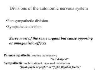
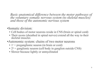
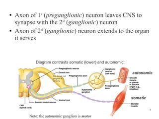
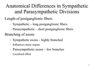
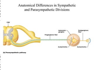
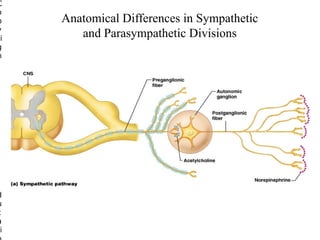
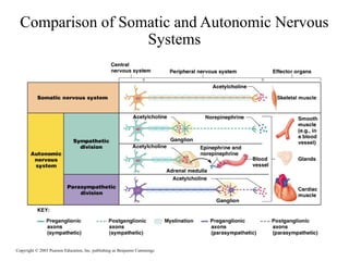
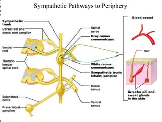
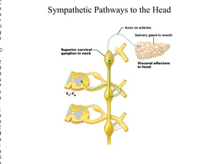
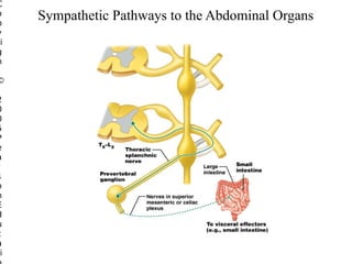
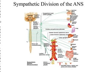
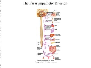
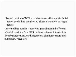
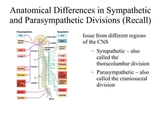
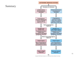
The Autonomic Nervous System

Moderator
Dr. Gangaram
Introduction
The critical component of central network involved in
homeostasis & adaptation
3 subdivisions :
●Sympathetic Nervous System
●Parasympathetic Nervous System
●Enteric Nervous System
To repeat…

ANS is the subdivision of the peripheral nervous system that
regulates body activities that are generally not under
conscious control
●Visceral motor innervates non-skeletal (non-somatic)
muscles
●Composed of a special group of neurons serving:
●

●
●
●
●

Cardiac muscle (the heart)
Smooth muscle (walls of viscera and blood vessels)
Internal organs
Skin

4
Divisions of the autonomic nervous system
Parasympathetic division
●
Sympathetic division
●

Serve most of the same organs but cause opposing
or antagonistic effects
Parasysmpathetic: routine maintenance
“rest &digest”
Sympathetic: mobilization & increased metabolism
“fight, flight or fright” or “fight, flight or freeze”

5
Basic anatomical difference between the motor pathways of
the voluntary somatic nervous system (to skeletal muscles)
and those of the autonomic nervous system
Somatic division:

●

●
●

Cell bodies of motor neurons reside in CNS (brain or spinal cord)
Their axons (sheathed in spinal nerves) extend all the way to their
skeletal muscles

Autonomic system: chains of two motor neurons

●

●
●
●

1st = preganglionic neuron (in brain or cord)
2nd = ganglionic neuron (cell body in ganglion outside CNS)
Slower because lightly or unmyelinated

6
●

●

Axon of 1st (preganglionic) neuron leaves CNS to
synapse with the 2nd (ganglionic) neuron
Axon of 2nd (ganglionic) neuron extends to the organ
it serves

Diagram contrasts somatic (lower) and autonomic:
autonomic
this dorsal root
ganglion is
sensory

somatic
7

Note: the autonomic ganglion is motor
Anatomical Differences in Sympathetic
and Parasympathetic Divisions
Length of postganglionic fibers
–

Sympathetic – long postganglionic fibers

–

Parasympathetic – short postganglionic fibers

Branching of axons
–

Sympathetic axons – highly branched

–

Influences many organs

–

Parasympathetic axons – few branches

–

Localized effect

8
C
o
p
y
ri
g
h

©

2
0
0
5
P
e
a
r
s
o
n
E
d
u
c
a
i

Anatomical Differences in Sympathetic
and Parasympathetic Divisions
C
o
p
y
ri
g
h

©

2
0
0
5
P
e
a
r
s
o
n
E
d
u
c
a
i

Anatomical Differences in Sympathetic
and Parasympathetic Divisions
Comparison of Somatic and Autonomic Nervous
Systems

Copyright © 2003 Pearson Education, Inc. publishing as Benjamin Cummings
Sympathetic nervous system continued
Regardless of target, all begin same
●
Preganglionic axons exit spinal
cord through ventral root and enter
spinal nerve
●
Exit spinal nerve via
communicating ramus
●
Enter sympathetic trunk/chain
where postganglionic neurons are
●
Has three options…
●

12
Options of preganglionic axons in
sympathetic trunk
Synapse on postganglionic neuron in chain ganglion then
return to spinal nerve and follow its branch to the skin
●
Ascend or descend within sympathetic trunk, synapse with a
posganglionic neuron within a chain ganglion, and return to
spinal nerve at that level and follow branches to skin
●
Enter sympathetic chain, pass through without synapsing, form
a splanchnic nerve that passes toward thoracic or abdominal
organs
●
These synapses in prevertebral ganglion in front of aorta
●
Postganglionic axons follow arteries to organs
●

13
SPINAL SEGMENTS

INNERVATION

T1 – T3

HEAD

T1 – T6

UPPER EXTREMITIES
THORACIC VISCERA

T5 – T11

ABDOMINAL VISCERA

T11 – L2

LOWER EXTREMITIES
PELVIC & PERINEAL ORGANS
C
o
p
y
ri
g
h

Sympathetic Pathways to Periphery

©

2
0
0
5
P
e
a
r
s
o
n
E
d
u
c
a
i

Figure 15.9
C
o
p
y
ri
g
h

©

2
0
0
5
P
e
a
r
s
o
n
E
d
u
c
a
i

Sympathetic Pathways to the Head
C
o
p
y
ri
g
h

©

2
0
0
5
P
e
a
r
s
o
n
E
d
u
c
a
i

Sympathetic Pathways to the Abdominal Organs
C
o
p
y
ri
g
h

©

2
0
0
5
P
e
a
r
s
o
n
E
d
u
c
a
i

Sympathetic Pathways to the Pelvic Organs
The Role of the Adrenal Medulla in the
Sympathetic Division
Major organ of the sympathetic nervous system
Secretes great quantities epinephrine (a little
norepinephrine)
Stimulated to secrete by preganglionic sympathetic
fibers
C
o
p
y
ri
g
h

©

2
0
0
5
P
e
a
r
s
o
n
E
d
u
c
a
i

The Adrenal Medulla
C
o
p
y
ri
g
h

©

2
0
0
5
P
e
a
r
s
o
n
E
d
u
c
a
i

Sympathetic Division of the ANS
C
o
p
y
ri
g
h

©

2
0
0
5
P
e
a
r
s
o
n
E
d
u
c
a
i

The Parasympathetic Division
Cranial Outflow
Preganglionic fibers run via:
–

Oculomotor nerve (III)

–

Facial nerve (VII)

–

Glossopharyngeal nerve (IX)

–

Vagus nerve (X)

Cell bodies located in cranial nerve nuclei in the brain
stem
Westphal nucleus is a part of occulomotor complex in
midbrain – sends preganglionic axons that occupy peripheral
portion of the occulomotor nerve and synapse on the neurons
of ciliary ganglion in the orbit
●

These neurons innervate iris and ciliary muscles

●

Eliciting pupil constriction, accomodation of eye

●
Superior salivatory nucleus –

●

In pons

●

projects via facial nerve to sphenopalatine ganglion, which
innervates - lacrimal gland ( lacrimation )
cerebral blood vessels ( vasodilatation )

●

to submandibular ganglion – secretomotor & vasodilator
inputs to corresponding salivary glands

●
Inferior salivatory nucleus –

●

In medulla

●

Sends axons via glossopharyngeal nerve

●

Synapse on Otic ganglion

●

Stimulate parotid gland secretion

●
Outflow via the Vagus Nerve (X)
Fibers innervate visceral organs of the
thorax and most of the abdomen
Stimulates - digestion, reduction in
heart rate and blood pressure
Preganglionic cell bodies
Located in dorsal motor nucleus
in the medulla
Ganglionic neurons
–

–

Confined within the walls of
organs being innervated
Vagus innervates heart, respiratory tract and entire
gastrointestinal tract except descending colon and rectum
●

Most vagal preganglionic neurons – situated in dorsal motor
nucleus of vagus provides input to git and respiratory
tracts, heart

●

Vagal preganglionic output to heart - Neurons in
ventrolateral portion of nucleus ambiguus
●

Vagus – cardioinhibitory, visceromotor and secretomotor
effects
●
9

Sacral Parasympathetic Outflow
• Consists of S2-S4.
• Pelvic splanchnic nerves
Sacral preganglionic output –
Arises from neurons of sacral preganglionic nucleus located
in lateral gray matter of spinal segments S2 & S3
●

Their axons pass via ventral roots of pelvic splanchnic nerves
which join inferior hypogastric plexus
●

Innervate colon, bladder, sexual organs

●

Parasympathetic output – contraction of bladder detrussor
muscle & circular smooth muscle of rectum.
●
Sacral parasympathetic output elicits
Vasodilatation of cavernous tissue of penis
required for penile erection ,
whereas sympathetic output controls ejaculation

●
2

Autonomic Plexuses
• A network of sympathetic and parasympathetic axons.
• Cardiac plexus- heart.
• Pulmonary plexus- the bronchial tree.
• Celiac plexus- largest. Supplies the stomach, spleen,
pancreas, liver, gallbladder, and adrenal medullae.
3

Autonomic Plexuses (Cont’d)..
• Superior mesenteric plexus- small intestine and proximal
colon.
• Inferior mesenteric plexus- distal colon and rectum.
• Hypogastric plexus- urinary bladder and genital organs.
• Renal plexus- kidneys and ureters.
Enteric Nervous System
Includes several types of sensory neurons, inter-neurons,
motor neurons – which form integrative local reflex circuits
●

Controlling motility, secretion, blood flow throughout the gut

●

The activity of enteric nervous system is largely independent
of extrinsic innervation but is modulated by both vagal inputs
from dorsal vagal nucleus and sympathetic inputs from
prevertebral ganglia
●
5

Integration and Control of Autonomic
Functions
• Direct innervation- brain stem and spinal cord.
• Hypothalamus is the major control and integration center of
the ANS.
• It receives input from the limbic system.
Central control of the
Autonomic NS

Amygdala: main limbic region
for emotions
-Stimulates sympathetic activity,
especially previously learned fearrelated behavior
-Can be voluntary when decide to recall
frightful experience - cerebral cortex
acts through amygdala
-Some people can regulate some
autonomic activities by gaining
extraordinary control over their
emotions

Hypothalamus: main integration
center
Reticular formation: most direct
influence over autonomic
function
36
Components
Telencephalic structures –insular cortex
ant cingulate cortex
Amygdala
●
Insular cortex - integration of bodily sensation, emotion,
decision making
●
anterior cingulate cortex – motivation, goal directed
behavior
●
amygdala – conditioned fear responses
●
Hypothalamus – central role in integrating autonomic and
endocrine responses
●
Hypothalmus – critical role in adaptation to internal /
external stimuli, while maintaining homeostasis
●
3 zones
●
Paraventricular zone – neuroendocrine control via
connections to pituitary
●
Medial zone – thermoregulation, osmoregulation, food
intake, response to stress
●
Lateral zone – arousal behaviour, sleep wake cycle
●
Brain Stem Components –
●
Periaqueductal gray –
●
integration of autonomic, somatic and antinociceptive
responses to external stress.
●
coordinates cardiovascular, respiratory, thermoregulatory,
urinary, reproductive and pain control systems
●
Parabrachial nucleus – taste, salivation, gi activity,
cardiorespiratory activity, thermoregulation
●
NTS – 1st relay center for taste and visceral afferent
information carried in CN VII,IX,X
And also for all medullary, cvs, rs, gi reflexes
●
Visceral Afferents
Inform CNS about Mechanical & Chemical events in internal
organs
●
This information is conveyed to produce conscious visceral
sensation and initiate visceral reflex responses
●

Spinal visceral afferents innervate all peripheral organs
●
Their cell bodies are in dorsal root ganglion
●
Brain stem visceral afferents are carried primarily by
glossopharyngeal and vagus nerves
●
Cell bodies in petrosal & nodose ganglia
●
All brain stem visceral afferent nerves relay in nucleus of
solitary tract ( NTS )
●
NTS is a major site of information integration of many
bodily functions
●
Rostral portion of NTS – receives taste afferents via facial
nerve( geniculate ganglion ) , glossopharyngeal & vagus
nerves
●
Intermediate portion – receives gastrointestinal afferents
●
Caudal portion of the NTS recieves afferent information
from baroreceptors, cardioreceptors, chemoreceptors and
pulmonary receptors
●
Anatomical Differences in Sympathetic
and Parasympathetic Divisions (Recall)
Issue from different regions
of the CNS
–

Sympathetic – also
called the
thoracolumbar division

–

Parasympathetic – also
called the craniosacral
division
TARGET

SYMPATHETIC

PARASYMPATHETIC

PUPIL

DILATATION ( A 1 )

CONSTRICTION (M3)

CILIARY MUSCLE

…

ACCOMODATION (M3)

SALIVARY &
LACRIMAL GLANDS

INHIBITION

STIMULATION (M3)

HEART

STIMULATION (B1)

INHIBITION (A2)

BRONCHI

DILATATION (B1)

CONSTRICTION (M3)

SWEAT GLANDS

STIMULATION (M3)

GI MOTILITY

INHIBITION (B2)

CONTRACTION (M3)
RELAXATION (NO,VIP)

BLADDER DETRUSSOR INHIBITION (B2)

STIMULATION (M3,M2)

RECTAL SM

INHIBITION (B2)

STIMULATION (M3)

ERECTILE TISSUE

CONSTRTICTION (A1)

DILATATION (NO)
Horner's syndrome
●

Interruption of sympathetic supply to head and neck

●

Constriction of pupil

●

Drooping of upper eyelid

●

Reduced prominance of eye(enophthalmous)

●

anhydrosis
Dysautonomic polyneuropathy
●

●

Rare, both sympathetic and parasympathetic are affected at
post ganglionic level
Somatosensory and motor nerves spared
Orthostatic hypotension
Primary
●

●

●

Middle aged
Post-ganglionic
sympathetic

Secondary
●

●

Parasympathetic spared
●

ANS impairment as a part
of peripheral neuropathy
Afferent pathways or post
ganglionic sympathetic
fibres in spinal nerves
Eg GBS, porphyrias, DM,
alcoholic, nutritional

Anhydrosis, orthostatic hypotension, impotency, atonicity of
bladder
Summary

52
Thank you
Dr. Shirisha
Autonomic lab tests :

●

impaired sudomotor axon reflex response
reduced variation of heart rate to deep breathing
reduced valsalva ratio
orthostatic hypotension
gi motility – delayed gastric emptying
usg post void urine – 250 cc
Autonomic Nervous System

Autonomic Nervous System

  • 1.
    The Autonomic NervousSystem Moderator Dr. Gangaram
  • 3.
    Introduction The critical componentof central network involved in homeostasis & adaptation 3 subdivisions : ●Sympathetic Nervous System ●Parasympathetic Nervous System ●Enteric Nervous System
  • 4.
    To repeat… ANS isthe subdivision of the peripheral nervous system that regulates body activities that are generally not under conscious control ●Visceral motor innervates non-skeletal (non-somatic) muscles ●Composed of a special group of neurons serving: ● ● ● ● ● Cardiac muscle (the heart) Smooth muscle (walls of viscera and blood vessels) Internal organs Skin 4
  • 5.
    Divisions of theautonomic nervous system Parasympathetic division ● Sympathetic division ● Serve most of the same organs but cause opposing or antagonistic effects Parasysmpathetic: routine maintenance “rest &digest” Sympathetic: mobilization & increased metabolism “fight, flight or fright” or “fight, flight or freeze” 5
  • 6.
    Basic anatomical differencebetween the motor pathways of the voluntary somatic nervous system (to skeletal muscles) and those of the autonomic nervous system Somatic division: ● ● ● Cell bodies of motor neurons reside in CNS (brain or spinal cord) Their axons (sheathed in spinal nerves) extend all the way to their skeletal muscles Autonomic system: chains of two motor neurons ● ● ● ● 1st = preganglionic neuron (in brain or cord) 2nd = ganglionic neuron (cell body in ganglion outside CNS) Slower because lightly or unmyelinated 6
  • 7.
    ● ● Axon of 1st(preganglionic) neuron leaves CNS to synapse with the 2nd (ganglionic) neuron Axon of 2nd (ganglionic) neuron extends to the organ it serves Diagram contrasts somatic (lower) and autonomic: autonomic this dorsal root ganglion is sensory somatic 7 Note: the autonomic ganglion is motor
  • 8.
    Anatomical Differences inSympathetic and Parasympathetic Divisions Length of postganglionic fibers – Sympathetic – long postganglionic fibers – Parasympathetic – short postganglionic fibers Branching of axons – Sympathetic axons – highly branched – Influences many organs – Parasympathetic axons – few branches – Localized effect 8
  • 9.
  • 10.
  • 11.
    Comparison of Somaticand Autonomic Nervous Systems Copyright © 2003 Pearson Education, Inc. publishing as Benjamin Cummings
  • 12.
    Sympathetic nervous systemcontinued Regardless of target, all begin same ● Preganglionic axons exit spinal cord through ventral root and enter spinal nerve ● Exit spinal nerve via communicating ramus ● Enter sympathetic trunk/chain where postganglionic neurons are ● Has three options… ● 12
  • 13.
    Options of preganglionicaxons in sympathetic trunk Synapse on postganglionic neuron in chain ganglion then return to spinal nerve and follow its branch to the skin ● Ascend or descend within sympathetic trunk, synapse with a posganglionic neuron within a chain ganglion, and return to spinal nerve at that level and follow branches to skin ● Enter sympathetic chain, pass through without synapsing, form a splanchnic nerve that passes toward thoracic or abdominal organs ● These synapses in prevertebral ganglion in front of aorta ● Postganglionic axons follow arteries to organs ● 13
  • 14.
    SPINAL SEGMENTS INNERVATION T1 –T3 HEAD T1 – T6 UPPER EXTREMITIES THORACIC VISCERA T5 – T11 ABDOMINAL VISCERA T11 – L2 LOWER EXTREMITIES PELVIC & PERINEAL ORGANS
  • 15.
    C o p y ri g h Sympathetic Pathways toPeriphery © 2 0 0 5 P e a r s o n E d u c a i Figure 15.9
  • 16.
  • 17.
  • 18.
  • 19.
    The Role ofthe Adrenal Medulla in the Sympathetic Division Major organ of the sympathetic nervous system Secretes great quantities epinephrine (a little norepinephrine) Stimulated to secrete by preganglionic sympathetic fibers
  • 20.
  • 21.
  • 22.
  • 23.
    Cranial Outflow Preganglionic fibersrun via: – Oculomotor nerve (III) – Facial nerve (VII) – Glossopharyngeal nerve (IX) – Vagus nerve (X) Cell bodies located in cranial nerve nuclei in the brain stem
  • 24.
    Westphal nucleus isa part of occulomotor complex in midbrain – sends preganglionic axons that occupy peripheral portion of the occulomotor nerve and synapse on the neurons of ciliary ganglion in the orbit ● These neurons innervate iris and ciliary muscles ● Eliciting pupil constriction, accomodation of eye ●
  • 25.
    Superior salivatory nucleus– ● In pons ● projects via facial nerve to sphenopalatine ganglion, which innervates - lacrimal gland ( lacrimation ) cerebral blood vessels ( vasodilatation ) ● to submandibular ganglion – secretomotor & vasodilator inputs to corresponding salivary glands ●
  • 26.
    Inferior salivatory nucleus– ● In medulla ● Sends axons via glossopharyngeal nerve ● Synapse on Otic ganglion ● Stimulate parotid gland secretion ●
  • 27.
    Outflow via theVagus Nerve (X) Fibers innervate visceral organs of the thorax and most of the abdomen Stimulates - digestion, reduction in heart rate and blood pressure Preganglionic cell bodies Located in dorsal motor nucleus in the medulla Ganglionic neurons – – Confined within the walls of organs being innervated
  • 28.
    Vagus innervates heart,respiratory tract and entire gastrointestinal tract except descending colon and rectum ● Most vagal preganglionic neurons – situated in dorsal motor nucleus of vagus provides input to git and respiratory tracts, heart ● Vagal preganglionic output to heart - Neurons in ventrolateral portion of nucleus ambiguus ● Vagus – cardioinhibitory, visceromotor and secretomotor effects ●
  • 29.
    9 Sacral Parasympathetic Outflow •Consists of S2-S4. • Pelvic splanchnic nerves
  • 30.
    Sacral preganglionic output– Arises from neurons of sacral preganglionic nucleus located in lateral gray matter of spinal segments S2 & S3 ● Their axons pass via ventral roots of pelvic splanchnic nerves which join inferior hypogastric plexus ● Innervate colon, bladder, sexual organs ● Parasympathetic output – contraction of bladder detrussor muscle & circular smooth muscle of rectum. ●
  • 31.
    Sacral parasympathetic outputelicits Vasodilatation of cavernous tissue of penis required for penile erection , whereas sympathetic output controls ejaculation ●
  • 32.
    2 Autonomic Plexuses • Anetwork of sympathetic and parasympathetic axons. • Cardiac plexus- heart. • Pulmonary plexus- the bronchial tree. • Celiac plexus- largest. Supplies the stomach, spleen, pancreas, liver, gallbladder, and adrenal medullae.
  • 33.
    3 Autonomic Plexuses (Cont’d).. •Superior mesenteric plexus- small intestine and proximal colon. • Inferior mesenteric plexus- distal colon and rectum. • Hypogastric plexus- urinary bladder and genital organs. • Renal plexus- kidneys and ureters.
  • 34.
    Enteric Nervous System Includesseveral types of sensory neurons, inter-neurons, motor neurons – which form integrative local reflex circuits ● Controlling motility, secretion, blood flow throughout the gut ● The activity of enteric nervous system is largely independent of extrinsic innervation but is modulated by both vagal inputs from dorsal vagal nucleus and sympathetic inputs from prevertebral ganglia ●
  • 35.
    5 Integration and Controlof Autonomic Functions • Direct innervation- brain stem and spinal cord. • Hypothalamus is the major control and integration center of the ANS. • It receives input from the limbic system.
  • 36.
    Central control ofthe Autonomic NS Amygdala: main limbic region for emotions -Stimulates sympathetic activity, especially previously learned fearrelated behavior -Can be voluntary when decide to recall frightful experience - cerebral cortex acts through amygdala -Some people can regulate some autonomic activities by gaining extraordinary control over their emotions Hypothalamus: main integration center Reticular formation: most direct influence over autonomic function 36
  • 37.
    Components Telencephalic structures –insularcortex ant cingulate cortex Amygdala ● Insular cortex - integration of bodily sensation, emotion, decision making ● anterior cingulate cortex – motivation, goal directed behavior ● amygdala – conditioned fear responses ●
  • 38.
    Hypothalamus – centralrole in integrating autonomic and endocrine responses ● Hypothalmus – critical role in adaptation to internal / external stimuli, while maintaining homeostasis ● 3 zones ● Paraventricular zone – neuroendocrine control via connections to pituitary ● Medial zone – thermoregulation, osmoregulation, food intake, response to stress ● Lateral zone – arousal behaviour, sleep wake cycle ●
  • 39.
    Brain Stem Components– ● Periaqueductal gray – ● integration of autonomic, somatic and antinociceptive responses to external stress. ● coordinates cardiovascular, respiratory, thermoregulatory, urinary, reproductive and pain control systems ● Parabrachial nucleus – taste, salivation, gi activity, cardiorespiratory activity, thermoregulation ● NTS – 1st relay center for taste and visceral afferent information carried in CN VII,IX,X And also for all medullary, cvs, rs, gi reflexes ●
  • 40.
    Visceral Afferents Inform CNSabout Mechanical & Chemical events in internal organs ● This information is conveyed to produce conscious visceral sensation and initiate visceral reflex responses ● Spinal visceral afferents innervate all peripheral organs ● Their cell bodies are in dorsal root ganglion ●
  • 41.
    Brain stem visceralafferents are carried primarily by glossopharyngeal and vagus nerves ● Cell bodies in petrosal & nodose ganglia ● All brain stem visceral afferent nerves relay in nucleus of solitary tract ( NTS ) ● NTS is a major site of information integration of many bodily functions ●
  • 42.
    Rostral portion ofNTS – receives taste afferents via facial nerve( geniculate ganglion ) , glossopharyngeal & vagus nerves ● Intermediate portion – receives gastrointestinal afferents ● Caudal portion of the NTS recieves afferent information from baroreceptors, cardioreceptors, chemoreceptors and pulmonary receptors ●
  • 43.
    Anatomical Differences inSympathetic and Parasympathetic Divisions (Recall) Issue from different regions of the CNS – Sympathetic – also called the thoracolumbar division – Parasympathetic – also called the craniosacral division
  • 44.
    TARGET SYMPATHETIC PARASYMPATHETIC PUPIL DILATATION ( A1 ) CONSTRICTION (M3) CILIARY MUSCLE … ACCOMODATION (M3) SALIVARY & LACRIMAL GLANDS INHIBITION STIMULATION (M3) HEART STIMULATION (B1) INHIBITION (A2) BRONCHI DILATATION (B1) CONSTRICTION (M3) SWEAT GLANDS STIMULATION (M3) GI MOTILITY INHIBITION (B2) CONTRACTION (M3) RELAXATION (NO,VIP) BLADDER DETRUSSOR INHIBITION (B2) STIMULATION (M3,M2) RECTAL SM INHIBITION (B2) STIMULATION (M3) ERECTILE TISSUE CONSTRTICTION (A1) DILATATION (NO)
  • 49.
    Horner's syndrome ● Interruption ofsympathetic supply to head and neck ● Constriction of pupil ● Drooping of upper eyelid ● Reduced prominance of eye(enophthalmous) ● anhydrosis
  • 50.
    Dysautonomic polyneuropathy ● ● Rare, bothsympathetic and parasympathetic are affected at post ganglionic level Somatosensory and motor nerves spared
  • 51.
    Orthostatic hypotension Primary ● ● ● Middle aged Post-ganglionic sympathetic Secondary ● ● Parasympatheticspared ● ANS impairment as a part of peripheral neuropathy Afferent pathways or post ganglionic sympathetic fibres in spinal nerves Eg GBS, porphyrias, DM, alcoholic, nutritional Anhydrosis, orthostatic hypotension, impotency, atonicity of bladder
  • 52.
  • 53.
  • 54.
    Autonomic lab tests: ● impaired sudomotor axon reflex response reduced variation of heart rate to deep breathing reduced valsalva ratio orthostatic hypotension gi motility – delayed gastric emptying usg post void urine – 250 cc