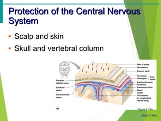
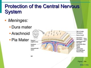
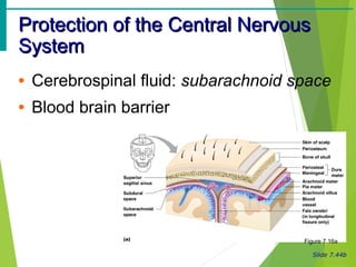
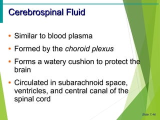
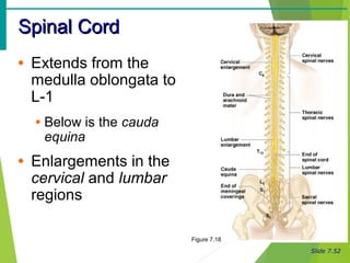
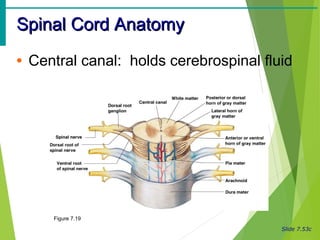
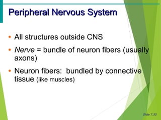
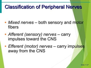
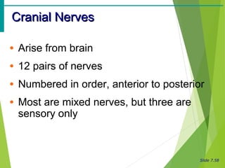
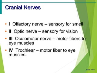
Slide 2.1
Human AnatomyHuman Anatomy
NERVOUS SYSTEMNERVOUS SYSTEM
pavemedicine.compavemedicine.com
Slide 7.43a
CerebellumCerebellum
• Two hemispheres with convoluted
surfaces
• Provides involuntary coordination of
body movements
Slide 7.43b
CerebellumCerebellum
Figure 7.15a
Slide 7.44a
Protection of the Central NervousProtection of the Central Nervous
SystemSystem
• Scalp and skin
• Skull and vertebral column
Figure 7.16a
Slide 7.44a
Protection of the Central NervousProtection of the Central Nervous
SystemSystem
• Meninges:
• Dura mater
• Arachnoid
• Pia Mater
Figure 7.16a
Slide 7.44b
Protection of the Central NervousProtection of the Central Nervous
SystemSystem
• Cerebrospinal fluid: subarachnoid space
• Blood brain barrier
Figure 7.16a
Slide 7.45a
MeningesMeninges
• Dura mater
• Double-layered external covering
• Periosteum – attached to surface of the
skull
• Meningeal layer – outer covering of the
brain
• Folds inward to support hemispheres
Slide 7.45b
MeningesMeninges
• Arachnoid layer
• Middle layer
• Web-like
• Pia mater
• Internal layer
• Clings to the surface of the brain
Slide 7.46
Cerebrospinal FluidCerebrospinal Fluid
• Similar to blood plasma
• Formed by the choroid plexus
• Forms a watery cushion to protect the
brain
• Circulated in subarachnoid space,
ventricles, and central canal of the
spinal cord
Slide 7.47a
Ventricles and Location of theVentricles and Location of the
Cerebrospinal FluidCerebrospinal Fluid
Figure 7.17a
Slide 7.47b
Ventricles and Location of theVentricles and Location of the
Cerebrospinal FluidCerebrospinal Fluid
Figure 7.17b
Slide 7.48
Blood Brain BarrierBlood Brain Barrier
• The least permeable capillaries of the
body + astrocyte processes
• Excludes many potentially harmful
substances
• Useless against some substances
• Fat soluble substances
• Anesthetics
Slide 7.48
Blood Brain BarrierBlood Brain Barrier
• “Good news”: keeps out most harmful
substances
• Alcohol, viruses, some drugs
• “Bad news”: keeps out most helpful
substances
• Antibiotics, chemotherapy drugs
Slide 7.49
Traumatic Brain InjuriesTraumatic Brain Injuries
• Concussion
• Slight brain injury
• Usually no permanent damage
• Contusion
• Nervous tissue destruction occurs
• Nervous tissue does not regenerate
• Cerebral edema
• May compress and kill brain tissue
Slide 7.50
Cerebrovascular Accident (CVA)Cerebrovascular Accident (CVA)
• Commonly called a stroke
• The result of a ruptured blood vessel
• Brain tissue supplied with oxygen from
that blood source dies
• Loss of some functions or death may
result
Slide 7.51
Alzheimer’s DiseaseAlzheimer’s Disease
• Progressive degenerative brain disease
• Mostly seen in the elderly, but may
begin in middle age
• Structural changes in the brain due to
abnormal proteins
• Symptoms: memory loss, irritability,
confusion, ultimately hallucinations and
death
Slide 7.52
Spinal CordSpinal Cord
• Extends from the
medulla oblongata to
L-1
• Below is the cauda
equina
• Enlargements in the
cervical and lumbar
regions
Figure 7.18
Slide 7.53a
Spinal Cord AnatomySpinal Cord Anatomy
• White mater – conduction tracts
• Superficially located
Figure 7.19
Slide 7.53b
Spinal Cord AnatomySpinal Cord Anatomy
• Gray matter - mostly cell bodies, deep
• Dorsal (posterior) gray horns
• Ventral (anterior) gray horns
Figure 7.19
Slide 7.53c
Spinal Cord AnatomySpinal Cord Anatomy
• Central canal: holds cerebrospinal fluid
Figure 7.19
Slide 7.54
Spinal Cord AnatomySpinal Cord Anatomy
• Meninges: same as brain
• Spinal Nerves attached to each spinal
segment
• Dorsal root: sensory
• Associated with the dorsal root ganglia
• Ventral root: motor
Slide 7.55
Peripheral Nervous SystemPeripheral Nervous System
• All structures outside CNS
• Nerve = bundle of neuron fibers (usually
axons)
• Neuron fibers: bundled by connective
tissue (like muscles)
Slide 7.56
Structure of a NerveStructure of a Nerve
• Endoneurium
surrounds each fiber
• Groups of fibers are
fascicles
• Coverd by
perineurium
• Fascicles bound by
epineurium
Figure 7.20
Slide 7.54
Nerves (PNS) vs. Tracts (CNS)Nerves (PNS) vs. Tracts (CNS)
• Tract: a bundle of neuron fibers inside
the CNS
• Usually axons
• Wrapped by CT
• Continuation of “nerve” from PNS
• Long tracts in spinal cord
• Short tracts in brain
Slide 7.57
Classification of Peripheral NervesClassification of Peripheral Nerves
• Mixed nerves – both sensory and motor
fibers
• Afferent (sensory) nerves – carry
impulses toward the CNS
• Efferent (motor) nerves – carry impulses
away from the CNS
Slide 7.58
Cranial NervesCranial Nerves
• Arise from brain
• 12 pairs of nerves
• Numbered in order, anterior to posterior
• Most are mixed nerves, but three are
sensory only
Slide 7.60
Cranial NervesCranial Nerves
• I Olfactory nerve – sensory for smell
• II Optic nerve – sensory for vision
• III Oculomotor nerve – motor fibers to
eye muscles
• IV Trochlear – motor fiber to eye
muscles
Slide 7.61
Cranial NervesCranial Nerves
• V Trigeminal nerve – sensory for the
face; motor fibers to chewing muscles
• VI Abducens nerve –
motor fibers to eye muscles
• VII Facial nerve – sensory for taste;
motor fibers to the face
• VIII Vestibulocochlear nerve –
sensory for balance and hearing
Slide 7.62
Cranial NervesCranial Nerves
• IX Glossopharyngeal nerve – sensory
for taste; motor fibers to the pharynx
• X Vagus nerves – sensory and motor
fibers for pharynx, larynx, and viscera
• XI Accessory nerve – motor fibers to
neck and upper back
• XII Hypoglossal nerve – motor fibers to
tongue

Cns, pns

  • 1.
    Slide 2.1 Human AnatomyHumanAnatomy NERVOUS SYSTEMNERVOUS SYSTEM pavemedicine.compavemedicine.com
  • 2.
    Slide 7.43a CerebellumCerebellum • Twohemispheres with convoluted surfaces • Provides involuntary coordination of body movements
  • 3.
  • 4.
    Slide 7.44a Protection ofthe Central NervousProtection of the Central Nervous SystemSystem • Scalp and skin • Skull and vertebral column Figure 7.16a
  • 5.
    Slide 7.44a Protection ofthe Central NervousProtection of the Central Nervous SystemSystem • Meninges: • Dura mater • Arachnoid • Pia Mater Figure 7.16a
  • 6.
    Slide 7.44b Protection ofthe Central NervousProtection of the Central Nervous SystemSystem • Cerebrospinal fluid: subarachnoid space • Blood brain barrier Figure 7.16a
  • 7.
    Slide 7.45a MeningesMeninges • Duramater • Double-layered external covering • Periosteum – attached to surface of the skull • Meningeal layer – outer covering of the brain • Folds inward to support hemispheres
  • 8.
    Slide 7.45b MeningesMeninges • Arachnoidlayer • Middle layer • Web-like • Pia mater • Internal layer • Clings to the surface of the brain
  • 9.
    Slide 7.46 Cerebrospinal FluidCerebrospinalFluid • Similar to blood plasma • Formed by the choroid plexus • Forms a watery cushion to protect the brain • Circulated in subarachnoid space, ventricles, and central canal of the spinal cord
  • 10.
    Slide 7.47a Ventricles andLocation of theVentricles and Location of the Cerebrospinal FluidCerebrospinal Fluid Figure 7.17a
  • 11.
    Slide 7.47b Ventricles andLocation of theVentricles and Location of the Cerebrospinal FluidCerebrospinal Fluid Figure 7.17b
  • 12.
    Slide 7.48 Blood BrainBarrierBlood Brain Barrier • The least permeable capillaries of the body + astrocyte processes • Excludes many potentially harmful substances • Useless against some substances • Fat soluble substances • Anesthetics
  • 13.
    Slide 7.48 Blood BrainBarrierBlood Brain Barrier • “Good news”: keeps out most harmful substances • Alcohol, viruses, some drugs • “Bad news”: keeps out most helpful substances • Antibiotics, chemotherapy drugs
  • 14.
    Slide 7.49 Traumatic BrainInjuriesTraumatic Brain Injuries • Concussion • Slight brain injury • Usually no permanent damage • Contusion • Nervous tissue destruction occurs • Nervous tissue does not regenerate • Cerebral edema • May compress and kill brain tissue
  • 15.
    Slide 7.50 Cerebrovascular Accident(CVA)Cerebrovascular Accident (CVA) • Commonly called a stroke • The result of a ruptured blood vessel • Brain tissue supplied with oxygen from that blood source dies • Loss of some functions or death may result
  • 16.
    Slide 7.51 Alzheimer’s DiseaseAlzheimer’sDisease • Progressive degenerative brain disease • Mostly seen in the elderly, but may begin in middle age • Structural changes in the brain due to abnormal proteins • Symptoms: memory loss, irritability, confusion, ultimately hallucinations and death
  • 17.
    Slide 7.52 Spinal CordSpinalCord • Extends from the medulla oblongata to L-1 • Below is the cauda equina • Enlargements in the cervical and lumbar regions Figure 7.18
  • 18.
    Slide 7.53a Spinal CordAnatomySpinal Cord Anatomy • White mater – conduction tracts • Superficially located Figure 7.19
  • 19.
    Slide 7.53b Spinal CordAnatomySpinal Cord Anatomy • Gray matter - mostly cell bodies, deep • Dorsal (posterior) gray horns • Ventral (anterior) gray horns Figure 7.19
  • 20.
    Slide 7.53c Spinal CordAnatomySpinal Cord Anatomy • Central canal: holds cerebrospinal fluid Figure 7.19
  • 21.
    Slide 7.54 Spinal CordAnatomySpinal Cord Anatomy • Meninges: same as brain • Spinal Nerves attached to each spinal segment • Dorsal root: sensory • Associated with the dorsal root ganglia • Ventral root: motor
  • 22.
    Slide 7.55 Peripheral NervousSystemPeripheral Nervous System • All structures outside CNS • Nerve = bundle of neuron fibers (usually axons) • Neuron fibers: bundled by connective tissue (like muscles)
  • 23.
    Slide 7.56 Structure ofa NerveStructure of a Nerve • Endoneurium surrounds each fiber • Groups of fibers are fascicles • Coverd by perineurium • Fascicles bound by epineurium Figure 7.20
  • 24.
    Slide 7.54 Nerves (PNS)vs. Tracts (CNS)Nerves (PNS) vs. Tracts (CNS) • Tract: a bundle of neuron fibers inside the CNS • Usually axons • Wrapped by CT • Continuation of “nerve” from PNS • Long tracts in spinal cord • Short tracts in brain
  • 25.
    Slide 7.57 Classification ofPeripheral NervesClassification of Peripheral Nerves • Mixed nerves – both sensory and motor fibers • Afferent (sensory) nerves – carry impulses toward the CNS • Efferent (motor) nerves – carry impulses away from the CNS
  • 26.
    Slide 7.58 Cranial NervesCranialNerves • Arise from brain • 12 pairs of nerves • Numbered in order, anterior to posterior • Most are mixed nerves, but three are sensory only
  • 27.
    Slide 7.60 Cranial NervesCranialNerves • I Olfactory nerve – sensory for smell • II Optic nerve – sensory for vision • III Oculomotor nerve – motor fibers to eye muscles • IV Trochlear – motor fiber to eye muscles
  • 28.
    Slide 7.61 Cranial NervesCranialNerves • V Trigeminal nerve – sensory for the face; motor fibers to chewing muscles • VI Abducens nerve – motor fibers to eye muscles • VII Facial nerve – sensory for taste; motor fibers to the face • VIII Vestibulocochlear nerve – sensory for balance and hearing
  • 29.
    Slide 7.62 Cranial NervesCranialNerves • IX Glossopharyngeal nerve – sensory for taste; motor fibers to the pharynx • X Vagus nerves – sensory and motor fibers for pharynx, larynx, and viscera • XI Accessory nerve – motor fibers to neck and upper back • XII Hypoglossal nerve – motor fibers to tongue