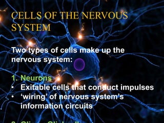
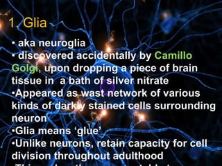
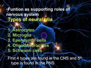
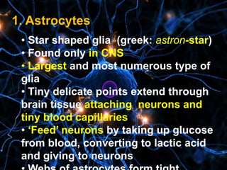
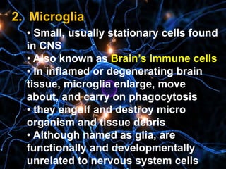
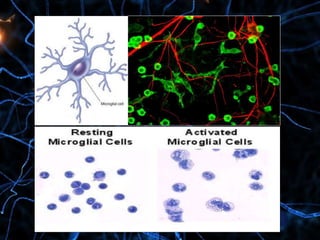
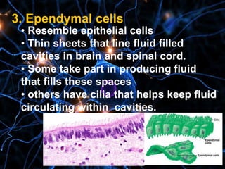
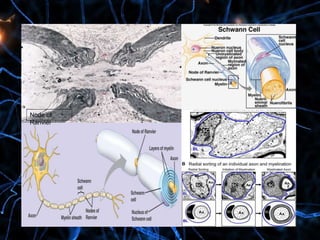
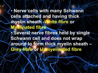
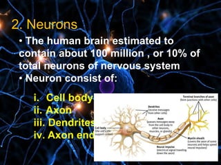
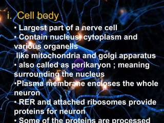
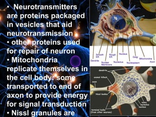
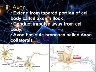
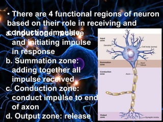
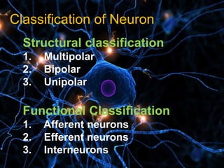
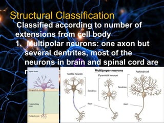
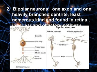
The document summarizes the structure and types of neurons and glial cells in the nervous system. It discusses that neurons are excitable cells that conduct nerve impulses, while glial cells provide support and insulation. The major types of glial cells are astrocytes, microglia, ependymal cells, oligodendrocytes, and Schwann cells. Neurons consist of a cell body, dendrites, and an axon. The axon conducts impulses away from the cell body and terminates in synaptic knobs. Neurons are classified structurally as multipolar, bipolar, or unipolar based on their processes, and functionally as afferent, efferent
This slides gives a brief description about the structure of neuron, information flow in neurons and also how transcription takesplace and protein synthesis in the cell. This presentation also explain the types of glia and non glial cells.
Structure of Neuron
Neuron contents ,neuron parts and processes
Classification of neurons according to function, according to
processes, according to shape
Histology of neuron
The central nervous system (CNS) is made up of the brain and spinal cord. The brain controls most body functions, including awareness, movements, sensations, thoughts, speech and memory. The spinal cord is connected to the brain at the brain stem and is covered by the vertebrae of the spine.
Introduction to nervous system, Divisions of Nervous System, Nervous System P...Shaista Jabeen
https://www.youtube.com/channel/UCrrAABI7QDRCJ1yMrQCip_w/videos
https://www.facebook.com/ShaistaJabeeen/
https://www.facebook.com/Human-Physiology-Lectures-100702741804409/
Introduction to nervous system, Divisions of Nervous System, Nervous System Physiology
Introduction to nervous system
Divisions of Nervous System
Nervous System Physiology
DIVISIONS OF NERVOUS SYSTEM
CENTRAL NERVOUS SYSTEM
PERIPHERAL NERVOUS SYSTEM
Short Notes
ppt pdf
“The right half of the brain controls the left half of the body. This means that only left handed people are in their right mind.”
Made up of brain and spinal cordActs as body’s control center, coordinates body’s activitiesImpulses travel through the neurons in your body to reach the brainCentral Nervous System is yellow in this diagram.
Made up of all the nerves that carry messages to and from the central nervous system.Similar to telephone wires that connect all of our houses in the communityCentral Nervous System and Peripheral Nervous System work together to make rapid changes in your body in response to stimuli.Peripheral Nervous System is green in this diagram.
Somatic Nervous SystemRelay information between skin, skeletal muscles and central nervous systemYou consciously control this pathway by deciding whether or not to move muscles (except reflexes)Reflexes: Automatic response to stimulusAutonomic Nervous SystemRelay information from central nervous system to organsInvoluntary: You do not consciously control theseSympathetic Nervous System: controls in times of stress, such as the flight or fight responseParasympathetic Nervous System: controls body in times of rest
This presentation based on a broad overview to the human central nervous system focusing over the parts of the system, different cell types present in the system, and special terminology used in the system.
The action potential travels from one location in the cell to another, but ion flow across the membrane occurs only at the nodes of Ranvier. As a result, the action potential signal jumps along the axon, from node to node, rather than propagating smoothly, as they do in axons that lack a myelin sheath.
Function of Dendrites. In order for neurons to become active, they must receive action potentials or other stimuli. Dendrites are the structures on the neuron that receive electrical messages. These messages come in two basic forms: excitatory and inhibitory.
The Cell body (soma) is the factory of the neuron. It produces all the proteins for the dendrites, axons and synaptic terminals and contains specialized organelles such as the mitochondria, Golgi apparatus, endoplasmic reticulum, secretory granules, ribosomes and polysomes to provide energy and make the parts, as well as a production line to assemble the parts into completed products.
Structures of Axon Terminals and Presynaptic Membrane
Presynaptic axon terminal has a definite intact membrane known as presynaptic
membrane.
Axon terminal has two important structures:
i. Mitochondria, which help in the synthesis of neurotransmitter substance
ii. Synaptic vesicles, which store neurotransmitter substanceMain function of the synapse is to transmit the
impulses, i.e. action potential from one neuron to
Another
1. Excitatory synapses
2. Inhibitory synapses,
Introduction to AI for Nonprofits with Tapp NetworkTechSoup
Dive into the world of AI! Experts Jon Hill and Tareq Monaur will guide you through AI's role in enhancing nonprofit websites and basic marketing strategies, making it easy to understand and apply.
How to Make a Field invisible in Odoo 17Celine George
It is possible to hide or invisible some fields in odoo. Commonly using “invisible” attribute in the field definition to invisible the fields. This slide will show how to make a field invisible in odoo 17.
Operation “Blue Star” is the only event in the history of Independent India where the state went into war with its own people. Even after about 40 years it is not clear if it was culmination of states anger over people of the region, a political game of power or start of dictatorial chapter in the democratic setup.
The people of Punjab felt alienated from main stream due to denial of their just demands during a long democratic struggle since independence. As it happen all over the word, it led to militant struggle with great loss of lives of military, police and civilian personnel. Killing of Indira Gandhi and massacre of innocent Sikhs in Delhi and other India cities was also associated with this movement.
The French Revolution, which began in 1789, was a period of radical social and political upheaval in France. It marked the decline of absolute monarchies, the rise of secular and democratic republics, and the eventual rise of Napoleon Bonaparte. This revolutionary period is crucial in understanding the transition from feudalism to modernity in Europe.
For more information, visit-www.vavaclasses.com
Embracing GenAI - A Strategic ImperativePeter Windle
Artificial Intelligence (AI) technologies such as Generative AI, Image Generators and Large Language Models have had a dramatic impact on teaching, learning and assessment over the past 18 months. The most immediate threat AI posed was to Academic Integrity with Higher Education Institutes (HEIs) focusing their efforts on combating the use of GenAI in assessment. Guidelines were developed for staff and students, policies put in place too. Innovative educators have forged paths in the use of Generative AI for teaching, learning and assessments leading to pockets of transformation springing up across HEIs, often with little or no top-down guidance, support or direction.
This Gasta posits a strategic approach to integrating AI into HEIs to prepare staff, students and the curriculum for an evolving world and workplace. We will highlight the advantages of working with these technologies beyond the realm of teaching, learning and assessment by considering prompt engineering skills, industry impact, curriculum changes, and the need for staff upskilling. In contrast, not engaging strategically with Generative AI poses risks, including falling behind peers, missed opportunities and failing to ensure our graduates remain employable. The rapid evolution of AI technologies necessitates a proactive and strategic approach if we are to remain relevant.
Model Attribute Check Company Auto PropertyCeline George
In Odoo, the multi-company feature allows you to manage multiple companies within a single Odoo database instance. Each company can have its own configurations while still sharing common resources such as products, customers, and suppliers.
Normal Labour/ Stages of Labour/ Mechanism of LabourWasim Ak
Normal labor is also termed spontaneous labor, defined as the natural physiological process through which the fetus, placenta, and membranes are expelled from the uterus through the birth canal at term (37 to 42 weeks
Macroeconomics- Movie Location
This will be used as part of your Personal Professional Portfolio once graded.
Objective:
Prepare a presentation or a paper using research, basic comparative analysis, data organization and application of economic information. You will make an informed assessment of an economic climate outside of the United States to accomplish an entertainment industry objective.
2. CELLS OF THE NERVOUS
SYSTEM
Two types of cells make up the
nervous system:
1. Neurons
• Exitable cells that conduct impulses
• ‘wiring’ of nervous system’s
information circuits
3. 1
1. Glia
• aka neuroglia
• discovered accidentally by Camillo
Golgi, upon dropping a piece of brain
tissue in a bath of silver nitrate
•Appeared as wast network of various
kinds of darkly stained cells surrounding
neuron
•Glia means ‘glue’
•Unlike neurons, retain capacity for cell
division throughout adulthood
4. •Funtion as supporting roles of
nervous system
Types of neural glia
1. Astrocytes
2. Microglia
3. Ependymal cells
4. Oligodendrocytes
5. Schwann cells
First 4 types are found in the CNS and 5th
type is found in the PNS
5. 1. Astrocytes
• Star shaped glia (greek: astron-star)
• Found only in CNS
• Largest and most numerous type of
glia
• Tiny delicate points extend through
brain tissue attaching neurons and
tiny blood capillaries
• ‘Feed’ neurons by taking up glucose
from blood, converting to lactic acid
and giving to neurons
6. • The BBB is a double barrier made up of
astrocyte feet and endothelial cells of
blood capillary walls
• Small molecules (oxygen, carbon
dioxide, water) diffuse rapidly to reach
brain neurons and other glia, larger
molecules penetrate slowly or not at all
7. 2. Microglia
• Small, usually stationary cells found
in CNS
• Also known as Brain’s immune cells
• In inflamed or degenerating brain
tissue, microglia enlarge, move
about, and carry on phagocytosis
• they engulf and destroy micro
organism and tissue debris
• Although named as glia, are
functionally and developmentally
unrelated to nervous system cells
8.
9. 3. Ependymal cells
• Resemble epithelial cells
• Thin sheets that line fluid filled
cavities in brain and spinal cord.
• Some take part in producing fluid
that fills these spaces
• others have cilia that helps keep fluid
circulating within cavities.
10. 4. Oligodendrocytes
• Smaller than astrocytes and have
fewer processes
• Oligo-few, dendro-branch, cyte-cell :
meaning cells with fewer branches
• Some lie clustered around nerve cell
body, some arranged in rows between
nerve fibre in brain and spinal cord
•Help hold nerve fibre together
• Important function : produce fatty
myelin sheath around nerve fibre of
CNS
12. 5. Schwann cells
• Found only in PNS
• Also called as ‘Neurolemmocytes’
•Sometimes called satellite cells
•Functional equivalents of
oligodendrocytes
• Support nerve fibre and form myelin
sheath around them
• Many schwann cells wrap around
single neuron
• Myelin sheath is formed by layers of
13. • Microscopic gaps in the sheath,
between adjecent Schwann cell-
Node of Ranvier or myelin sheath
gaps
•The myelin sheath and gaps in
nerve fibre are important for transfer
of nerve impulses
• Schwann cell’s nucleus and
cytoplasm are squeezed to perimeter
to form neurilemma.
• neurilemma important for
regeneration of injured nerve fibres.
15. • Nerve cells with many Schwann
cells attached and having thick
myelin sheath -White fibre or
Myelinated fibre
• Several nerve fibres held by single
Schwann cell and does not wrap
around to form thick myelin sheath –
Grey fibre or Unmyelinated fibre
16. 2. Neurons
• The human brain estimated to
contain about 100 million , or 10% of
total neurons of nervous system
• Neuron consist of:
i. Cell body
ii. Axon
iii. Dendrites
iv. Axon ends
17. i. Cell body
• Largest part of a nerve cell
• Contain nucleus, cytoplasm and
various organells
like mitochondria and golgi apparatus
• also called as perikaryon ; meaning
surrounding the nucleus
•Plasma membrane encloses the whole
neuron
• RER and attached ribosomes provide
proteins for neuron
18. • Neurotransmitters
are proteins packaged
in vesicles that aid
neurotransmission
• other proteins used
for repair of neuron
• Mitochondria
replicate themselves in
the cell body: some
transported to end of
axon to provide energy
for signal transduction
• Nissl granules are
19. ii. Dendrites
• Branch extensively from
cell body like tiny tree
• Greek word : tree
• Distal end of sensory
neurons called receptors :
receive stimuli that initiate
nerve signals
• some dendrites in brain
have knoblike dendritic
spines: serve as
connection point of other
20.
21. iii. Axon.
• Extend from tapered portion of cell
body called axon hillock
• Conduct impulse away from cell
body.
• Axon has side branches called Axon
collaterals
• Distal ends of axon form branches
called telodentria and each terminate
in a synaptic knob
22. • Some axons have vericosities or
swellings, and act as point of contact
with other cells or smooth muscle fibres
• Axons vary in size from a meter long to
just millimeters long
• Axon diameter also vary: larger the
diameter, greater the conduction
• Myelination of axon also affects
conductance
• Only axons have myelin and not
dendrites
23.
24. • Neurofibrills arebundles of
intermediate filaments called
neurofilaments extending through
cytoplasm of the neuron
• Microtubules and microfilaments also
present
25. • Along with providing structural
support, act as railway for rapid
transport of small organelles
• Small motor molecules attach to
vesicles and mitochondria , carry them
to end of axon
• Used vesicles and transmitters
returned by same process to the cell
body : Axonal transport
26. • There are 4 functional regions of neuron
based on their role in receiving and
conducting impulsea. Input zone: receive
and initiating impulse
in response
b. Summation zone:
adding together all
impulse received
c. Conduction zone:
conduct impulse to end
of axon
d. Output zone: release
28. Structural Classificationl
Classified according to number of
extensions from cell body
1. Multipolar neurons: one axon but
several dentrites, most of the
neurons in brain and spinal cord are
multipolar
29. 2. Bipolar neurons: one axon and one
heavily branched dentrite, least
nemerous kind and found in retina ,
inner ear and olfactory pathway
30. 3. Unipolar neuron : single process
extending from the cell body, which
branches into central process
(towards CNS) and peripheral process
(away from CNS), process forms a
single axon and conducting impulse
away from dentrite, they are always
sensory neurons.
31. Functional Classification
Classified based on direction of
conduction of nerve impulse.
1. Afferent neuron:
to spinal cord or
brain
2. Efferent neuron:
away from brain
or spinal cord to
muscle or gland
3. Interneurons:
from afferent