Development of nervous system
•
17 likes•5,235 views

well describes the development of nervous system from basic to advanced concept including neural tube defects. the concepts are presented in graphical form for easy understanding of concepts.
Report
Share
Report
Share
Download to read offline

Recommended
Embryology of nervous system
- Human embryology involves the study of development in the first 8 weeks after fertilization.
- The neural tube develops from the ectoderm and gives rise to the central nervous system. Neural crest cells form from the neural tube tips and develop into much of the peripheral nervous system.
- The brain and spinal cord develop from the enlarged cranial and caudal parts of the neural tube, respectively. The brain forms three primary vesicles that later develop into the distinct brain regions.
- Neurulation is the process of neural tube formation from the ectoderm through the thickening, elevation and fusion of the neural folds. This forms the cylindrical neural tube detached from the surface ectoderm.
Cerebellum and its connections
The cerebellum controls posture and voluntary movement through precise coordination of muscle activity. It receives input from the cerebral cortex, spinal cord, and vestibular system and influences motor control through output to the cerebral cortex and brainstem. The cerebellum consists of gray matter forming a cortex of three layers and white matter containing nerve fibers that connect its different regions to each other and to other parts of the brain and spinal cord.
Anatomy of cerebellum
The cerebellum has three main parts - the vermis, two hemispheres, and four lobes. It receives sensory input from the spinal cord, brainstem, and cerebral cortex. There are three layers in the cerebellar cortex - molecular layer, purkinje cell layer, and granular layer. The cerebellum is connected to the brainstem via three cerebellar peduncles and plays a role in motor coordination and balance.
The brainstem
The brainstem consists of three parts - the midbrain, pons, and medulla. It connects the spinal cord to the forebrain and contains nuclei that control vital functions like breathing and heart rate. It also contains tracts that relay signals between the spinal cord and higher brain centers. The reticular formation is a network of fibers and neurons throughout the brainstem that plays roles in motor control, sensory processing, autonomic functions, and maintaining alertness. Important structures in the brainstem include the cranial nerve nuclei, pyramidal tract, olives, and red nucleus.
Thalamus
The thalamus is a paired, oval structure located in the diencephalon that serves as a relay center for sensory and motor signals to and from the cerebral cortex. It is divided into several nuclei that process different sensory modalities. The thalamus receives input from various areas and projects to specific regions of the cortex. Damage to certain thalamic nuclei can disrupt motor control, sensory processing, and cause syndromes like thalamic pain. Surgical procedures targeting thalamic nuclei have been used to treat chronic pain conditions.
Sensory and motor pathways
This document discusses sensory and motor pathways in the human body. It begins by listing the key learning outcomes, which include describing sensory receptors, pathways in the spinal cord and brain, motor neurons and tracts, and the corticospinal tract. It then discusses myelinated and non-myelinated nerve fibers, how they conduct impulses, and their roles. The document proceeds to explain sensory pathways from receptors to the brain, motor pathways from the brain to muscles, and provides diagrams of sensory and motor tracts in the spinal cord. It concludes by describing functions of sensory and motor neurons.
CNS Development
The document summarizes key stages of central nervous system development from neurulation through embryonic development of the brain and spinal cord. It discusses:
- Formation of the neural tube from ectoderm and neural crest cells by the 4th week
- Development of the brain into 5 vesicles and their adult structures by the 5th week
- Formation of the ventricular system and spinal cord plates, horns, and central canal
- Development of the meninges and pituitary gland from surrounding tissues
Anatomy of cerebellum
The cerebellum is located behind the brain stem and is divided into three lobes - anterior, posterior, and flocculonodular. It receives input from the spinal cord, vestibular system, and cerebral cortex. The cerebellar cortex consists of molecular, purkinje, and granular layers. Purkinje cells are the sole output, projecting to deep cerebellar nuclei which connect to motor and premotor areas. The cerebellum is involved in coordination, precision of movement, and maintaining balance and posture.
Embryology of brain
The human brain is the most complex organ on Earth. It develops from the neural tube which forms the basic structure of the central nervous system. As the brain develops from early embryonic stages through fetal development, different regions form including the rhombencephalon (medulla and pons), mesencephalon, and prosencephalon (telencephalon and diencephalon). Precisely regulated molecular processes guide the formation, migration and differentiation of neurons and glial cells in each brain region. Malformations can occur if these processes are disrupted, leading to neurological disorders.
Thalamus ppt
The thalamus is a structure located in the middle of the brain between the cerebral cortex and midbrain. It is the largest component of the diencephalon. The thalamus acts as a relay station for sensory information (except smell) sending signals to the appropriate areas of the cerebral cortex. It is divided into nuclei that each have distinct functions and connections related to motor control, sensory processing and integration, arousal, memory and cognition. Damage to different thalamic nuclei can disrupt various functions and result in sensory deficits, movement problems or changes to consciousness.
Detailed Anatomy of Medulla Oblongata ppt
1.Anatomy of the Medulla
2. Introduction to Brainstem Anatomy of the brainstem includes ( midbrain-pons-medulla ) is very complicated !! •It connects spinal cord to the cerebrum. • The mid brain pons, and medulla are connected to cerebellum posteriorly. •1 - ascending an descending tracts that connect brain to spinal cord. •2 - cranial nerves nuclei and their connections •3 - Reticular formation •4 - others e.g (olivarynucleus in MO tapizusbody in pons and red nucleus in MB )
3. Medulla oblongata •The medulla oblongata is the part of the brainstem between the pons and spinal cord •It extends through the foramen magnum to the level of the atlas. •Medulla is vital for our function, without medulla we die. •Above the foramen magnum it is embraced dorsally by the cerebellar hemispheres. 1.The lower end which contains the upward continuation of the central canal of the spinal cord is the ‘closed part of the medulla’, 2.The upper end, where the canal comes to the surface as the lower part of the floor of the fourth ventricle, is the ‘open part’.
4. Medulla contd….. MO is lowest 3 cm of the brainstem •it extend from the ponto- medullary junction until plane below foramina magnum for about 0.5 cm. •Medulla spinalis have a central canal which prolonged into its lower half to open in the fourth ventricle at its upper half. •CSF is encircle the MO from outside ( subarachnoid space ) and inside ( central canal ). •MO is between the two lobes of cerebellum ( anterior cerebellar notch )
5. EXTERNAL FEATURES AND RELATIONS • 3Cm long. • Located at the caudal portion of brainstem • Upper limit is cerebello-pontine angle • Transverse plane that above C1 (suboccipital) intersects upper border of atlas dorsally and centre of dens ventrally marks lower limit
6. VENTRAL SURFACE • Ventral median fissure extends from foramen coecum to caudal end of pyramid decussation • Lateral to median fissure is pyramid • Lat to pyramid is the ventrolateral sulcus (VLS) • Hypoglossal nerve rootlets emerge from VLS • Lat to VLS is olive which contains inf olivary nucleus • Inferior cerebellar peduncle connects medulla with cerebellum and forms side wall of caudal half of fourth ventricle
7. Ventral Surface Pyramid: Swelling on each side of anterior median fissure. • Composed of bundles of nerve fibers, (corticospinal fibers) originate from the precentral gyrus of the cerebral cortex. • The pyramids taper inferiorly and majority of the descending fibers decussate to the opposite side. Olive: • Olives are the anterolateral oval elevations produced by the underlying inferior olivary nuclei. • From the groove between the pyramid and the olive, the rootlets of the hypoglossal nerve emerge
8. LATERAL ASPECT • Roots of glossopharyngeal , vagus and cranial division of accessory nerves are attached to the medulla dorsal to olive.
9. Dorsal surface At dorsal surface of closed part of medulla, gracile and cuneate fasciculi continue from the spinal
Basal ganglia physiology
The basal ganglia consist of several structures including the caudate nucleus, putamen, globus pallidus, substantia nigra, and subthalamic nucleus. They are located within the cerebral hemispheres and are involved in motor control and cognition. Two main circuits exist - the putamen circuit for executing movements and the caudate circuit for cognitive motor control. Diseases that impact the basal ganglia like Parkinson's and Huntington's result from dysfunction of neurotransmitter pathways and can cause both hyperkinetic and hypokinetic movement disorders. Common treatments involve replacing dopamine or modifying basal ganglia circuitry.
Cerebellum-Connections and Functions
The document discusses the anatomy and functions of the cerebellum. It describes the cerebellum's connections to other parts of the brain and its divisions. The cerebellum receives input from various pathways and sends output through several nuclei to control muscle tone, coordinate movement, balance, equilibrium, and speech. It plays an important role in motor learning and planning sequential movements.
Basal ganglia
The document provides an overview of the basal ganglia. It discusses the physiological anatomy and components of the basal ganglia, including the caudate nucleus, putamen, globus pallidus, substantia nigra, and subthalamic nucleus. It describes the connections and functional neuronal circuits of the basal ganglia. The functions of the basal ganglia in motor control and disorders such as Parkinson's disease, chorea, athetosis, and Huntington's disease are summarized.
Spinal cord
The document provides an overview of the anatomy, internal structure, blood supply, and functional organization of the spinal cord. Key points include:
- The spinal cord extends from the foramen magnum to L1-L2 and is segmented into cervical, thoracic, lumbar, and sacral regions.
- It has a butterfly-shaped gray matter H-shape surrounded by white matter tracts that contain ascending and descending nerve fibers.
- The principal blood supply comes from the anterior and posterior spinal arteries along with segmental radicular arteries like the artery of Adamkiewicz.
- The spinal cord has a segmental organization corresponding to 31 pairs of spinal nerves that mediate motor and sensory functions.
Cerebellum
The cerebellum and basal ganglia play important roles in motor control and coordination. The cerebellum helps control the timing, smoothness, and intensity of muscle movements. It receives sensory feedback and compares actual movements to planned movements, sending corrections back to the motor system. The basal ganglia help plan and control complex patterns of muscle movement. The cerebellum has distinct input and output pathways and its Purkinje cells provide inhibitory signals that help regulate the output of deep nuclear cells and coordinate movement.
Reticular formation
The reticular formation is a network of neurons located in the brainstem that serves important functions. It extends from the spinal cord up through the midbrain. The reticular formation receives input from various areas of the brain and spinal cord and sends output to many regions including the thalamus and cerebral cortex. It is involved in arousal, motor control, sensory processing, sleep-wake cycles and other vital functions through the ascending and descending reticular activating systems. Damage to or disruption of the reticular formation can impact consciousness, muscle tone, learning, and circadian rhythms.
Functional Anatomy & physiology of the Basal nuclei
Provides a good description of the functional anatomy & physiology of the basal nuclei/ basal ganglia for undergraduate medical students. It also describes disorders of the basal ganglia like parkinsonism & chorea.
Cerebellum parts and functions
The cerebellum is located behind the brain stem and contains half of the brain's neurons. It consists of a central vermis and two lateral lobes. The cerebellum is divided into three lobes - anterior, posterior, and flocculonodular. It receives input and sends output via three peduncles. The cerebellum coordinates movement, balance, and posture through feedforward processing in its modular circuitry and plays a role in motor learning. Damage impairs coordination, balance, and movement control.
EMBRYOLOGY OF BRAIN,NEW
The document discusses the embryology of the brain, including:
- The formation of the neural tube and its subdivisions into primary brain vesicles.
- The development of the medulla, pons, midbrain, cerebellum, and cerebral hemispheres from the brain vesicles.
- The formation of structures within each brain region like the cranial nerve nuclei in the medulla and pons.
- The timeline of important developmental events from the formation of the neural plate to myelination.
Development of brain
1. The brain develops from three primary vesicles and their outgrowths that appear during the 4th and 5th weeks of development. Brain flexures form and influence the shape and organization of brain regions.
2. Specific cell groups in the basal and alar plates of the hindbrain, midbrain, and developing brainstem give rise to various nuclei and nerve fibers that contribute to different functional components involved in motor and sensory functions.
3. Histogenesis occurs in the developing cerebellum, with neuronal and glial cells migrating and differentiating into the distinct layers that characterize the adult cerebellar cortex.
Anatomy of pons
The pons lies between the medulla oblongata and midbrain, connecting them. It contains motor and sensory nuclei for cranial nerves 5-8 and helps transmit signals between the cerebellum and cerebral cortex. The pons has anterior and posterior surfaces and contains fibers, nuclei, and tracts that process sensory information and coordinate motor functions. Damage to different areas can cause deficits like hemiplegia, hearing loss, or facial paralysis.
Corpus callosum with disconnection syndromes
This document discusses the anatomy and functions of the corpus callosum. It describes the corpus callosum as the wide bundle of neural fibers beneath the cortex that connects the left and right cerebral hemispheres. It summarizes that the corpus callosum is divided into five regions - the splenium, body, genu, rostrum, and isthmus. It also briefly discusses the blood supply, development, and functions of the different regions of the corpus callosum.
ASCENDING TRACTS
The document discusses the ascending tracts and posterior column pathway in the spinal cord. It provides details on:
1) The medial lemniscus system carries sensations for fine touch, pressure, and vibration from receptors through the dorsal roots and fasciculus gracilis and cuneatus tracts in the spinal cord.
2) Fibers from the tracts synapse in the medulla and cross over before ascending to the thalamus and primary sensory cortex.
3) The posterior column pathway conveys proprioception, vibration, discriminative touch, weight discrimination and stereognosis signals up the spinal cord within the posterior column tracts.
Basal ganglia
The basal ganglia are large masses of gray matter located in the cerebral hemispheres. They are comprised of the caudate nucleus, lentiform nucleus (putamen and globus pallidus), amygdaloid nuclear complex, and claustrum. The basal ganglia receive input from the cerebral cortex and thalamus and output mainly to the globus pallidus and substantia nigra. They are involved in motor control and planning through direct and indirect pathways that facilitate or inhibit motor activity. Disorders like Parkinson's and Huntington's result from disruptions to these circuits.
Neuroanatomy | 1. Introduction to Neuroanatomy
This is the first lecture in neuroanatomy presented and taught by Ahmed Eljack to second level medical students at Alneelain University.
This lecture discussed the basics of neuroanatomy regarding anatomical terms, planes of section, anatomical divisions of the nervous system, and cells of the nervous system and their major functions.
Anatomy of spinal cord
The document describes the anatomy and structure of the spinal cord. It details the external structure including its cylindrical shape and location within the vertebral canal surrounded by meninges. Internally, it describes the gray and white matter, including the ascending and descending tracts that transmit sensory and motor signals. It also discusses the blood supply, clinical correlations, and applications related to injuries and diseases of the spinal cord.
Evolution of Social Brains
- The document discusses the evolution of social brains in animals from different perspectives including differences in brain regions, genes, molecules, and behaviors between species.
- Key findings include that similar brain regions regulate social behaviors across species, including the mesolimbic reward system and social behavior network, and that similar genes and molecules like dopamine and vasopressin influence social behaviors in all animals.
- Differences in behaviors between species arise from quantitative differences in the activity of these shared neuromolecular systems, influenced by ecology and other factors, giving rise to diversity in mating systems and other social decisions.
Brain development
This document discusses brain development from infancy through adolescence. It notes that connections in the brain are strengthened through use and pruned away if not used. The brain grows rapidly in the first few years of life as connections are formed and then pruned away. Environmental influences shape brain development, with the earliest years seeing the most change as experiences bombard the developing brain. Brain growth continues through childhood and adolescence as different areas mature at different rates.
More Related Content
What's hot
Embryology of brain
The human brain is the most complex organ on Earth. It develops from the neural tube which forms the basic structure of the central nervous system. As the brain develops from early embryonic stages through fetal development, different regions form including the rhombencephalon (medulla and pons), mesencephalon, and prosencephalon (telencephalon and diencephalon). Precisely regulated molecular processes guide the formation, migration and differentiation of neurons and glial cells in each brain region. Malformations can occur if these processes are disrupted, leading to neurological disorders.
Thalamus ppt
The thalamus is a structure located in the middle of the brain between the cerebral cortex and midbrain. It is the largest component of the diencephalon. The thalamus acts as a relay station for sensory information (except smell) sending signals to the appropriate areas of the cerebral cortex. It is divided into nuclei that each have distinct functions and connections related to motor control, sensory processing and integration, arousal, memory and cognition. Damage to different thalamic nuclei can disrupt various functions and result in sensory deficits, movement problems or changes to consciousness.
Detailed Anatomy of Medulla Oblongata ppt
1.Anatomy of the Medulla
2. Introduction to Brainstem Anatomy of the brainstem includes ( midbrain-pons-medulla ) is very complicated !! •It connects spinal cord to the cerebrum. • The mid brain pons, and medulla are connected to cerebellum posteriorly. •1 - ascending an descending tracts that connect brain to spinal cord. •2 - cranial nerves nuclei and their connections •3 - Reticular formation •4 - others e.g (olivarynucleus in MO tapizusbody in pons and red nucleus in MB )
3. Medulla oblongata •The medulla oblongata is the part of the brainstem between the pons and spinal cord •It extends through the foramen magnum to the level of the atlas. •Medulla is vital for our function, without medulla we die. •Above the foramen magnum it is embraced dorsally by the cerebellar hemispheres. 1.The lower end which contains the upward continuation of the central canal of the spinal cord is the ‘closed part of the medulla’, 2.The upper end, where the canal comes to the surface as the lower part of the floor of the fourth ventricle, is the ‘open part’.
4. Medulla contd….. MO is lowest 3 cm of the brainstem •it extend from the ponto- medullary junction until plane below foramina magnum for about 0.5 cm. •Medulla spinalis have a central canal which prolonged into its lower half to open in the fourth ventricle at its upper half. •CSF is encircle the MO from outside ( subarachnoid space ) and inside ( central canal ). •MO is between the two lobes of cerebellum ( anterior cerebellar notch )
5. EXTERNAL FEATURES AND RELATIONS • 3Cm long. • Located at the caudal portion of brainstem • Upper limit is cerebello-pontine angle • Transverse plane that above C1 (suboccipital) intersects upper border of atlas dorsally and centre of dens ventrally marks lower limit
6. VENTRAL SURFACE • Ventral median fissure extends from foramen coecum to caudal end of pyramid decussation • Lateral to median fissure is pyramid • Lat to pyramid is the ventrolateral sulcus (VLS) • Hypoglossal nerve rootlets emerge from VLS • Lat to VLS is olive which contains inf olivary nucleus • Inferior cerebellar peduncle connects medulla with cerebellum and forms side wall of caudal half of fourth ventricle
7. Ventral Surface Pyramid: Swelling on each side of anterior median fissure. • Composed of bundles of nerve fibers, (corticospinal fibers) originate from the precentral gyrus of the cerebral cortex. • The pyramids taper inferiorly and majority of the descending fibers decussate to the opposite side. Olive: • Olives are the anterolateral oval elevations produced by the underlying inferior olivary nuclei. • From the groove between the pyramid and the olive, the rootlets of the hypoglossal nerve emerge
8. LATERAL ASPECT • Roots of glossopharyngeal , vagus and cranial division of accessory nerves are attached to the medulla dorsal to olive.
9. Dorsal surface At dorsal surface of closed part of medulla, gracile and cuneate fasciculi continue from the spinal
Basal ganglia physiology
The basal ganglia consist of several structures including the caudate nucleus, putamen, globus pallidus, substantia nigra, and subthalamic nucleus. They are located within the cerebral hemispheres and are involved in motor control and cognition. Two main circuits exist - the putamen circuit for executing movements and the caudate circuit for cognitive motor control. Diseases that impact the basal ganglia like Parkinson's and Huntington's result from dysfunction of neurotransmitter pathways and can cause both hyperkinetic and hypokinetic movement disorders. Common treatments involve replacing dopamine or modifying basal ganglia circuitry.
Cerebellum-Connections and Functions
The document discusses the anatomy and functions of the cerebellum. It describes the cerebellum's connections to other parts of the brain and its divisions. The cerebellum receives input from various pathways and sends output through several nuclei to control muscle tone, coordinate movement, balance, equilibrium, and speech. It plays an important role in motor learning and planning sequential movements.
Basal ganglia
The document provides an overview of the basal ganglia. It discusses the physiological anatomy and components of the basal ganglia, including the caudate nucleus, putamen, globus pallidus, substantia nigra, and subthalamic nucleus. It describes the connections and functional neuronal circuits of the basal ganglia. The functions of the basal ganglia in motor control and disorders such as Parkinson's disease, chorea, athetosis, and Huntington's disease are summarized.
Spinal cord
The document provides an overview of the anatomy, internal structure, blood supply, and functional organization of the spinal cord. Key points include:
- The spinal cord extends from the foramen magnum to L1-L2 and is segmented into cervical, thoracic, lumbar, and sacral regions.
- It has a butterfly-shaped gray matter H-shape surrounded by white matter tracts that contain ascending and descending nerve fibers.
- The principal blood supply comes from the anterior and posterior spinal arteries along with segmental radicular arteries like the artery of Adamkiewicz.
- The spinal cord has a segmental organization corresponding to 31 pairs of spinal nerves that mediate motor and sensory functions.
Cerebellum
The cerebellum and basal ganglia play important roles in motor control and coordination. The cerebellum helps control the timing, smoothness, and intensity of muscle movements. It receives sensory feedback and compares actual movements to planned movements, sending corrections back to the motor system. The basal ganglia help plan and control complex patterns of muscle movement. The cerebellum has distinct input and output pathways and its Purkinje cells provide inhibitory signals that help regulate the output of deep nuclear cells and coordinate movement.
Reticular formation
The reticular formation is a network of neurons located in the brainstem that serves important functions. It extends from the spinal cord up through the midbrain. The reticular formation receives input from various areas of the brain and spinal cord and sends output to many regions including the thalamus and cerebral cortex. It is involved in arousal, motor control, sensory processing, sleep-wake cycles and other vital functions through the ascending and descending reticular activating systems. Damage to or disruption of the reticular formation can impact consciousness, muscle tone, learning, and circadian rhythms.
Functional Anatomy & physiology of the Basal nuclei
Provides a good description of the functional anatomy & physiology of the basal nuclei/ basal ganglia for undergraduate medical students. It also describes disorders of the basal ganglia like parkinsonism & chorea.
Cerebellum parts and functions
The cerebellum is located behind the brain stem and contains half of the brain's neurons. It consists of a central vermis and two lateral lobes. The cerebellum is divided into three lobes - anterior, posterior, and flocculonodular. It receives input and sends output via three peduncles. The cerebellum coordinates movement, balance, and posture through feedforward processing in its modular circuitry and plays a role in motor learning. Damage impairs coordination, balance, and movement control.
EMBRYOLOGY OF BRAIN,NEW
The document discusses the embryology of the brain, including:
- The formation of the neural tube and its subdivisions into primary brain vesicles.
- The development of the medulla, pons, midbrain, cerebellum, and cerebral hemispheres from the brain vesicles.
- The formation of structures within each brain region like the cranial nerve nuclei in the medulla and pons.
- The timeline of important developmental events from the formation of the neural plate to myelination.
Development of brain
1. The brain develops from three primary vesicles and their outgrowths that appear during the 4th and 5th weeks of development. Brain flexures form and influence the shape and organization of brain regions.
2. Specific cell groups in the basal and alar plates of the hindbrain, midbrain, and developing brainstem give rise to various nuclei and nerve fibers that contribute to different functional components involved in motor and sensory functions.
3. Histogenesis occurs in the developing cerebellum, with neuronal and glial cells migrating and differentiating into the distinct layers that characterize the adult cerebellar cortex.
Anatomy of pons
The pons lies between the medulla oblongata and midbrain, connecting them. It contains motor and sensory nuclei for cranial nerves 5-8 and helps transmit signals between the cerebellum and cerebral cortex. The pons has anterior and posterior surfaces and contains fibers, nuclei, and tracts that process sensory information and coordinate motor functions. Damage to different areas can cause deficits like hemiplegia, hearing loss, or facial paralysis.
Corpus callosum with disconnection syndromes
This document discusses the anatomy and functions of the corpus callosum. It describes the corpus callosum as the wide bundle of neural fibers beneath the cortex that connects the left and right cerebral hemispheres. It summarizes that the corpus callosum is divided into five regions - the splenium, body, genu, rostrum, and isthmus. It also briefly discusses the blood supply, development, and functions of the different regions of the corpus callosum.
ASCENDING TRACTS
The document discusses the ascending tracts and posterior column pathway in the spinal cord. It provides details on:
1) The medial lemniscus system carries sensations for fine touch, pressure, and vibration from receptors through the dorsal roots and fasciculus gracilis and cuneatus tracts in the spinal cord.
2) Fibers from the tracts synapse in the medulla and cross over before ascending to the thalamus and primary sensory cortex.
3) The posterior column pathway conveys proprioception, vibration, discriminative touch, weight discrimination and stereognosis signals up the spinal cord within the posterior column tracts.
Basal ganglia
The basal ganglia are large masses of gray matter located in the cerebral hemispheres. They are comprised of the caudate nucleus, lentiform nucleus (putamen and globus pallidus), amygdaloid nuclear complex, and claustrum. The basal ganglia receive input from the cerebral cortex and thalamus and output mainly to the globus pallidus and substantia nigra. They are involved in motor control and planning through direct and indirect pathways that facilitate or inhibit motor activity. Disorders like Parkinson's and Huntington's result from disruptions to these circuits.
Neuroanatomy | 1. Introduction to Neuroanatomy
This is the first lecture in neuroanatomy presented and taught by Ahmed Eljack to second level medical students at Alneelain University.
This lecture discussed the basics of neuroanatomy regarding anatomical terms, planes of section, anatomical divisions of the nervous system, and cells of the nervous system and their major functions.
Anatomy of spinal cord
The document describes the anatomy and structure of the spinal cord. It details the external structure including its cylindrical shape and location within the vertebral canal surrounded by meninges. Internally, it describes the gray and white matter, including the ascending and descending tracts that transmit sensory and motor signals. It also discusses the blood supply, clinical correlations, and applications related to injuries and diseases of the spinal cord.
What's hot (20)
Functional Anatomy & physiology of the Basal nuclei
Functional Anatomy & physiology of the Basal nuclei
Viewers also liked
Evolution of Social Brains
- The document discusses the evolution of social brains in animals from different perspectives including differences in brain regions, genes, molecules, and behaviors between species.
- Key findings include that similar brain regions regulate social behaviors across species, including the mesolimbic reward system and social behavior network, and that similar genes and molecules like dopamine and vasopressin influence social behaviors in all animals.
- Differences in behaviors between species arise from quantitative differences in the activity of these shared neuromolecular systems, influenced by ecology and other factors, giving rise to diversity in mating systems and other social decisions.
Brain development
This document discusses brain development from infancy through adolescence. It notes that connections in the brain are strengthened through use and pruned away if not used. The brain grows rapidly in the first few years of life as connections are formed and then pruned away. Environmental influences shape brain development, with the earliest years seeing the most change as experiences bombard the developing brain. Brain growth continues through childhood and adolescence as different areas mature at different rates.
Slide Share Assignment
This document discusses physical, sensory, and perceptual development from infancy through early childhood. It covers topics such as brain development, motor skills, reflexes, vision, hearing, taste, and Erikson's first stage of development. Charts provide information on locomotor, nonlocomotor and manipulative skills attained at different ages. The importance of understanding child development for career goals in education and childcare is also addressed.
Brain training – dual n back
Leon Wurfel evaluated claims that dual n-back brain training can increase IQ scores. He conducted dual n-back training over 20 sessions, finding it very difficult. Of 12 participants, only 2 completed the training. These 2 participants saw IQ increases of 14 and 9 points respectively. While the training was hard, Wurfel concluded it was worth it due to potential lifelong cognitive benefits even from small IQ increases.
The Enchanted Loom reviews Sharon Begley's book, Train Your Mind, Change Your...
Sharon Begley goes into great detail about the many research findings having to do with neuroplasticity in the brain. She is a strong advocate for how possibilities for change that few of us have imagined can be the result of consistent, disciplined mind training, mostly involving contemplative processes.
Basic Brain Function and Injury
The document provides an overview of the basic parts and functions of the human brain. It discusses the major lobes of the cerebrum including the frontal, parietal, temporal and occipital lobes and their responsibilities. It also outlines the key components of the brain stem including the medulla oblongata, pons and midbrain. The document then covers the diencephalon and its structures like the thalamus, hypothalamus, pineal gland and pituitary gland. It concludes with a brief discussion of traumatic brain injuries and concussions, noting the differences between them and providing examples.
ActiveMemory.com - Big Data and Personalised Brain Training
Active Memory is a uniquely personalised, scientifically-designed online brain training program developed by the Australian Broadcasting Corporation in partnership with the University of Melbourne and Florey Institute of Neuroscience. It utilises big data (gameplay data) and a groundbreaking Bayesian statistical algorithm to create personalised training programs for its subscribers. This presentation outlines some of the considerations and challenges of using big data that were overcome in the course of product development.
Use Your Mind to Change Your Brain: Tools for Cultivating Happiness, Love an...
Tools for well-being, grounded in cutting-edge science and the wisdom of the world’s contemplative traditions.
More resources, freely offered at http://www.rickhanson.net
Neurofeeedback for Concussions 05 2015 *FINAL DRAFT*
Final Draft of Concussion PPT for my presentation to 3 ROP sports medicine classes at Central East, May 26, 2015.
Lesson 1 the structure and function of the brain 2015
The document summarizes the main lobes of the brain - frontal, parietal, temporal, and occipital lobes. It describes the key functions of each lobe, including motor control and planning in the frontal lobe, somatosensory processing in the parietal lobe, auditory processing and language comprehension in the temporal lobe, and visual processing in the occipital lobe. Within each lobe, it distinguishes between primary areas that receive sensory input and association areas that integrate information for higher-level functions like speech, memory, and visual recognition.
Insr conference 9 10 1.1
Patt lind-Kyle's workshop slides at the Denver INSR conference. Her workshop title: "Meditation, EEG, and Brain Connectivity Training for Creativity, Peace and Presence"
Lesson 1 plasticity
The brain has the ability to change its structure and function through experience, such as the development of myelin, the formation of new connections between neurons, and synaptic pruning. The frontal lobe is an area of the brain that exhibits plasticity. Brain plasticity has implications for learning, memory, development, and recovery from injury.
Chapter 05: Development & Plasticity of the Brain
The document summarizes several aspects of brain development including:
1) The brain begins forming in the embryo around 2 weeks as the neural tube, which later develops into the hindbrain, midbrain and forebrain.
2) Neuron development involves proliferation, migration, differentiation, myelination and synaptogenesis.
3) Experience and activity influence brain development and plasticity through processes like synapse formation and strengthening.
4) The developing brain is vulnerable to various insults but can recover through mechanisms like collateral sprouting and reorganization.
Brain function
The document discusses brain functions and lateralization of functions between the left and right hemispheres. It covers:
1) Early studies by Gall and phrenology localized brain functions, while Flourens argued functions were distributed. Broca and Wernicke's studies of aphasia patients localized language production and comprehension areas.
2) The left hemisphere is specialized for language in most people. Damage can cause speech problems. The right hemisphere handles visuospatial tasks, and can take over some language functions after left damage.
3) Split-brain patients, whose corpus callosum is cut, have independent hand use and respond differently to each visual field, showing lateralization of functions between hemispheres.
Evolution of human brain & mind
1) Early hominid fossils date back around 3.7 million years ago in Tanzania belonging to Australopithecus, a small-brained bipedal species that later diversified and may have given rise to the genus Homo.
2) According to Paul MacLean's triune brain theory, the human brain evolved in three stages - the reptilian brain, old limbic system, and newer neocortex - with each newer section building upon the previous ones.
3) Genetic evidence suggests that genes related to brain development evolved more rapidly in human lineages compared to other primates or mammals, indicating strong natural selection for increased brain size and complexity unique to humans.
Training with your brain martijn nas
Small workshop in how you can make your training even more effective using the 6 brain principles.
Martijn Nas
Information processing theories
The document discusses information processing theories of memory. It describes memory as having three main stages: encoding, where information enters the memory system; storage, where encoded information is retained over time; and retrieval, where information is taken out of storage. The standard model of the information processing system has three components: sensory memory, which processes external stimuli; working/short-term memory, which temporarily stores new information; and long-term memory, which is a permanent store of facts and skills. Information moves from sensory memory to working memory and can then be forgotten or transferred to long-term memory through rehearsal and elaboration.
How does an Child\'s Environment Effect their Development
examining how divorce/ separation, witnessing domestic abuse, abuse, and witnessing repeated community violence and show how the interaction between family, community, and society can stimulate and influence a child’s development; looking at the externalizing and internalizing behaviors,within the child’s psychological, emotional, behavioral, cognitive processes, school competence and performance, and relationships with others.
Neuroplasticity and related concepts in Cognition
This presentation is about Neuroplasticity, its types and introduction to how memory is built and one Cognition disorder with Plasticity, Synesthesia
Epliepsy
This document provides information about epilepsy including:
- The parts of the brain involved in epilepsy like the cerebral cortex, basal ganglia, diencephalon, brain stem, and cerebellum.
- The causes of epilepsy which can be structural, chemical, physiological, genetic factors, trauma, tumors, circulatory disorders, infections, and toxicity.
- The types and phases of seizures including generalized, partial, simple partial, complex partial, tonic-clonic, absence, myoclonic and atonic seizures.
- Common treatments for epilepsy including anti-epileptic medications like carbamazepine, phenytoin, phenobarbital and valproate.
Viewers also liked (20)
The Enchanted Loom reviews Sharon Begley's book, Train Your Mind, Change Your...
The Enchanted Loom reviews Sharon Begley's book, Train Your Mind, Change Your...
ActiveMemory.com - Big Data and Personalised Brain Training
ActiveMemory.com - Big Data and Personalised Brain Training
Use Your Mind to Change Your Brain: Tools for Cultivating Happiness, Love an...
Use Your Mind to Change Your Brain: Tools for Cultivating Happiness, Love an...
Neurofeeedback for Concussions 05 2015 *FINAL DRAFT*
Neurofeeedback for Concussions 05 2015 *FINAL DRAFT*
Lesson 1 the structure and function of the brain 2015
Lesson 1 the structure and function of the brain 2015
How does an Child\'s Environment Effect their Development
How does an Child\'s Environment Effect their Development
Similar to Development of nervous system
Introduction to neurology Walid Reda Ashour
Walid M. Reda Ashour
M.D Neurology, Lecturer of Neurology, Faculty of Medicine, Zagazig University, Egypt
walidashour2000@yahoo.com
Nervous system anatomy and physiology
The nervous system can be divided into the central nervous system (CNS) and peripheral nervous system (PNS). The CNS consists of the brain and spinal cord and is responsible for integrating sensory information and coordinating motor commands. The PNS has two parts - the cerebrospinal part made up of cranial and spinal nerves, and the autonomic nervous system. There are two main cell types in the nervous system - neurons, which transmit signals, and neuroglia, which provide support and insulation. The autonomic nervous system has two divisions - the parasympathetic and sympathetic systems, which work in opposition to activate "rest and digest" and "fight or flight" responses respectively.
Anatomy of spine and autonomic ns
The spine is made up of 33 stacked bones that are connected by ligaments and muscles. It provides structure and support for the body, allowing for movement. Within the spinal canal runs the spinal cord, which connects the brain to the rest of the body and enables movement and sensation. The spine has natural curves that absorb shock and help maintain balance. Key regions include the cervical, thoracic, lumbar, sacral and coccyx vertebrae. Between each vertebrae is an intervertebral disc that acts as a shock absorber and allows for movement. The autonomic nervous system controls involuntary functions and is composed of the sympathetic and parasympathetic nervous systems, which work in opposition to regulate various organs and glands
Neurologic nursing
Perception involves awareness of sensory stimuli through mental processes like memory and interpretation. Coordination requires perception of necessary movements and completion of actions via muscles working together. The central nervous system, peripheral nervous system, autonomic nervous system, and neuroendocrine system control coordination and perception. [Neurons, neuroglia, neurotransmitters, and nerves are the basic structures involved. The brain and spinal cord make up the central nervous system. The peripheral nervous system includes cranial and spinal nerves. The autonomic nervous system regulates involuntary functions and has sympathetic and parasympathetic divisions.]
Introduction to neurology walid
This document provides an introduction to neurology. It discusses that neurology deals with diseases of the nervous system. The major divisions are the central nervous system and peripheral nervous system. The central nervous system consists of the brain and spinal cord. The brain is divided into the cerebrum, brain stem, and cerebellum. The brain stem contains motor nuclei that control cranial nerves. The spinal cord ends at the L1 vertebra and contains ascending and descending tracts. The peripheral nervous system includes the peripheral nerves.
Nervous system: part I
The document discusses the structure and function of the human nervous system. It describes that the nervous system consists of the central nervous system (CNS), which includes the brain and spinal cord, and the peripheral nervous system (PNS), which carries information into and out of the CNS. Neurons are the basic functional units of the nervous system that communicate via electrical and chemical signals to integrate sensory input and coordinate motor output.
INTRODUCTION TO NEURO NURSING
The document discusses the structure and function of the nervous system. It describes the central nervous system including the brain and spinal cord. It discusses the lobes of the cerebrum and their functions. It also describes the peripheral nervous system, autonomic nervous system, neurons, neuroglia cells, neurotransmitters, and the 12 pairs of cranial nerves.
Introduction to Neurology
Introduction to neurology for medical students
brain, spinal cord,
Olfactory, Optic , Oculomotor , Trochlear • 5 Trigeminal , 6 Abducent , Facial , Vestibulocochlear , Glossopharyngeal , Vagus, Accessory , Hypoglossal, cranial nerves, CEREBELLUM, MEDULLA OBLONGATA, SPINAL CORD, CAUDA EQUINA, THE MOTOR SYSTEM, UPPER MOTOR NEURON, pyramidal system, LOWER MOTOR NEURON, EXTRA PYRAMIDAL SYSTEM,BASAL GANGLIA
Anatomy lab. 7 nervous sys.
The nervous system controls and coordinates the body's activities. It has two main parts:
1. The central nervous system (CNS) consisting of the brain and spinal cord which acts as the control center.
2. The peripheral nervous system (PNS) consisting of nerves that connect the CNS to the rest of the body and allows communication.
The nervous system receives sensory input, integrates the information, and causes motor responses via output from the CNS to muscles and glands. It works through neurons, which are specialized cells that transmit electrochemical signals, and neurotransmitters which allow communication between neurons.
Anatomy and physiology wcu [autosaved]
This document provides an overview of anatomy and physiology topics related to the cranial nerves. It discusses the 12 pairs of cranial nerves, their functions, and how to test them. Key points include that CN V is responsible for sensation in the face and mastication. CN VII innervates facial muscles and assists with taste. CN X innervates the larynx, pharynx, and visceral organs. The document also covers embryological development of the nervous system, structures of the brain, and orientation terminology.
Introduction to neurology
This document provides an introduction to neurology. It discusses that neurology deals with diseases of the nervous system, which is divided into the central nervous system and peripheral nervous system. The central nervous system comprises the brain and spinal cord. The brain is divided into the cerebrum, brain stem, and cerebellum. The cerebrum contains two hemispheres. The brain stem is formed of the midbrain, pons, and medulla. It also contains 12 pairs of cranial nerves. The peripheral nervous system includes nerves that connect the central nervous system to other parts of the body.
Nervous System
The document summarizes key aspects of the nervous system, including its main components and functions. It describes the main parts of neurons and how they transmit electrical signals. It also outlines the main divisions and structures of the central and peripheral nervous systems, including the brain, spinal cord, nerves, and reflex arcs. The central nervous system is responsible for processing sensory input and coordinating motor responses, while the peripheral nervous system connects to sensory receptors and muscles.
6. nervous system
The document discusses the functions of the cerebral hemispheres. It states that the cerebral hemispheres are responsible for sensory functions like touch, vision, and hearing through specialized sensory areas. They are also responsible for motor functions through areas like the primary motor cortex. The cerebral hemispheres enable conscious awareness and functions like language, emotions, and memory which are mediated by structures like the limbic system. Association areas allow the integration and interpretation of sensory information. In most people, the left hemisphere dominates functions like language and logic while the right hemisphere dominates spatial skills, intuition, and artistic appreciation.
Central Nervous System Physiology
Carlos Darcy Alves Bersot is a medical doctor who is responsible for the anesthesia department at the Federal Hospital of Lagoa in Rio de Janeiro, Brazil. He is also an anesthesiologist at Pedro Ernesto University Hospital, which is affiliated with the State University of Rio de Janeiro. The document provides biographical information about Dr. Bersot and his medical roles and affiliations.
Autonomic Nervous System.pptx
It is very useful for you in your study as well as your education and it will definitely improve your knowledge and it is helpful for your study
Peripheral nervous system
The document discusses the peripheral nervous system. It describes the classification of the PNS into the somatic, autonomic, and enteric nervous systems. The autonomic nervous system is further divided into the sympathetic and parasympathetic nervous systems. The structure and functions of the sympathetic and parasympathetic nervous systems are explained. The spinal cord, spinal nerves, and plexuses are also described.
Nervous System
The document describes the structure and function of the nervous system. It discusses the central nervous system (CNS), which includes the brain and spinal cord, and the peripheral nervous system (PNS), which includes nerves and ganglia outside the CNS. The nervous system has sensory and motor divisions, with sensory receiving information and motor transmitting it. Neurons conduct nerve impulses while glial cells support neurons. The brain regions include the cerebrum, diencephalon, brain stem, and cerebellum. The spinal cord extends from the brain stem and is divided into sections.
5) The Nervous System.pdf and anatomy ok
The document discusses the nervous system, including its major divisions of the central nervous system and peripheral nervous system. It describes the roles and components of the nervous system, including neurons, neuroglia, the spinal cord and spinal nerves, the somatic nervous system, and the autonomic nervous system. It also discusses the protective structures of the brain and spinal cord, the main divisions of the brain, and the twelve pairs of cranial nerves.
Central nervous system
The document discusses the nervous system, including:
- It is divided into the central nervous system (CNS) comprising the brain and spinal cord, and the peripheral nervous system (PNS) connecting to the CNS.
- The CNS contains over 1 trillion neurons and quadrillions of connections, and is responsible for functions like perception, movement, and cognition.
- The brain is the control center and is divided into the cerebrum, cerebellum, and brain stem, each with distinct functions like learning, coordination, and regulating vital functions.
- The spinal cord transmits signals between the brain and body and coordinates reflexes through ascending and descending tracts.
Neuroanatomy & Neurophysiology DCP 1105.ppt
Overview of the general anatomy & physiology
( Skeletal, Muscular, Endocrine, GIT, Urinary, CVS, & Reproductive Systems, Special senses, Physiology of the immune system)
2. Neuro anatomy (structure of the CNS, Brain, Spinal Cord, Application of Neuro anatomy)
3. Structure of the ANS, Parasympathetic & Sympathetic NS
4. Neurophysiology-Endocrine system, CNS, ANS, RAS, Limbic system, application of Neurophysiology
Similar to Development of nervous system (20)
More from Dr Laxman Khanal
introduction to genetics
1. The document discusses principles of genetics including concepts like heredity, variation, Mendelian genetics, and branches of genetics like cytogenetics and molecular genetics.
2. It summarizes Gregor Mendel's experiments with pea plants from 1856-1863 which led to his principles of segregation, independent assortment, and dominance and the rediscovery of his work in 1900.
3. Key genetics terminology is defined including genes, alleles, genotype and phenotype, and symbols and concepts used in pedigrees like dominant/recessive alleles and Punnett squares are explained.
General consideration, muscles
describes the general features and histological features of skeletal muscle, smooth muscle and cardiac muscles.
Gross anatomy and histology of extrahepatic biliary apparatus
This document summarizes the gross anatomy and histology of the extrahepatic biliary apparatus. It describes the components and functions of the extrahepatic biliary apparatus, which includes the right and left hepatic ducts, common hepatic duct, gallbladder, cystic duct, and common bile duct. Key anatomical structures like Calot's triangle and their clinical significance in conditions like cholelithiasis and surgical procedures like cholecystectomy are discussed. The microanatomy and blood supply of the gallbladder are also outlined.
Mediastinum
it describes the mediastinum with respect to its different parts and different contents of it. some of the clinical aspects are also included.
Introduction of GIT and stomach
The document provides an overview of the gastrointestinal tract (GIT) and stomach. It discusses that the GIT performs digestion, absorption and waste excretion. The stomach is the widest part of the GIT and is J-shaped. The stomach is located in the epigastric, umbilical and left hypochondriac regions. The wall of the stomach has a lumen, mucosa, submucosa, muscularis layer and serosa. The stomach receives blood supply from branches of the coeliac trunk and is innervated by the vagus nerve. Peptic ulcers are commonly caused by H. pylori infection or NSAIDs and can form in the stomach or duodenum.
Histology of skin and its appendages.
it describes the microanatomy of skin and its appendages in a concise format. it will give the overview of the integumentary system of our body and largest organ of our body.
Arm and cubital fossa
The arm contains important neurovascular structures and muscles. The brachial artery and musculocutaneous nerve are located on the medial side of the arm. The biceps and triceps are large flexor and extensor muscles. The cubital fossa, located at the elbow, contains the brachial artery and median nerve surrounded by important superficial structures that form the roof over the fossa.
Facial nerve
The document discusses the facial nerve (cranial nerve VII), including its anatomy and function. It has both sensory and motor components. The motor root innervates the muscles of facial expression, while the sensory root (nervus intermedius) carries preganglionic fibers for lacrimation, salivation, and taste. The facial nerve exits the skull through the stylomastoid foramen and gives off branches in the parotid gland to innervate the face. Common injuries include Bell's palsy which causes paralysis of the lower half of the ipsilateral face.
Development of eye.
The document summarizes eye development from day 22 of gestation. Key steps include formation of the optic vesicle and stalk from the forebrain, which induce the surface ectoderm to form the lens placode. The optic vesicle then invaginates to form the optic cup. Mesoderm enters through the choroidal fissure to form hyaloid vessels and vitreous humor. The optic cup develops into the two layers of the retina while the optic stalk forms the optic nerve. PAX6 is the master regulatory gene for eye development. By 7 weeks, the optic nerve is myelinated and structures such as the iris, ciliary body, sclera, choroid, cornea and conjunctiva have
Anatomy of larynx
The larynx is located in the anterior midline of the neck. It functions in phonation and plays a role in deglutition. The larynx contains cartilages such as the thyroid, cricoid, and arytenoid cartilages connected by intrinsic and extrinsic ligaments. It is innervated by the recurrent laryngeal nerve and its muscles are involved in vocal fold movement and tension. Blood supply and lymphatic drainage occurs above and below the vocal folds.
Anatomy of TMJ
this presentation describes the detail anatomy of Temporo-mandibular joint with respect to its articulating surfaces, ligaments, muscles and blood and nerve supply.
Antomy of Thalamus and hypothalamus
this presentation summaries the basic anatomy of thalamus and hypothalamus with respect to their position, nuclei and their functions.
Anatomy of Midbrain and pons
well describes the external and internal anatomy of upper brain stem. slide also explain some lesion disorders of pons and midbrain.
Anterior triangle of neck
1) The anterior neck contains two groups of muscles - suprahyoid and infrahyoid. The suprahyoid muscles include the geniohyoid, mylohyoid, digastric and stylohyoid, while the infrahyoid muscles attach below the hyoid bone.
2) The carotid triangle contains important structures like the common carotid artery, internal and external carotid arteries, the vagus nerve (CN X), spinal accessory nerve (CN XI), and hypoglossal nerve (CN XII). The internal jugular vein also passes through this triangle.
3) The ansa cervicalis lies anterior to the carotid sheath and supplies all the infrahyoid muscles except for the thyrohy
Modes of autosomal and sex linked inheritance
This document discusses different modes of inheritance including autosomal dominant, autosomal recessive, X-linked recessive, and X-linked dominant inheritance. Autosomal dominant inheritance involves a 50% risk of passing on a trait with each pregnancy, affects both sexes equally, and shows no generational gaps. Autosomal recessive inheritance typically involves unaffected carrier parents who have a 25% risk of passing on the recessive trait with each pregnancy. X-linked recessive traits mainly affect males and carrier females have a 50% risk of passing the trait to sons or daughters, respectively. X-linked dominant traits affect females more than males.
Gross anatomy and Histology of urinary system.
description of gross anatomy of Kidney, ureter, urinary bladder and urethra long with the microscopic anatomy of all the parts of urinary system.
Histology of male reproductive system
very well describes the different male reproductive tracts on histology aspects and their importance.
Suboccipital region and muscles of back
This document provides information about muscles in the back and suboccipital region. It begins with multiple choice questions then provides details about:
- Muscles of the back including the superficial group (trapezius, levator scapulae) and deep group (erector spinae, semispinalis, suboccipital muscles)
- Structures in the suboccipital region such as the suboccipital triangle bounded by the atlas, occiput and mastoid process
- Nerves that supply muscles including the suboccipital nerve, a branch of C1
- Procedures like cistern puncture that are done through the suboccipital triangle
Pituitary gland- Anatomy, histology and embryology
The pituitary gland is a pea-sized structure located in the sella tursica bone that is connected to the hypothalamus by the pituitary stalk. It is made up of the anterior and posterior lobes. The anterior lobe secretes hormones that control other endocrine glands while the posterior lobe stores and releases hormones involved in water balance and milk release. During development, the pituitary forms from an invagination in the roof of the mouth called Rathke's pouch. Disorders of the pituitary can result in too much or too little hormone secretion and impact growth, metabolism and other functions.
Anatomy of Back of fore arm and dorsum of hand
describes all the features of back of forearm and dorsum of hand. also describes the clinical importance of the structures.
More from Dr Laxman Khanal (20)
Gross anatomy and histology of extrahepatic biliary apparatus
Gross anatomy and histology of extrahepatic biliary apparatus
Pituitary gland- Anatomy, histology and embryology
Pituitary gland- Anatomy, histology and embryology
Recently uploaded
Local Advanced Lung Cancer: Artificial Intelligence, Synergetics, Complex Sys...
Overall life span (LS) was 1671.7±1721.6 days and cumulative 5YS reached 62.4%, 10 years – 50.4%, 20 years – 44.6%. 94 LCP lived more than 5 years without cancer (LS=2958.6±1723.6 days), 22 – more than 10 years (LS=5571±1841.8 days). 67 LCP died because of LC (LS=471.9±344 days). AT significantly improved 5YS (68% vs. 53.7%) (P=0.028 by log-rank test). Cox modeling displayed that 5YS of LCP significantly depended on: N0-N12, T3-4, blood cell circuit, cell ratio factors (ratio between cancer cells-CC and blood cells subpopulations), LC cell dynamics, recalcification time, heparin tolerance, prothrombin index, protein, AT, procedure type (P=0.000-0.031). Neural networks, genetic algorithm selection and bootstrap simulation revealed relationships between 5YS and N0-12 (rank=1), thrombocytes/CC (rank=2), segmented neutrophils/CC (3), eosinophils/CC (4), erythrocytes/CC (5), healthy cells/CC (6), lymphocytes/CC (7), stick neutrophils/CC (8), leucocytes/CC (9), monocytes/CC (10). Correct prediction of 5YS was 100% by neural networks computing (error=0.000; area under ROC curve=1.0).
Artificial Intelligence Symposium (THAIS)
Artificial Intelligence Symposium (THAIS). Parc Taulí. Sabadell
Histololgy of Female Reproductive System.pptx
Dive into an in-depth exploration of the histological structure of female reproductive system with this comprehensive lecture. Presented by Dr. Ayesha Irfan, Assistant Professor of Anatomy, this presentation covers the Gross anatomy and functional histology of the female reproductive organs. Ideal for students, educators, and anyone interested in medical science, this lecture provides clear explanations, detailed diagrams, and valuable insights into female reproductive system. Enhance your knowledge and understanding of this essential aspect of human biology.
Promoting Wellbeing - Applied Social Psychology - Psychology SuperNotes
A proprietary approach developed by bringing together the best of learning theories from Psychology, design principles from the world of visualization, and pedagogical methods from over a decade of training experience, that enables you to: Learn better, faster!
Hiranandani Hospital Powai News [Read Now].pdf
Hiranandani Hospital in Powai, Mumbai, is a premier healthcare institution that has been serving the community with exceptional medical care since its establishment. As a part of the renowned Hiranandani Group, the hospital is committed to delivering world-class healthcare services across a wide range of specialties, including kidney transplantation. With its state-of-the-art facilities, advanced medical technology, and a team of highly skilled healthcare professionals, Hiranandani Hospital has earned a reputation as a trusted name in the healthcare industry. The hospital's patient-centric approach, coupled with its focus on innovation and excellence, ensures that patients receive the highest standard of care in a compassionate and supportive environment.
NARCOTICS- POLICY AND PROCEDURES FOR ITS USE
This document outlines policies and procedures for handling narcotic and controlled drugs in NABH accredited hospitals.
Top Travel Vaccinations in Manchester
Travel vaccination in Manchester offers comprehensive immunization services for individuals planning international trips. Expert healthcare providers administer vaccines tailored to your destination, ensuring you stay protected against various diseases. Conveniently located clinics and flexible appointment options make it easy to get the necessary shots before your journey. Stay healthy and travel with confidence by getting vaccinated in Manchester. Visit us: www.nxhealthcare.co.uk
Ketone bodies and metabolism-biochemistry
This slide consists of all the topics of ketone . This can be used for exam purpose for writing about Diabetic keto acidosis etc . Thank you
Post-Menstrual Smell- When to Suspect Vaginitis.pptx
Do you have abnormal smell after periods? It may be vaginitis. Do not worry, herbal medicine Fuyan Pill can help you get a cure.
Hemodialysis: Chapter 5, Dialyzers Overview - Dr.Gawad
- Video recording of this lecture in English language: https://youtu.be/Pt1nA32sdHQ
- Video recording of this lecture in Arabic language: https://youtu.be/uFdc9F0rlP0
- Link to download the book free: https://nephrotube.blogspot.com/p/nephrotube-nephrology-books.html
- Link to NephroTube website: www.NephroTube.com
- Link to NephroTube social media accounts: https://nephrotube.blogspot.com/p/join-nephrotube-on-social-media.html
Efficacy of Avartana Sneha in Ayurveda
Avartana Sneha is a unique method of Preparation of Sneha Kalpana in Ayurveda, mainly it is indicated for the Vataja rogas.
share - Lions, tigers, AI and health misinformation, oh my!.pptx
• Pitfalls and pivots needed to use AI effectively in public health
• Evidence-based strategies to address health misinformation effectively
• Building trust with communities online and offline
• Equipping health professionals to address questions, concerns and health misinformation
• Assessing risk and mitigating harm from adverse health narratives in communities, health workforce and health system
8 Surprising Reasons To Meditate 40 Minutes A Day That Can Change Your Life.pptx
8 Surprising Reasons To Meditate 40 Minutes A Day That Can Change Your Life.pptxHolistified Wellness
We’re talking about Vedic Meditation, a form of meditation that has been around for at least 5,000 years. Back then, the people who lived in the Indus Valley, now known as India and Pakistan, practised meditation as a fundamental part of daily life. This knowledge that has given us yoga and Ayurveda, was known as Veda, hence the name Vedic. And though there are some written records, the practice has been passed down verbally from generation to generation.Recently uploaded (20)
CHEMOTHERAPY_RDP_CHAPTER 6_Anti Malarial Drugs.pdf
CHEMOTHERAPY_RDP_CHAPTER 6_Anti Malarial Drugs.pdf
Local Advanced Lung Cancer: Artificial Intelligence, Synergetics, Complex Sys...
Local Advanced Lung Cancer: Artificial Intelligence, Synergetics, Complex Sys...
Tests for analysis of different pharmaceutical.pptx
Tests for analysis of different pharmaceutical.pptx
Promoting Wellbeing - Applied Social Psychology - Psychology SuperNotes
Promoting Wellbeing - Applied Social Psychology - Psychology SuperNotes
Post-Menstrual Smell- When to Suspect Vaginitis.pptx
Post-Menstrual Smell- When to Suspect Vaginitis.pptx
Ear and its clinical correlations By Dr. Rabia Inam Gandapore.pptx
Ear and its clinical correlations By Dr. Rabia Inam Gandapore.pptx
Hemodialysis: Chapter 5, Dialyzers Overview - Dr.Gawad
Hemodialysis: Chapter 5, Dialyzers Overview - Dr.Gawad
share - Lions, tigers, AI and health misinformation, oh my!.pptx
share - Lions, tigers, AI and health misinformation, oh my!.pptx
8 Surprising Reasons To Meditate 40 Minutes A Day That Can Change Your Life.pptx
8 Surprising Reasons To Meditate 40 Minutes A Day That Can Change Your Life.pptx
Development of nervous system
- 1. To MBBS 2nd year Dr Laxman Khanal Assistant Professor, Department of Anatomy BPKIHS, Dharan, Nepal 12-02-2017 Development of nervous system
- 2. Cranial nerves 1. Motor parts 2. Sensory parts 3. Autonomic parts Parasympathetic Spinal nerves 1. Motor parts 2. Sensory parts 3. Autonomic parts A. Sympathetic (thoracic and lumbar segment) B. Parasympathetic (sacral segment) Parts of nervous system 1. CNS- brain and spinal cord 2. PNS- cranial and spinal nerve 3. ANS- contributed by both CNS and PNS ANS -cardiac muscle, smooth muscle and glands.
- 3. Notochord acts as an inducer for development of nervous system. BMP • Ectoderm • Neuroectoderm • Neural plate • Neural fold • Neural groove • Neural tube Neuroectoderm Neural tube Edge of Neuroectoderm and embryonic ectoderm Neural crest cells Embryonic ectoderm Ectodermal derivatives
- 4. Development of neural crest cells Epithelial Mesenchymal E- Cadherin
- 5. Pharyngeal arch derivatives • Skeletal component • Nerves and ganglia of each arch • Dermis overlying each arch • Blood vessels of face. Migration of neural crest cells
- 6. Cranial neuropore Closure at 25th day Caudal neuropore Closure at 27th day Brain SC Alpha feto protein (AFP) increases in maternal AF if neuropores fail to close (neural tube defects).
- 7. Anencephaly Craniorachischisis Neural tube defects of cranial part of neural tube
- 8. Neural tube defects of caudal part of neural tube Rachischisis Myelomeningocele Spina bifida occulta
- 9. Prevented by maternal use of folic acid prior to and during pregnancy. Spina bifida occulta Meningocele Myelomeningocele Rachischisis
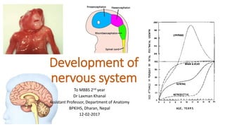
- 10. Anterior portion of neural tube forms three primary brain vesicles. 1. Prosencephalon: Telencephalon + Diencephalon 2. Mesencephalon: 3. Rhombencephalon: Metencephalon + Mylencephalon I & II CN III & IV CN V- XII CN
- 11. Development of ventricular system 4th
- 12. Development of spinal cord L TS neural tube Neuroepithelial cells L Neuroblast cell L Mantle cell layer Marginal cell layer L A B A B R F Anterior horn Posterior horn intermediate horn Sensory Motor Autonomic
- 13. Neuroblast Glioblast Microglial cells Ependymal cells Origin of various types of cells
- 14. Brain stem Arrangement of alar and basal plate is intact but the arrangement is different. Higher center Basal plate regress and alar plate accentuate. A B
- 15. G S E S V E G V E Development of functional columns in brain stem Basal plate – motor components Skeletal muscle 1. Somatic muscles - GSE 2. Pharyngeal arch muscles- SVE Smooth muscle- GVE (autonomic) CN supplying somatic skeletal muscles Motor nerve of pharyngeal arch CN with parasympathetic actions
- 16. G S E S V E G V E G S A S V A G V A S S A Development of functional columns in brain stem Alar plate- sensory column Senses • General senses • Special sense- taste • Senses from viscera • Special sense-hearing & Eqb Pain, touch, temperature, pressure Taste Sense of distension of viscera Hearing & equilibrium
- 17. Mesencephalic flexure Cervical flexure Pontine flexure Rhombic lip • Postero-lateral extension of alar plate of metencephalon. • Give rise to cerebellum. Rhombic lip
- 18. TS of caudal part of Pons Rhombic lip Cerebellar plate TS Mesencephalon Alar plate Basal plate Alar plate Sup colliculus Inf colliculus Basal plate Nuclei of 3rd CN Nucleus of 4th CN
- 19. Development of Prosencephalon Diencephalon 3rd ventricle Telencephalon 2 lateral ventricles 1. Thalamus 2. Hypothalamus 3. Epithalamus 4. Subthalamus 5. Metathalamus 6. Neurohypophysis
- 20. Summary • The CNS develops from a dorsal thickening of ectoderm-the neural plate, which appears around the middle of the third week. • The neural plate is induced by the underlying notochord and paraxial mesoderm to form neural tube. • The cranial end of the neural tube forms the brain and the remainder forms the spinal cord. • The neural canal, the lumen of the neural tube, becomes the ventricles of the brain and the central canal of the spinal cord. • Defects in the closure of the neural tube (NTDs) account for most severe anomalies of nervous system.