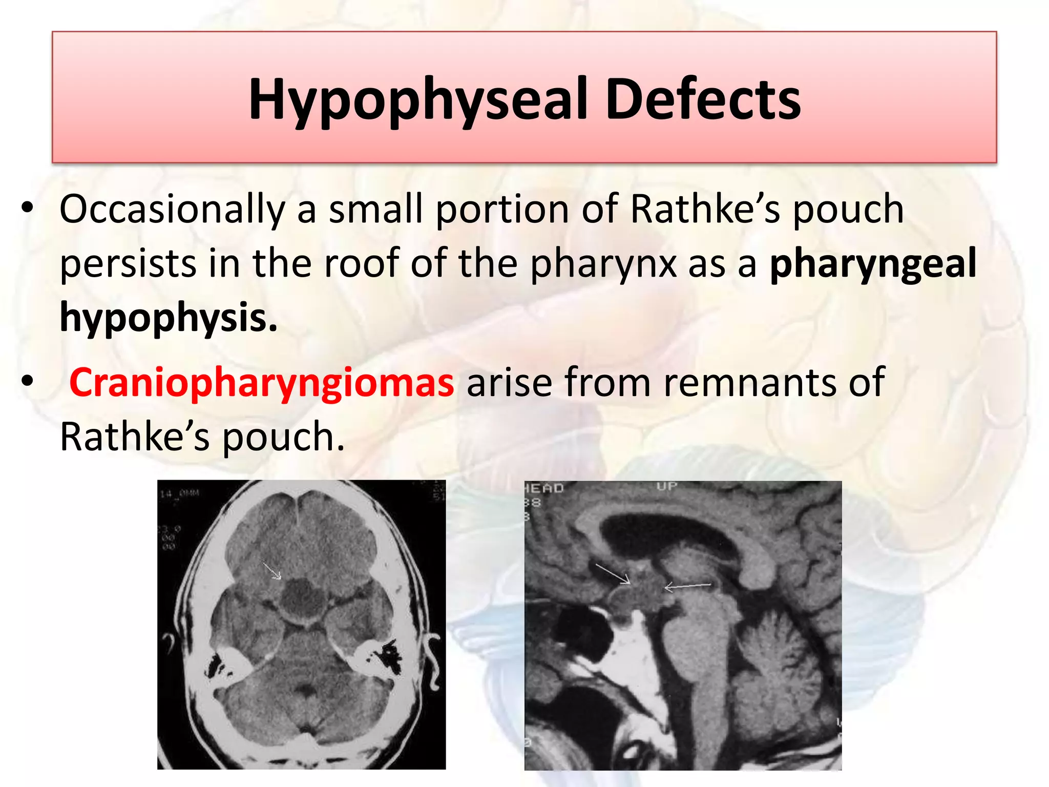
Downloaded 1,003 times


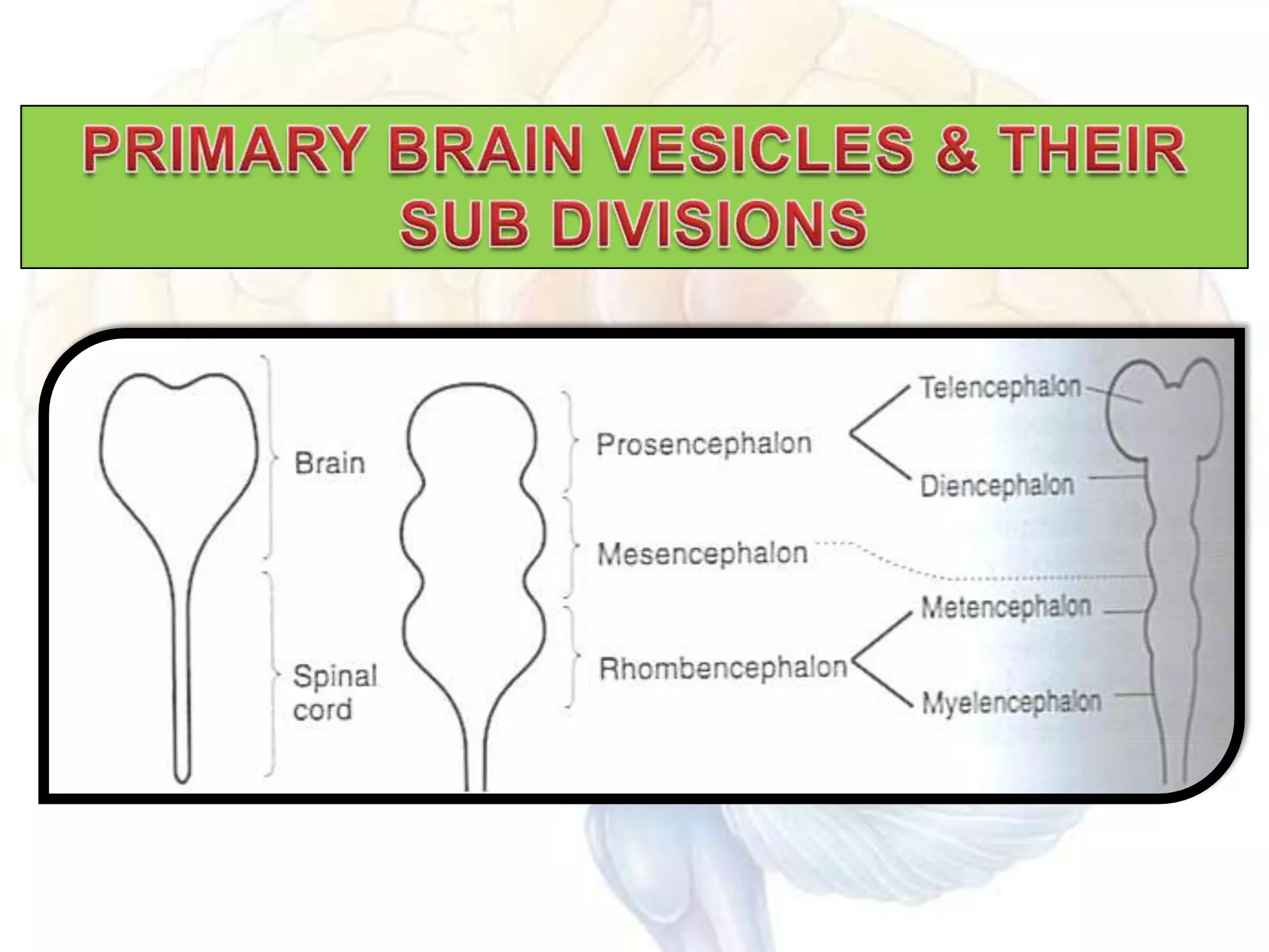
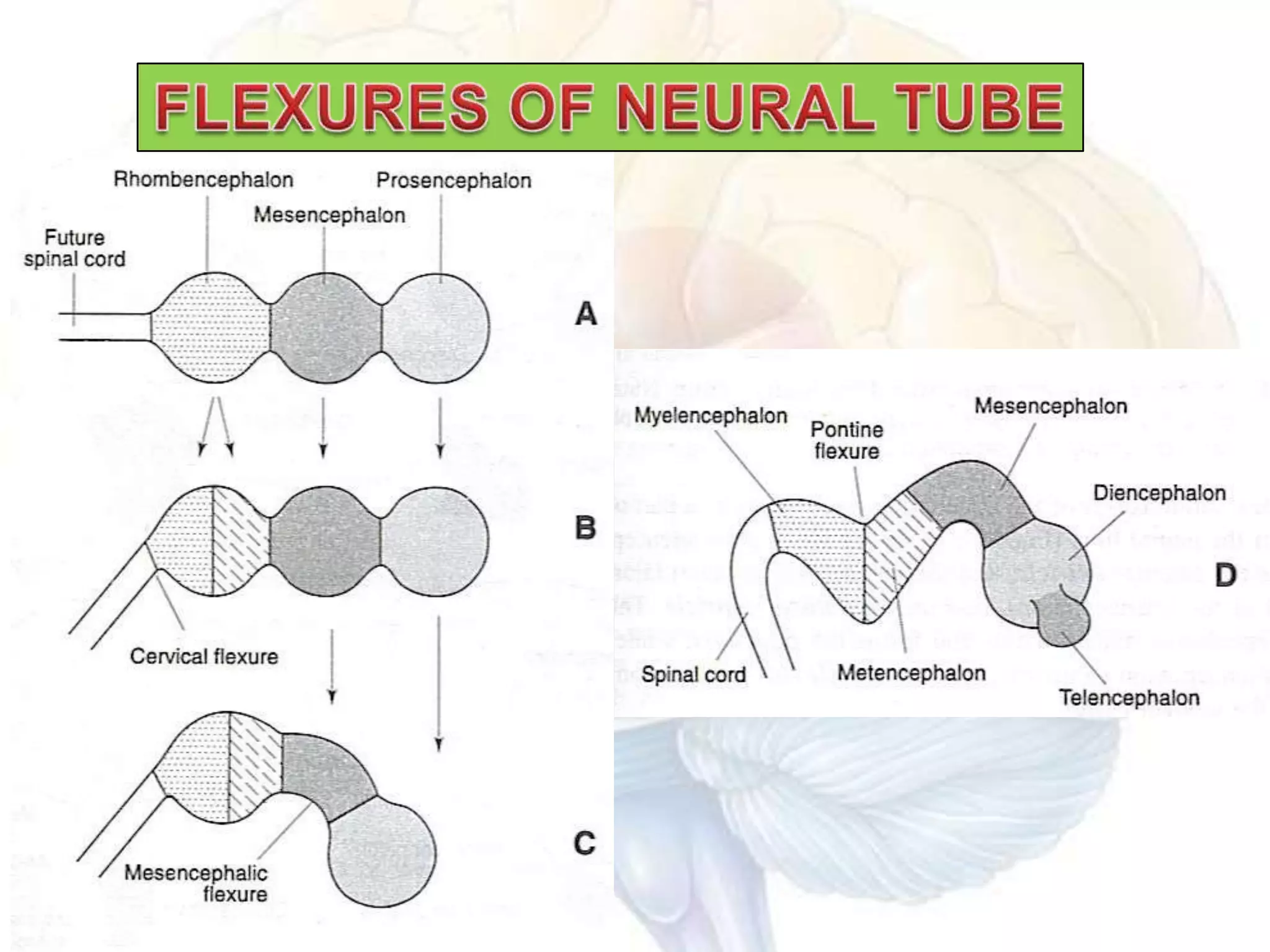
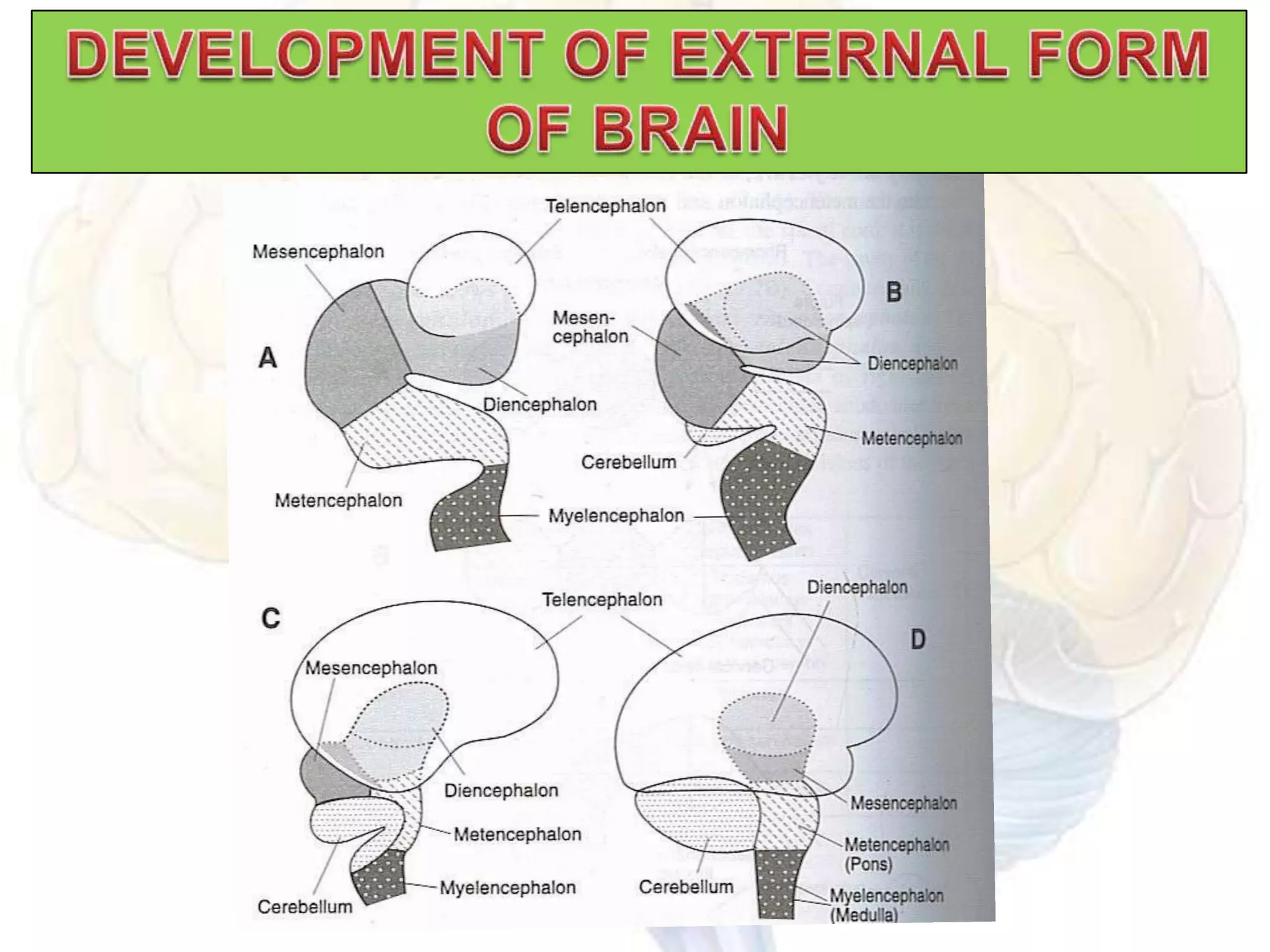


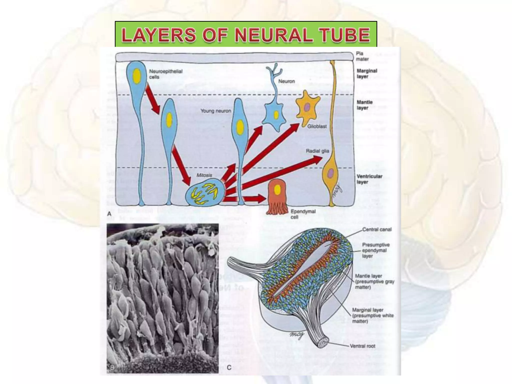


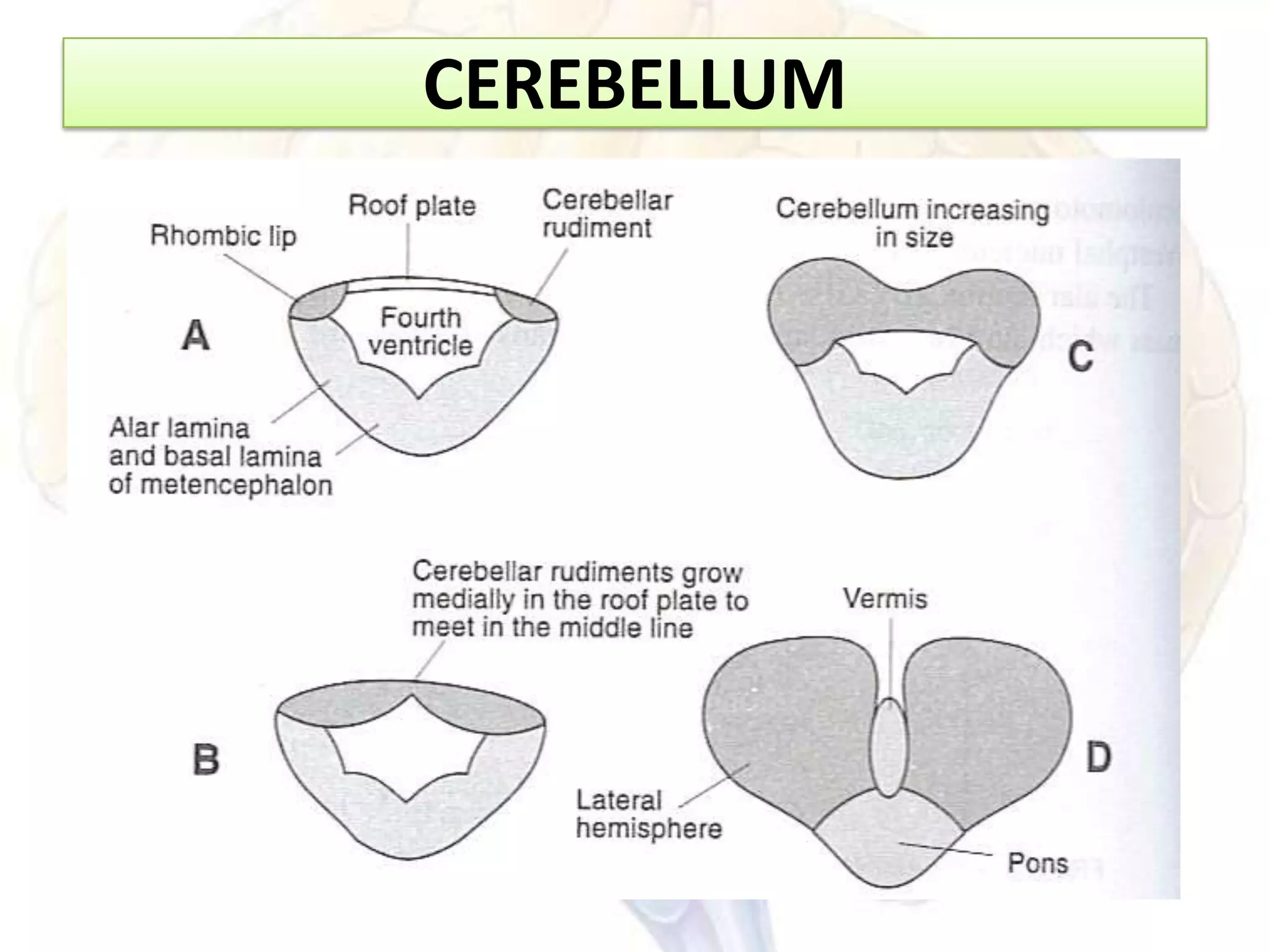
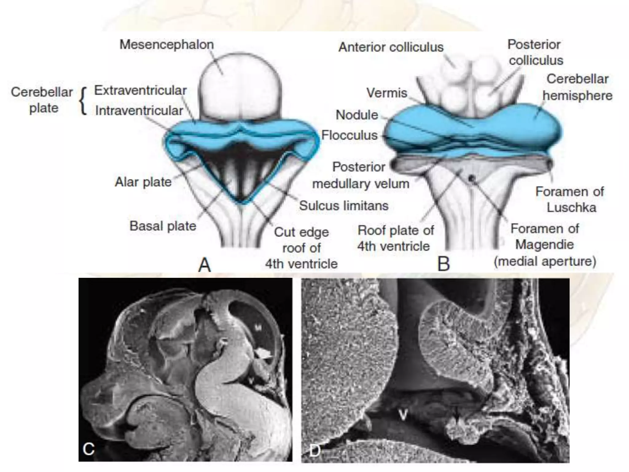

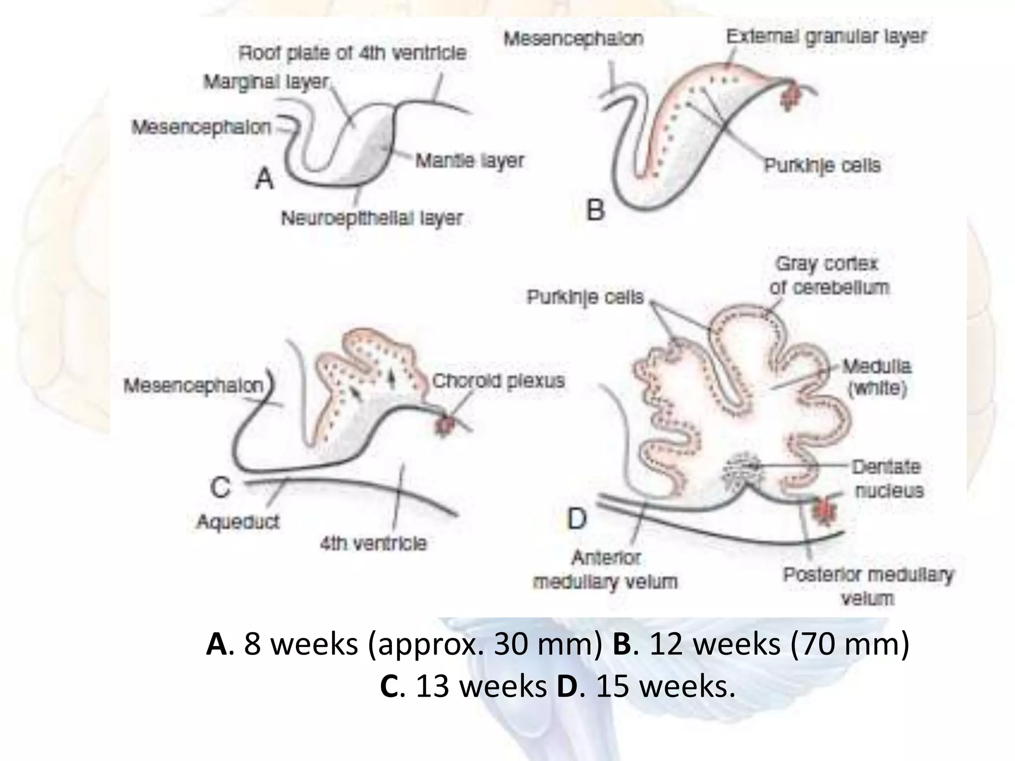

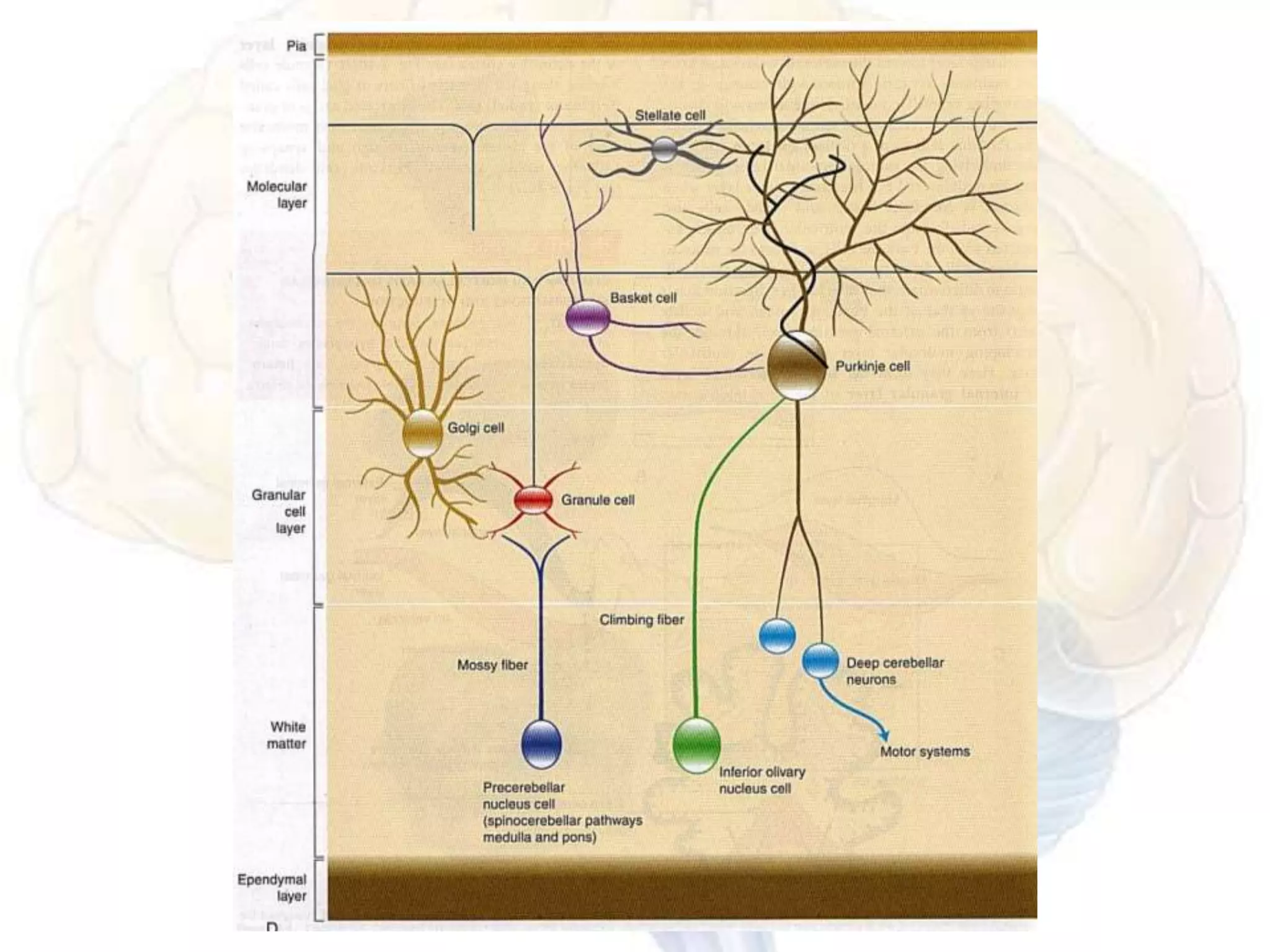


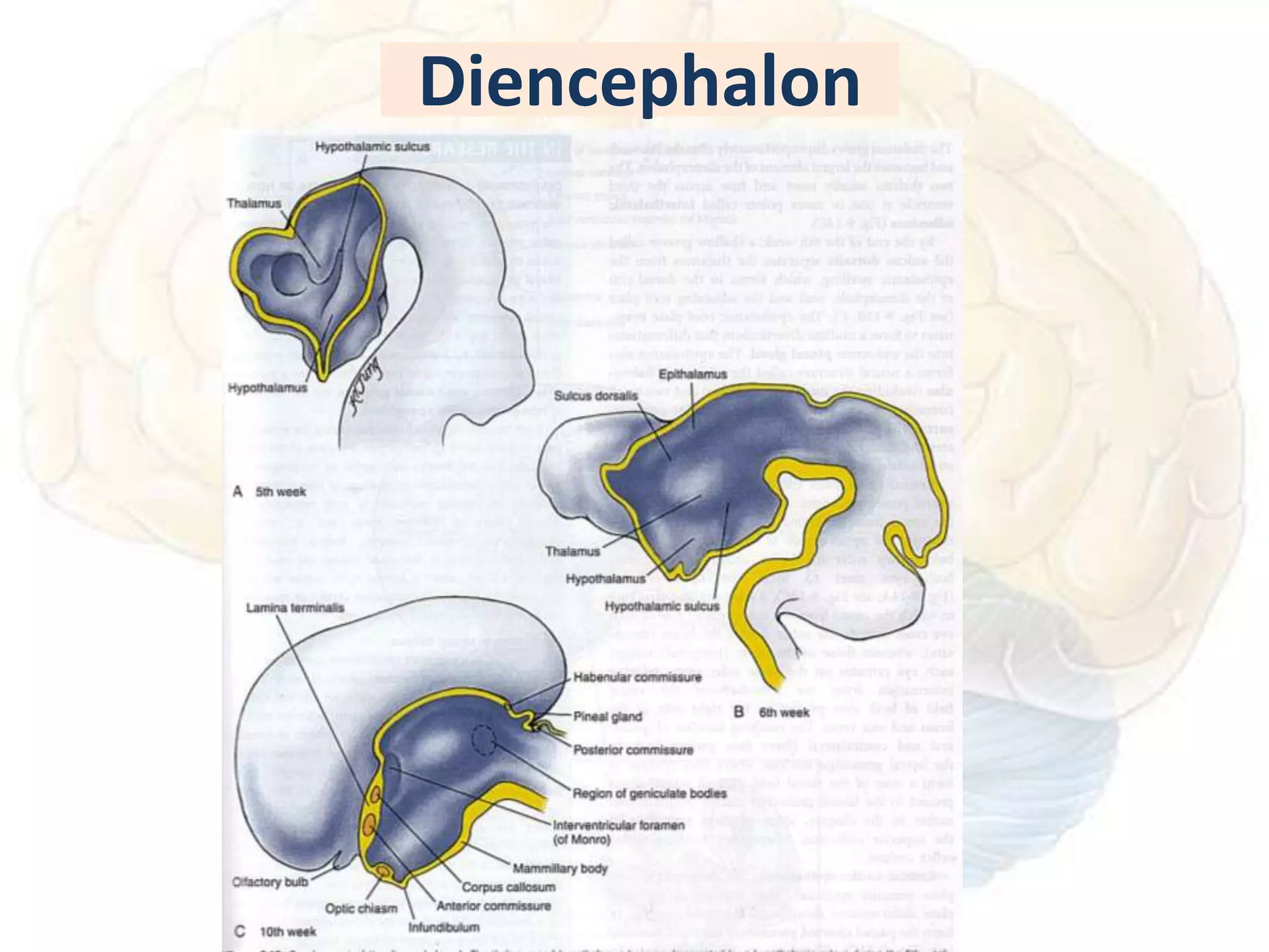

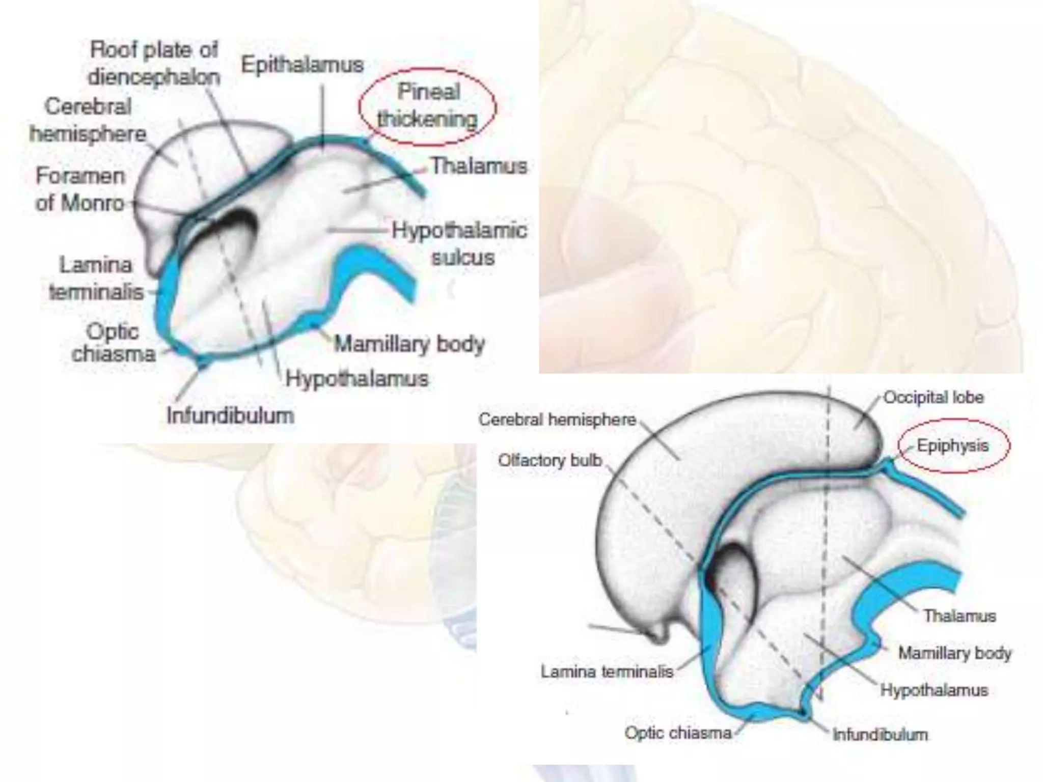


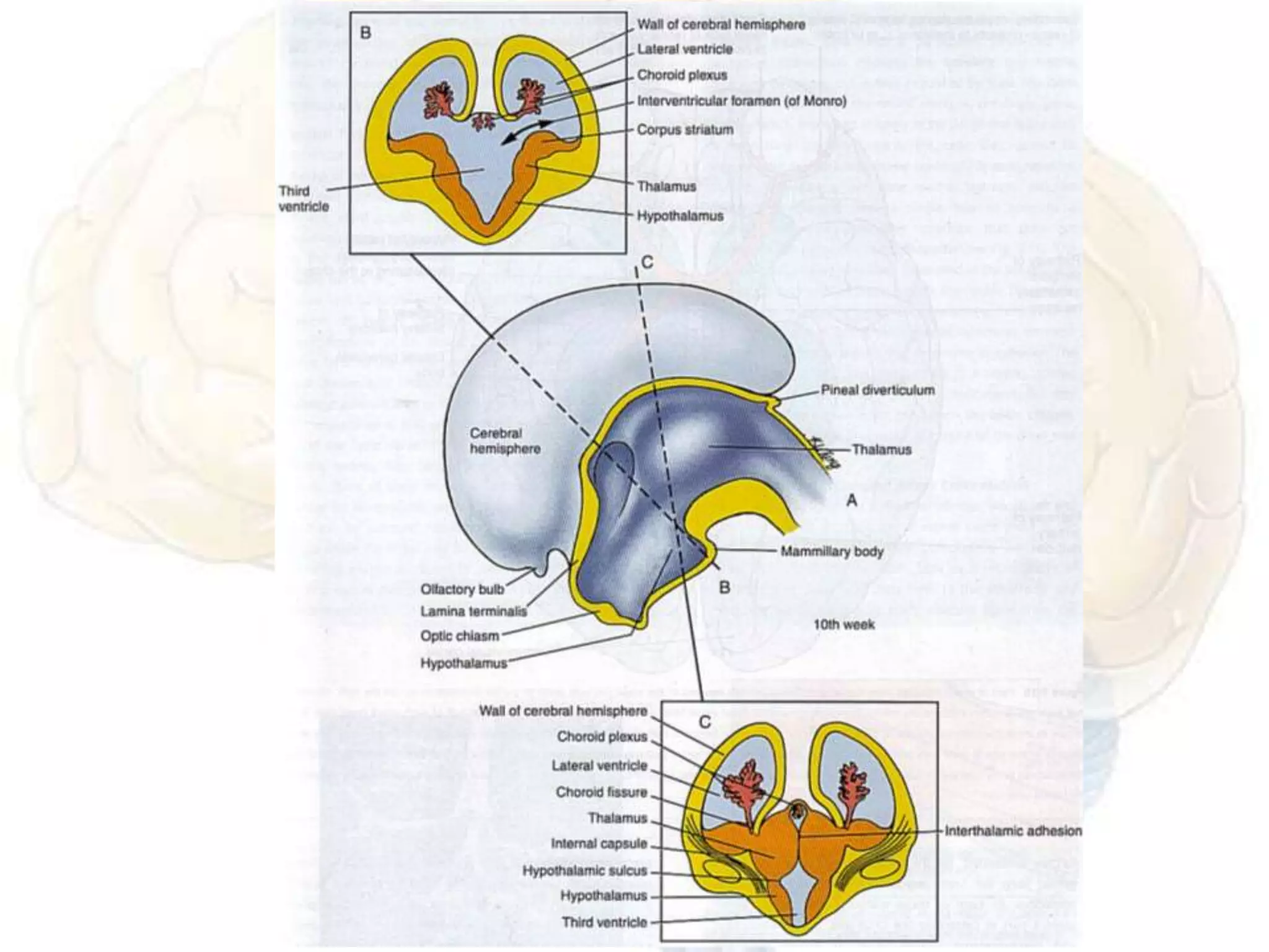


The human brain is the most complex organ on Earth. It develops from the neural tube which forms the basic structure of the central nervous system. As the brain develops from early embryonic stages through fetal development, different regions form including the rhombencephalon (medulla and pons), mesencephalon, and prosencephalon (telencephalon and diencephalon). Precisely regulated molecular processes guide the formation, migration and differentiation of neurons and glial cells in each brain region. Malformations can occur if these processes are disrupted, leading to neurological disorders.


