Downloaded 148 times


![Ion Movements in a Neuron
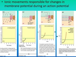
• K+
– [K+] higher inside cell than outside
– Attracted to fixed anions inside cell
– High membrane permeability
– Flows slowly out of cell
• Na+
– [Na+] higher outside cell than inside
– Attracted to fixed anions inside cell
– Low membrane permeability
– Flows slowly into cell](https://image.slidesharecdn.com/6-140503112043-phpapp02/85/6-nervous-system-108-320.jpg)
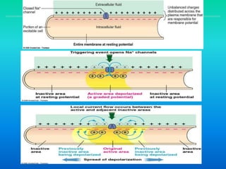
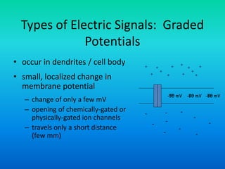
![Resting Potentials
• Resting potential
– Typical membrane potential for
cells
– Depends on concentration
gradients and membrane
permeabilities for different ions
involved
– -65 to -85 mV
– [Na+] and [K+] inside the cell are
maintained using Na+/K+ pumps
ICF (-)ECF (+)
Na/K pump](https://image.slidesharecdn.com/6-140503112043-phpapp02/85/6-nervous-system-109-320.jpg)
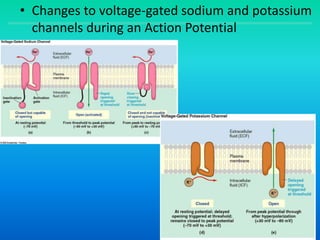
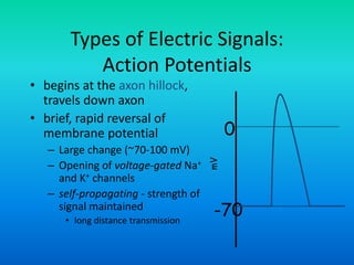
![mV
0
-70
+30
threshold
• voltage-gated Na+ channels open
– Na+ enters cell → further depolarization → more channels open → further
depolarization
• membrane reverses polarity (+30 mV)
• K+ channels close
• [Na+] and [K+] restored by the Na+-K+ pump
• K+ rushes out of the cell
– membrane potential restored
• Na+ channels close
• Delayed opening of voltage-gated K+ channels
Action Potential: Repolarization](https://image.slidesharecdn.com/6-140503112043-phpapp02/85/6-nervous-system-119-320.jpg)

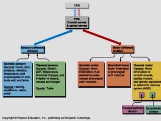
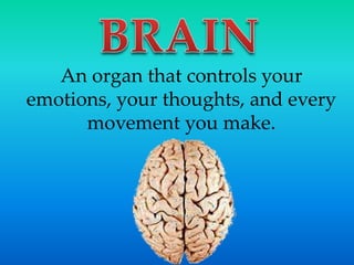
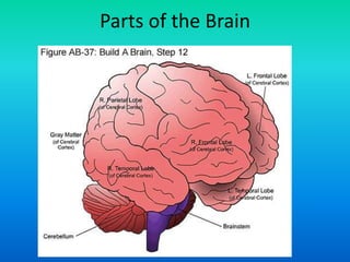
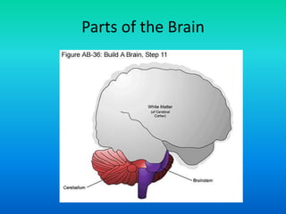
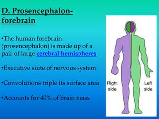
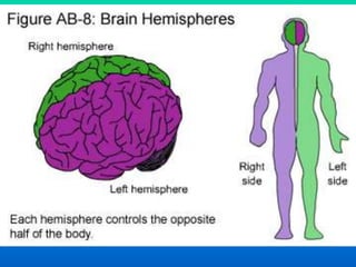
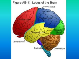
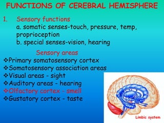
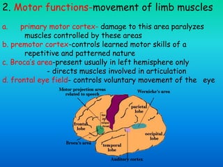
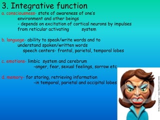
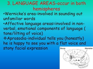
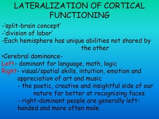
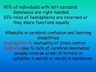
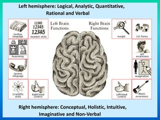
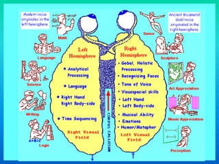
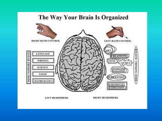
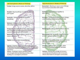
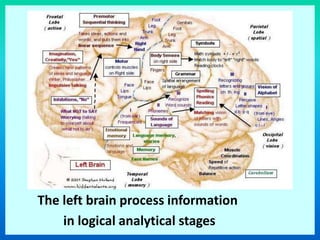
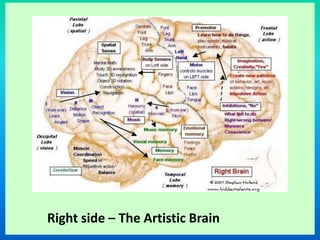
The document discusses the functions of the cerebral hemispheres. It states that the cerebral hemispheres are responsible for sensory functions like touch, vision, and hearing through specialized sensory areas. They are also responsible for motor functions through areas like the primary motor cortex. The cerebral hemispheres enable conscious awareness and functions like language, emotions, and memory which are mediated by structures like the limbic system. Association areas allow the integration and interpretation of sensory information. In most people, the left hemisphere dominates functions like language and logic while the right hemisphere dominates spatial skills, intuition, and artistic appreciation.


![Chapter 2[1]](https://cdn.slidesharecdn.com/ss_thumbnails/chapter21-150306090428-conversion-gate01-thumbnail.jpg?width=640&height=640&fit=bounds)
![Chapter 1[1]](https://cdn.slidesharecdn.com/ss_thumbnails/chapter11-150306090427-conversion-gate01-thumbnail.jpg?width=640&height=640&fit=bounds)
![Bio sci 8_lec_001[2]](https://cdn.slidesharecdn.com/ss_thumbnails/biosci8lec0012-150306090424-conversion-gate01-thumbnail.jpg?width=640&height=640&fit=bounds)
![3 lec metabolic_changes_in_drugs[1]](https://cdn.slidesharecdn.com/ss_thumbnails/3lecmetabolicchangesindrugs1-150306090419-conversion-gate01-thumbnail.jpg?width=640&height=640&fit=bounds)
![2 lab metabolic_changes_in_organic_medicinals[2]](https://cdn.slidesharecdn.com/ss_thumbnails/2labmetabolicchangesinorganicmedicinals2-150306090407-conversion-gate01-thumbnail.jpg?width=640&height=640&fit=bounds)
![1 lab physico-chemical_properties_of_drugs[1]](https://cdn.slidesharecdn.com/ss_thumbnails/1labphysico-chemicalpropertiesofdrugs1-150306090358-conversion-gate01-thumbnail.jpg?width=640&height=640&fit=bounds)


