Downloaded 2,327 times

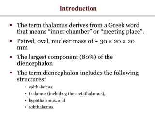
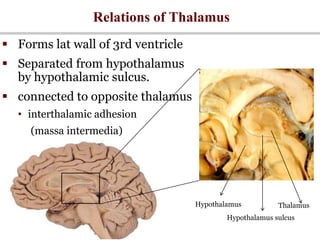
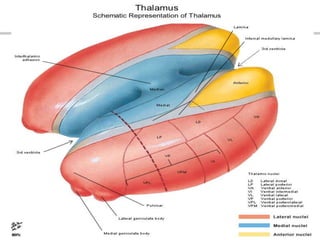
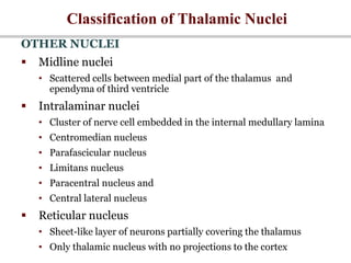
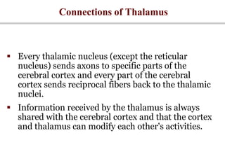
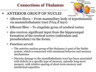
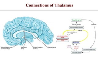
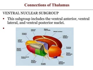


The thalamus is a paired, oval structure located in the diencephalon that serves as a relay center for sensory and motor signals to and from the cerebral cortex. It is divided into several nuclei that process different sensory modalities. The thalamus receives input from various areas and projects to specific regions of the cortex. Damage to certain thalamic nuclei can disrupt motor control, sensory processing, and cause syndromes like thalamic pain. Surgical procedures targeting thalamic nuclei have been used to treat chronic pain conditions.