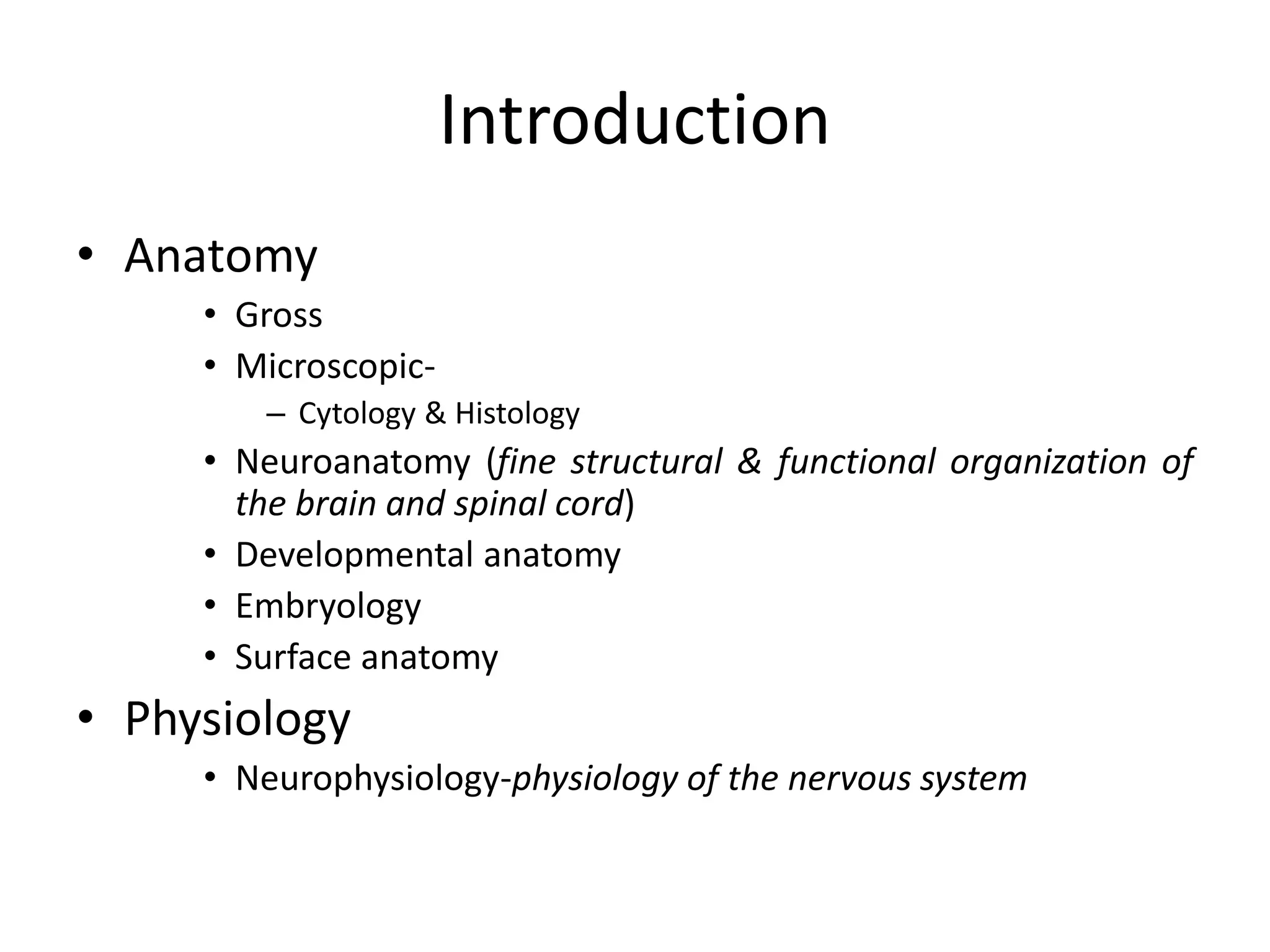
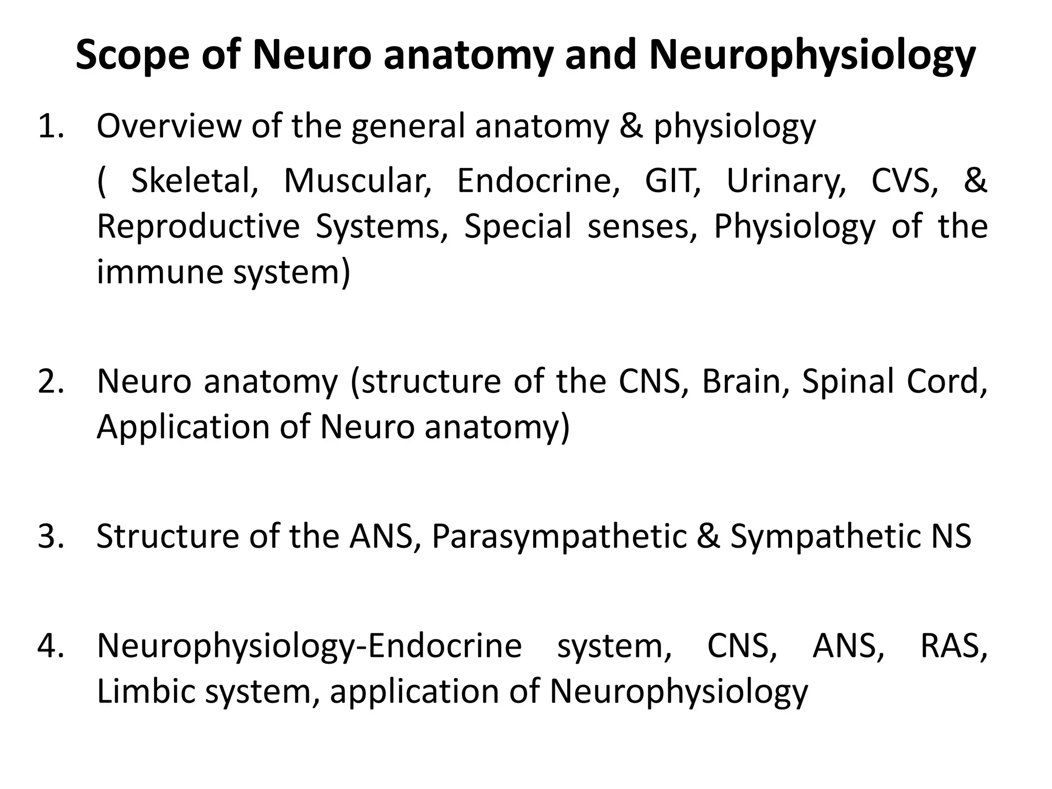
NEUROANATOMY & NEUROPHYSIOLOGY
By
Dr. Ssebadduka Daniel
Introduction
• Anatomy
• Gross
• Microscopic-
– Cytology & Histology
• Neuroanatomy (fine structural & functional organization of
the brain and spinal cord)
• Developmental anatomy
• Embryology
• Surface anatomy
• Physiology
• Neurophysiology-physiology of the nervous system
Scope of Neuro anatomy and Neurophysiology
1. Overview of the general anatomy & physiology
( Skeletal, Muscular, Endocrine, GIT, Urinary, CVS, &
Reproductive Systems, Special senses, Physiology of the
immune system)
2. Neuro anatomy (structure of the CNS, Brain, Spinal Cord,
Application of Neuro anatomy)
3. Structure of the ANS, Parasympathetic & Sympathetic NS
4. Neurophysiology-Endocrine system, CNS, ANS, RAS,
Limbic system, application of Neurophysiology
Major Divisions of the NS
The Skull: Anterior View
Lateral View of the Skull
Posterior View of the skull
Superior View of the Skull
Base of Skull: Exterior View
Base of Skull: Interior View
• Afferent (Sensory)-Toward
• Efferent (Motor)- Away from
• Dorsal - toward the back
• Ventral - toward the stomach
• Anterior & Posterior
– Rostral & Caudal ~
• Medial - toward the midline
• Lateral - away from the midline
• Ipsilateral - same side
• Contralateral - opposite side
• Decussate - crossover ~
Terminology
Medial
Anatomical Reference
Dorsal
Ventral
Rostral/
Anterior
Caudal/
Posterior
Lateral
Ipsilateral &
Contralateral
Dorsal -
Ventral
-
In reference to the BRAIN
Anterior - Posterior -
Rostral Caudal
Inferior
Superior
Brainstem &
spinal cord
Ventral - Dorsal -
Rostral
Caudal
Anterior Posterior
Superior
Inferior
Development of the Nervous System
• Embryo has 3 layers
– endoderm ---> internal organs
– mesoderm ---> muscles & skeleton
– ectoderm ---> nervous system & skin
• Neural plate (~3 weeks)
– sheet of cells
– neural groove
– folds rostral to caudal ---> tube ~
Devt of the NS
• Neural tube
– CNS develops from walls
– as tube develops dorsal portion pinches off &
forms...
• Neural crest
– develops into all neurons of PNS
• Mesoderm
– somites ---> vertebrae & muscles ~
Early development of nervous system in embryo
Neural Plate  Neural Groove  Neural Tube
Fuse Dorsally
Rostral
Caudal
Prosencephalon
(forebrain)
Mesencephalon
(midbrain)
Rhombencephalon
(hindbrain)
Telencephalon
(2 cerebral
hemespheres)
Diencephalon
(between brain)
Mesencephalon
(Midbain)
Development of nervous system in embryo
Telencephalon
Diencephalon
Corpus Callosum
Cerebral Cortex
Thalamus
Hypothalamus
Development of nervous system in embryo
Coronal Section
Lateral Ventricles &
Third Ventricle
Development of nervous system in embryo
Midbrain:
becomes Tectum (roof)
Tegmentum (floor)
Hindbrain:
becomes Cerebellum
Pons
Medulla
Stages of Cellular Activity
• 6 distinct stages
1. Neurogenesis
– mitosis
– nonneural cells
neurons do not divide
– develop into neurons or glia
glia produced throughout life ~
Stages of Cellular Activity
2. Cell migration
– to specific “brain” locations
– along radial glia
– later: along other neurons
3. Differentiation
– into specific neuron types
– by induction
influenced by surrounding cells
– or cell-autonomous ~
Stages of Cellular Activity
4. Synaptogenesis
– formation of synapses
– growth of axons & dendrites
growth cones
5. Neuronal cell death
– apoptosis: programmed cell death
– 20-80% of neurons in a region
– genetically programmed
– lack of neurotrophic factors ~
Stages of Cellular Activity
6. synaptic rearrangement
– elimination of synapse
– formation of new synapses
– dependent on neural activity ~
Differentiation
• Specialization of structures
• 3 primary vesicles
– rostral end of tube
– develops into brain
• Prosencephalon ---> forebrain
• Mesencephalon ---> midbrain
• Rhombencephalon ---> hindbrain ~
Prosencephalon
• Secondary vesicles form & separate
– optic ---> retinas
retina & optic nerve part of brain
not PNS
– telencephalic ---> telencephalon
– remainder ---> diencephalon ~
Other Primary Vesicles
• Mesencephalic ---> mesencephalon
– dorsal - tectum
– ventral - tegmentum
– tube - cerebral aqueduct
• Rhombencephalic
– rostral - metencephalon
– caudal - myelencephalon
– tube - 4th ventricle ~
Telencephalon
• Cortical areas
• Fiber systems
• Landmarks
– Fissures /Sulci
– Lobes
– Gyri ~
Superior surface
Longitudinal Fissure
(sulcus)
Central
Lateral
The Structures of the CNS
• The Cerebrum
• The Diencephalon
• The Mesencephalon
• The Spinal cord
Copyright © 2002 Pearson Education, Inc., publishing as Benjamin Cummings
Fig. 48.20
cerebrum corpus
callosum
thalamus
cerebellum
medulla
oblongata
hypothalamus
pituitary
pons
spinal cord
Pineal gland
Cerebrum
• Involved with higher brain functions.
• Processes sensory information.
• Initiates motor functions.
• Integrates information.
• The
cerebrum is
divided into
frontal,
temporal,
occipital,
and parietal
lobes.
Regions of the cerebrum are
specialized for different functions
• Frontal lobe.
– Contains the primary motor cortex.
• Parietal lobe.
– Contains the primary somatosensory cortex.
Copyright © 2002 Pearson Education, Inc., publishing as Benjamin Cummings
Fig. 48.25
• Integrative Function of the Association Areas.
–Much of the cerebrum is given over to
association areas.
• Areas where sensory information is integrated
and assessed and motor responses are
planned.
Copyright © 2002 Pearson Education, Inc., publishing as Benjamin Cummings
• The brain exhibits plasticity of function.
–For example, infants with intractable
epilepsy may have an entire cerebral
hemisphere removed.
• The remaining hemisphere can provide the
function normally provided by both
hemispheres.
Copyright © 2002 Pearson Education, Inc., publishing as Benjamin Cummings
• Lateralization of Brain Function.
–The left hemisphere.
• Specializes in language, math, logic operations, and
the processing of serial sequences of information, and
visual and auditory details.
• Specializes in detailed activities required for motor
control.
–The right hemisphere.
• Specializes in pattern recognition, spatial relationships,
nonverbal ideation, emotional processing, and the
parallel processing of information.
Copyright © 2002 Pearson Education, Inc., publishing as Benjamin Cummings
• Language and Speech.
–Broca’s area.
• Usually located in the left hemisphere’s frontal lobe
• Responsible for speech production.
–Wernicke’s area.
• Usually located in the right hemisphere’s temporal lobe
• Responsible for the comprehension of speech.
–Other speech areas are involved
generating verbs to match nouns, grouping
together related words, etc.
Copyright © 2002 Pearson Education, Inc., publishing as Benjamin Cummings
• Emotions.
– In mammals, the limbic system is composed
of the hippocampus, olfactory cortex, inner
portions of the cortex’s lobes, and parts of the
thalamus and hypothalamus.
• Mediates basic emotions (fear, anger), involved in
emotional bonding, establishes emotional memory
– For example,
the amygdala
is involved in
recognizing
the emotional
content of
facial expression.
• Memory and Learning.
–Short-term memory stored in the frontal
lobes.
–The establishment of long-term memory
involves the hippocampus.
• The transfer of information from short-term to
long-term memory.
– Is enhanced by repetition (remember that when you are
preparing for an exam).
– Influenced by emotional states mediated by the
amygdala.
– Influenced by association with previously stored
information.
Copyright © 2002 Pearson Education, Inc., publishing as Benjamin Cummings
–Different types of long-term memories are
stored in different regions of the brain.
–Memorization-type memory can be rapid.
• Primarily involves changes in the strength of
existing nerve connections.
–Learning of skills and procedures is slower.
• Appears to involve cellular mechanisms similar
to those involved in brain growth and
development.
Copyright © 2002 Pearson Education, Inc., publishing as Benjamin Cummings
• Human Consciousness.
–Brain imaging can show neural activity
associated with:
• Conscious perceptual choice
• Unconscious processing
• Memory retrieval
• Working memory.
–Consciousness appears to be a whole-
brain phenomenon.
Copyright © 2002 Pearson Education, Inc., publishing as Benjamin Cummings
• The mammalian PNS has the ability to repair
itself, the CNS does not.
–Research on nerve cell development and
neural stem cells may be the future of
treatment for damage to the CNS.
Research on neuron development
and neural stem cells may lead to
new approaches for treating CNS
injuries and diseases
Copyright © 2002 Pearson Education, Inc., publishing as Benjamin Cummings
• Neural Stem Cells.
–The adult human brain does produce new
nerve cells.
• New nerve cells have been found in the
hippocampus.
• Since mature human brain cells cannot
undergo cell division the new cells must have
arisen from stem cells.
Copyright © 2002 Pearson Education, Inc., publishing as Benjamin Cummings
Thalamus
• Relay center for sensory tracts
from the spinal cord to the
cerebrum.
• Contains centers for sensation
of pain, temperature, and touch.
• Involved with emotions and
alerting or arousal mechanisms.
• The Reticular System, Arousal, and Sleep.
–The reticular activating system (RAS) of
the reticular formation.
• Regulates sleep
and arousal.
• Acts as a
sensory filter.
Copyright © 2002 Pearson Education, Inc., publishing as Benjamin Cummings
Fig. 48.21
–Sleep and wakefulness produces patterns
of electrical activity in the brain that can be
recorded as an electroencephalogram
(EEG).
• Most dreaming
occurs during
REM (rapid
eye movement)
sleep.
Copyright © 2002 Pearson Education, Inc., publishing as Benjamin Cummings
Fig. 48.22b-d
Hypothalamus
• autonomic control center- blood pressure,
rate and force of heart contraction, center
for emotional response and behavior
• body temperature
• water balance and thirst
• sleep/wake cycles
• appetite
• sexual arousal
• control of endocrine functioning:
Acts on the pituitary gland through the
release of neurosecretions.
Regulates:
Hypothalamus
Pituitary
• Cerebellar peduncles
• Tectum
• Superior colliculi
• Inferior colliculi
• Substantia nigra
• Red nuclei
Midbrain
thalamus
Red nucleus
Substantia nigra
Posterior
Anterior
Midbrain
• Contains ascending and descending
tracts to the cerebrum and
thalamus.
• Reflex center for eye muscles.
• Also involved with processing visual
and auditory information (connects
head movements with visual and
auditory stimuli).
Pons
• Connects the two
halves of the
cerebellum.
• Regulates breathing.
Medulla Oblongata
• Composed of nerve tracts
to and from the brain
(these tracts cross over
left to right and right to left)
• May be regarded as an
extension of the spinal
cord
• Almost all of the cranial
nerves arise from this
region
Medulla Oblongata
Contains control centers for
many subconscious
activities
• Respiratory rate
• Heart rate
• Arteriole constriction
• Swallowing
• Hiccupping
• Coughing
• Sneezing
Cerebellum
• Controls and coordinates
muscular activity.
• Important in equilibrium,
posture and movement.

Neuroanatomy & Neurophysiology DCP 1105.ppt

  • 1.
  • 2.
    Introduction • Anatomy • Gross •Microscopic- – Cytology & Histology • Neuroanatomy (fine structural & functional organization of the brain and spinal cord) • Developmental anatomy • Embryology • Surface anatomy • Physiology • Neurophysiology-physiology of the nervous system
  • 3.
    Scope of Neuroanatomy and Neurophysiology 1. Overview of the general anatomy & physiology ( Skeletal, Muscular, Endocrine, GIT, Urinary, CVS, & Reproductive Systems, Special senses, Physiology of the immune system) 2. Neuro anatomy (structure of the CNS, Brain, Spinal Cord, Application of Neuro anatomy) 3. Structure of the ANS, Parasympathetic & Sympathetic NS 4. Neurophysiology-Endocrine system, CNS, ANS, RAS, Limbic system, application of Neurophysiology
  • 5.
  • 6.
  • 7.
    Lateral View ofthe Skull
  • 8.
  • 9.
  • 10.
    Base of Skull:Exterior View
  • 11.
    Base of Skull:Interior View
  • 12.
    • Afferent (Sensory)-Toward •Efferent (Motor)- Away from • Dorsal - toward the back • Ventral - toward the stomach • Anterior & Posterior – Rostral & Caudal ~ • Medial - toward the midline • Lateral - away from the midline • Ipsilateral - same side • Contralateral - opposite side • Decussate - crossover ~ Terminology
  • 13.
  • 14.
    Dorsal - Ventral - In referenceto the BRAIN Anterior - Posterior - Rostral Caudal Inferior Superior
  • 15.
    Brainstem & spinal cord Ventral- Dorsal - Rostral Caudal Anterior Posterior Superior Inferior
  • 16.
    Development of theNervous System • Embryo has 3 layers – endoderm ---> internal organs – mesoderm ---> muscles & skeleton – ectoderm ---> nervous system & skin • Neural plate (~3 weeks) – sheet of cells – neural groove – folds rostral to caudal ---> tube ~
  • 17.
    Devt of theNS • Neural tube – CNS develops from walls – as tube develops dorsal portion pinches off & forms... • Neural crest – develops into all neurons of PNS • Mesoderm – somites ---> vertebrae & muscles ~
  • 18.
    Early development ofnervous system in embryo Neural Plate  Neural Groove  Neural Tube Fuse Dorsally Rostral Caudal Prosencephalon (forebrain) Mesencephalon (midbrain) Rhombencephalon (hindbrain)
  • 19.
  • 20.
    Telencephalon Diencephalon Corpus Callosum Cerebral Cortex Thalamus Hypothalamus Developmentof nervous system in embryo Coronal Section Lateral Ventricles & Third Ventricle
  • 21.
    Development of nervoussystem in embryo Midbrain: becomes Tectum (roof) Tegmentum (floor) Hindbrain: becomes Cerebellum Pons Medulla
  • 22.
    Stages of CellularActivity • 6 distinct stages 1. Neurogenesis – mitosis – nonneural cells neurons do not divide – develop into neurons or glia glia produced throughout life ~
  • 23.
    Stages of CellularActivity 2. Cell migration – to specific “brain” locations – along radial glia – later: along other neurons 3. Differentiation – into specific neuron types – by induction influenced by surrounding cells – or cell-autonomous ~
  • 24.
    Stages of CellularActivity 4. Synaptogenesis – formation of synapses – growth of axons & dendrites growth cones 5. Neuronal cell death – apoptosis: programmed cell death – 20-80% of neurons in a region – genetically programmed – lack of neurotrophic factors ~
  • 25.
    Stages of CellularActivity 6. synaptic rearrangement – elimination of synapse – formation of new synapses – dependent on neural activity ~
  • 26.
    Differentiation • Specialization ofstructures • 3 primary vesicles – rostral end of tube – develops into brain • Prosencephalon ---> forebrain • Mesencephalon ---> midbrain • Rhombencephalon ---> hindbrain ~
  • 27.
    Prosencephalon • Secondary vesiclesform & separate – optic ---> retinas retina & optic nerve part of brain not PNS – telencephalic ---> telencephalon – remainder ---> diencephalon ~
  • 28.
    Other Primary Vesicles •Mesencephalic ---> mesencephalon – dorsal - tectum – ventral - tegmentum – tube - cerebral aqueduct • Rhombencephalic – rostral - metencephalon – caudal - myelencephalon – tube - 4th ventricle ~
  • 29.
    Telencephalon • Cortical areas •Fiber systems • Landmarks – Fissures /Sulci – Lobes – Gyri ~
  • 30.
  • 31.
  • 32.
    The Structures ofthe CNS • The Cerebrum • The Diencephalon • The Mesencephalon • The Spinal cord
  • 36.
    Copyright © 2002Pearson Education, Inc., publishing as Benjamin Cummings Fig. 48.20
  • 37.
  • 38.
    Cerebrum • Involved withhigher brain functions. • Processes sensory information. • Initiates motor functions. • Integrates information.
  • 39.
    • The cerebrum is dividedinto frontal, temporal, occipital, and parietal lobes. Regions of the cerebrum are specialized for different functions
  • 40.
    • Frontal lobe. –Contains the primary motor cortex. • Parietal lobe. – Contains the primary somatosensory cortex.
  • 41.
    Copyright © 2002Pearson Education, Inc., publishing as Benjamin Cummings Fig. 48.25
  • 42.
    • Integrative Functionof the Association Areas. –Much of the cerebrum is given over to association areas. • Areas where sensory information is integrated and assessed and motor responses are planned. Copyright © 2002 Pearson Education, Inc., publishing as Benjamin Cummings
  • 43.
    • The brainexhibits plasticity of function. –For example, infants with intractable epilepsy may have an entire cerebral hemisphere removed. • The remaining hemisphere can provide the function normally provided by both hemispheres. Copyright © 2002 Pearson Education, Inc., publishing as Benjamin Cummings
  • 44.
    • Lateralization ofBrain Function. –The left hemisphere. • Specializes in language, math, logic operations, and the processing of serial sequences of information, and visual and auditory details. • Specializes in detailed activities required for motor control. –The right hemisphere. • Specializes in pattern recognition, spatial relationships, nonverbal ideation, emotional processing, and the parallel processing of information. Copyright © 2002 Pearson Education, Inc., publishing as Benjamin Cummings
  • 45.
    • Language andSpeech. –Broca’s area. • Usually located in the left hemisphere’s frontal lobe • Responsible for speech production. –Wernicke’s area. • Usually located in the right hemisphere’s temporal lobe • Responsible for the comprehension of speech. –Other speech areas are involved generating verbs to match nouns, grouping together related words, etc. Copyright © 2002 Pearson Education, Inc., publishing as Benjamin Cummings
  • 46.
    • Emotions. – Inmammals, the limbic system is composed of the hippocampus, olfactory cortex, inner portions of the cortex’s lobes, and parts of the thalamus and hypothalamus. • Mediates basic emotions (fear, anger), involved in emotional bonding, establishes emotional memory – For example, the amygdala is involved in recognizing the emotional content of facial expression.
  • 47.
    • Memory andLearning. –Short-term memory stored in the frontal lobes. –The establishment of long-term memory involves the hippocampus. • The transfer of information from short-term to long-term memory. – Is enhanced by repetition (remember that when you are preparing for an exam). – Influenced by emotional states mediated by the amygdala. – Influenced by association with previously stored information. Copyright © 2002 Pearson Education, Inc., publishing as Benjamin Cummings
  • 48.
    –Different types oflong-term memories are stored in different regions of the brain. –Memorization-type memory can be rapid. • Primarily involves changes in the strength of existing nerve connections. –Learning of skills and procedures is slower. • Appears to involve cellular mechanisms similar to those involved in brain growth and development. Copyright © 2002 Pearson Education, Inc., publishing as Benjamin Cummings
  • 49.
    • Human Consciousness. –Brainimaging can show neural activity associated with: • Conscious perceptual choice • Unconscious processing • Memory retrieval • Working memory. –Consciousness appears to be a whole- brain phenomenon. Copyright © 2002 Pearson Education, Inc., publishing as Benjamin Cummings
  • 50.
    • The mammalianPNS has the ability to repair itself, the CNS does not. –Research on nerve cell development and neural stem cells may be the future of treatment for damage to the CNS. Research on neuron development and neural stem cells may lead to new approaches for treating CNS injuries and diseases Copyright © 2002 Pearson Education, Inc., publishing as Benjamin Cummings
  • 51.
    • Neural StemCells. –The adult human brain does produce new nerve cells. • New nerve cells have been found in the hippocampus. • Since mature human brain cells cannot undergo cell division the new cells must have arisen from stem cells. Copyright © 2002 Pearson Education, Inc., publishing as Benjamin Cummings
  • 52.
    Thalamus • Relay centerfor sensory tracts from the spinal cord to the cerebrum. • Contains centers for sensation of pain, temperature, and touch. • Involved with emotions and alerting or arousal mechanisms.
  • 53.
    • The ReticularSystem, Arousal, and Sleep. –The reticular activating system (RAS) of the reticular formation. • Regulates sleep and arousal. • Acts as a sensory filter. Copyright © 2002 Pearson Education, Inc., publishing as Benjamin Cummings Fig. 48.21
  • 54.
    –Sleep and wakefulnessproduces patterns of electrical activity in the brain that can be recorded as an electroencephalogram (EEG). • Most dreaming occurs during REM (rapid eye movement) sleep. Copyright © 2002 Pearson Education, Inc., publishing as Benjamin Cummings Fig. 48.22b-d
  • 55.
    Hypothalamus • autonomic controlcenter- blood pressure, rate and force of heart contraction, center for emotional response and behavior • body temperature • water balance and thirst • sleep/wake cycles • appetite • sexual arousal • control of endocrine functioning: Acts on the pituitary gland through the release of neurosecretions. Regulates:
  • 56.
  • 57.
  • 58.
    • Cerebellar peduncles •Tectum • Superior colliculi • Inferior colliculi • Substantia nigra • Red nuclei Midbrain thalamus Red nucleus Substantia nigra Posterior Anterior
  • 59.
    Midbrain • Contains ascendingand descending tracts to the cerebrum and thalamus. • Reflex center for eye muscles. • Also involved with processing visual and auditory information (connects head movements with visual and auditory stimuli).
  • 60.
    Pons • Connects thetwo halves of the cerebellum. • Regulates breathing.
  • 61.
    Medulla Oblongata • Composedof nerve tracts to and from the brain (these tracts cross over left to right and right to left) • May be regarded as an extension of the spinal cord • Almost all of the cranial nerves arise from this region
  • 62.
    Medulla Oblongata Contains controlcenters for many subconscious activities • Respiratory rate • Heart rate • Arteriole constriction • Swallowing • Hiccupping • Coughing • Sneezing
  • 63.
    Cerebellum • Controls andcoordinates muscular activity. • Important in equilibrium, posture and movement.