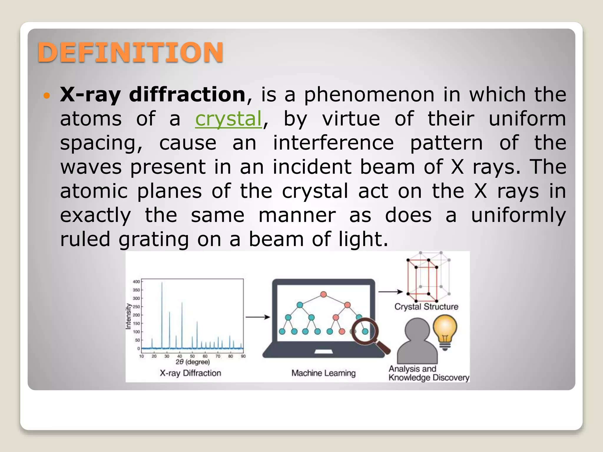
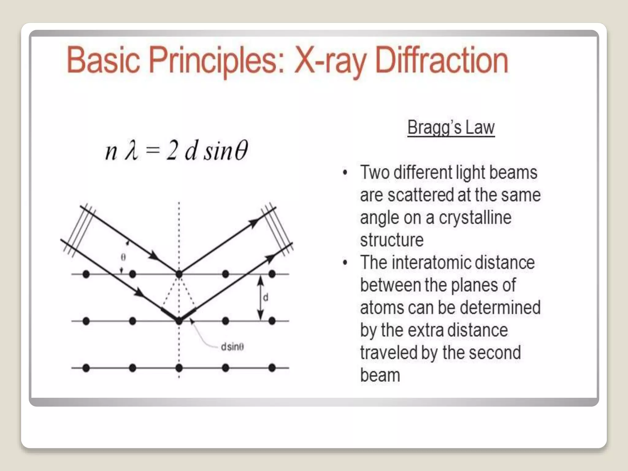
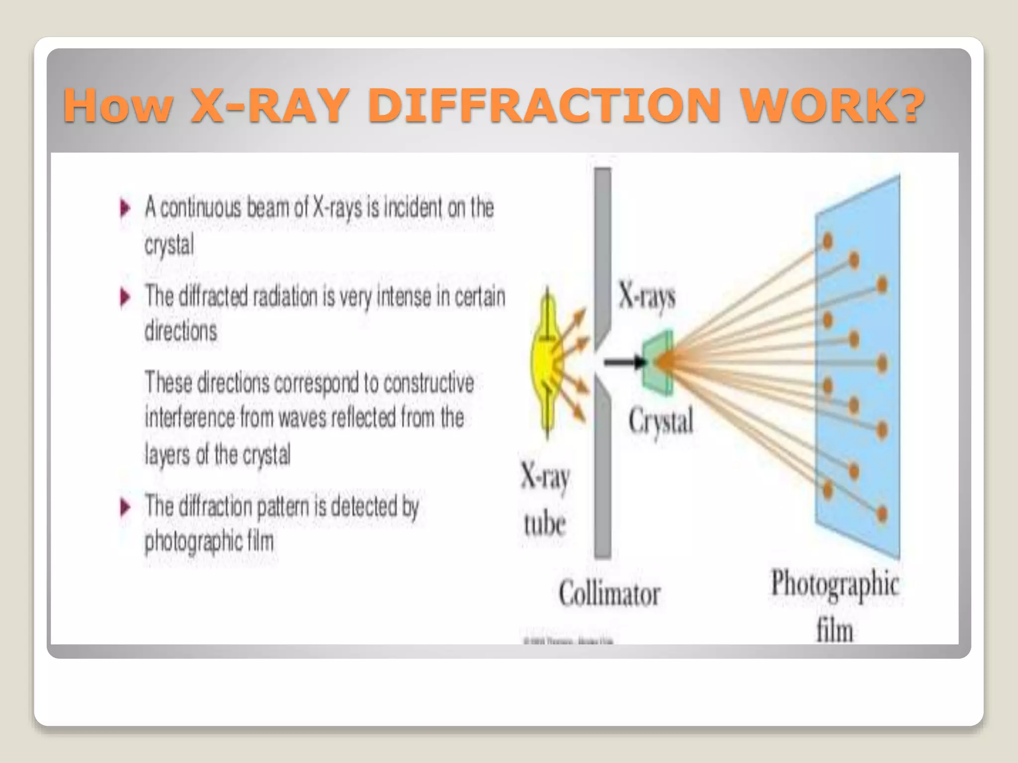
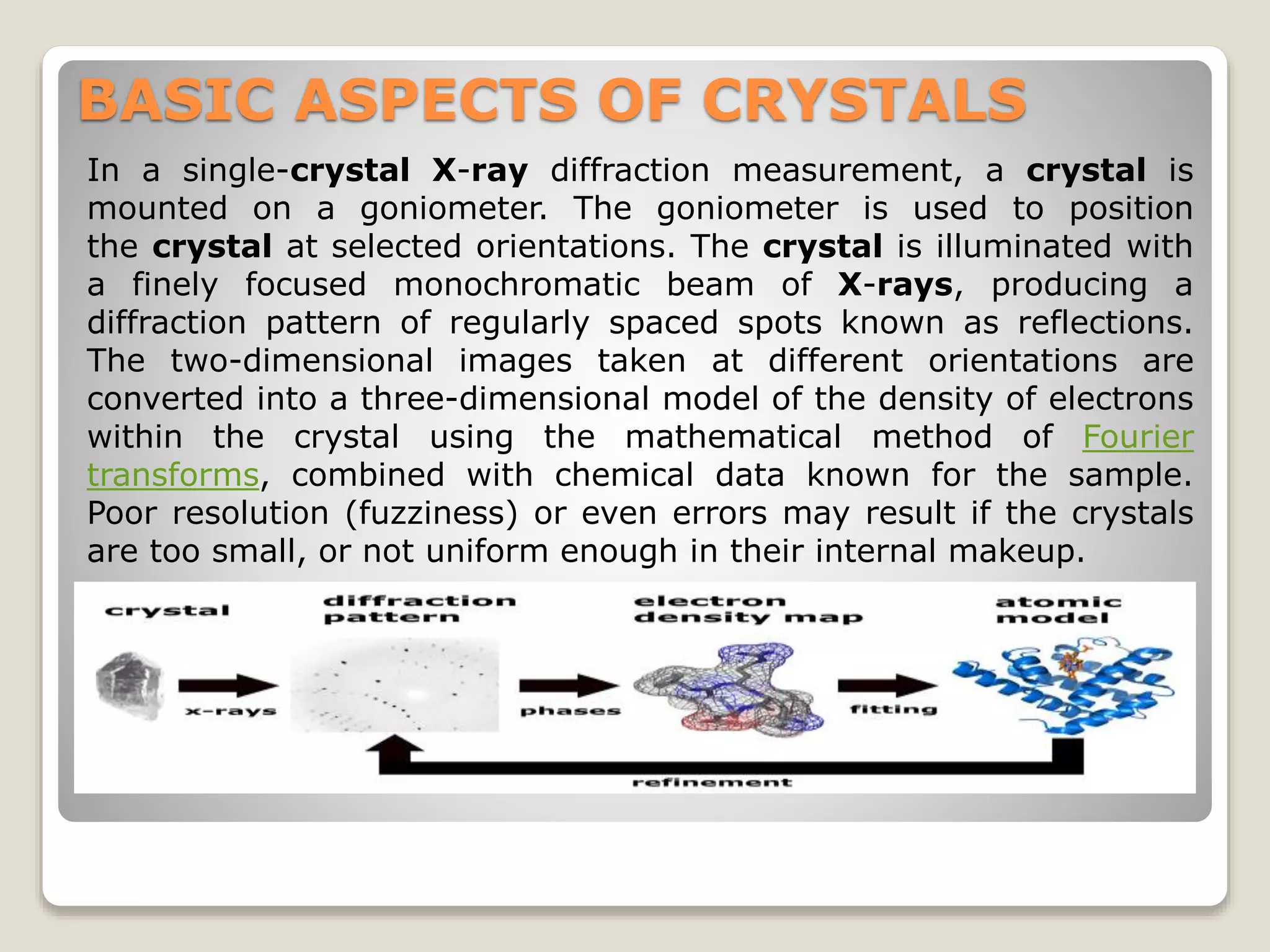
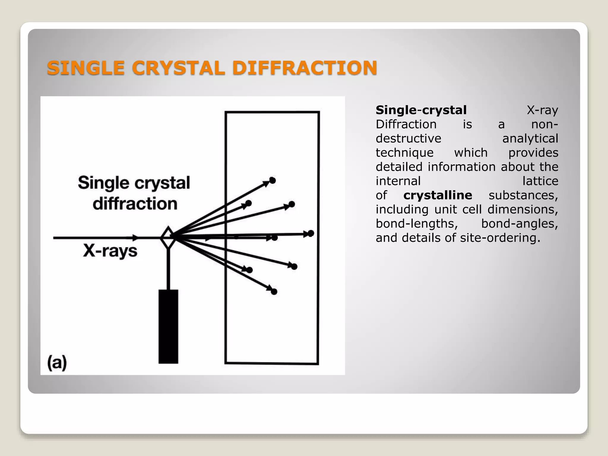
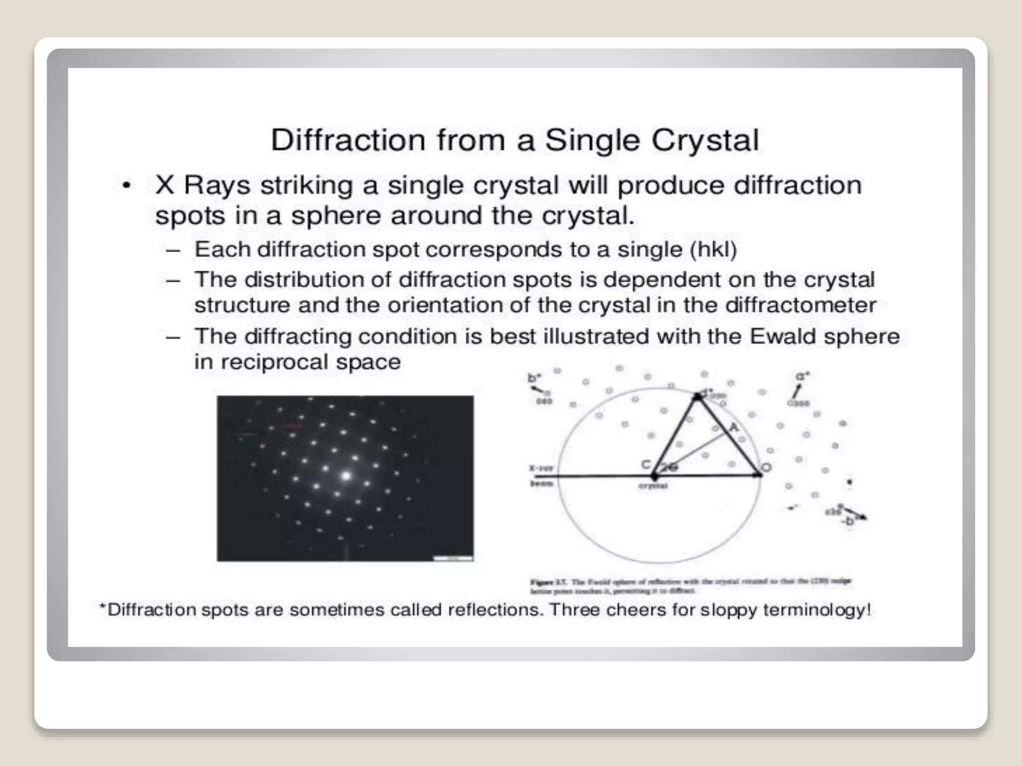
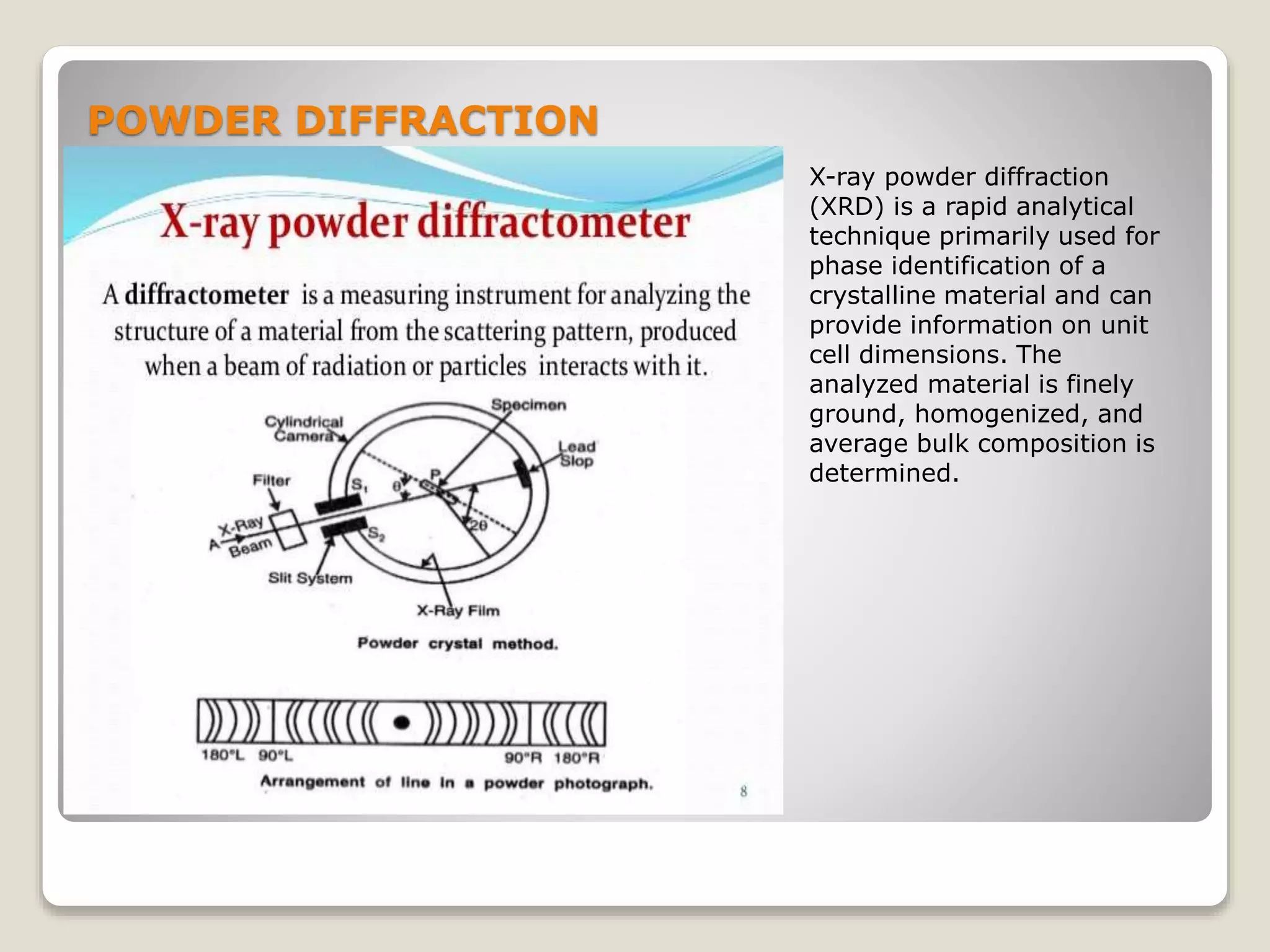
PRESENTED BY : ANANT NAG
ID: 17BPH005
BATCH : 17-2021
Semester 8th (4TH YEAR)
X-RAY DIFFRACTION
METHOD
DEFINITION
 X-ray diffraction, is a phenomenon in which the
atoms of a crystal, by virtue of their uniform
spacing, cause an interference pattern of the
waves present in an incident beam of X rays. The
atomic planes of the crystal act on the X rays in
exactly the same manner as does a uniformly
ruled grating on a beam of light.
X-RAY DIFFRACTION IS USE FOR
The incoming beam
(coming from upper
left) causes each
scatterer to re-radiate
a small portion of its
intensity as a spherical
wave. If scatterers are
arranged symmetrically
with a separation d,
these spherical waves
will be in sync (add
constructively) only in
directions where their
path-length difference
2d sin θ equals an
integer multiple of
the wavelength λ. In
that case, part of the
incoming beam is
deflected by an angle
2θ, producing
a reflection spot in
the diffraction pattern
ORIGIN OF X-RAY
How X-RAY DIFFRACTION WORK?
BASIC ASPECTS OF CRYSTALS
In a single-crystal X-ray diffraction measurement, a crystal is
mounted on a goniometer. The goniometer is used to position
the crystal at selected orientations. The crystal is illuminated with
a finely focused monochromatic beam of X-rays, producing a
diffraction pattern of regularly spaced spots known as reflections.
The two-dimensional images taken at different orientations are
converted into a three-dimensional model of the density of electrons
within the crystal using the mathematical method of Fourier
transforms, combined with chemical data known for the sample.
Poor resolution (fuzziness) or even errors may result if the crystals
are too small, or not uniform enough in their internal makeup.
X-RAY CRYSTALLOGRAPHY
 X-ray crystallography (XRC) is the experimental science
determining the atomic and molecular structure of
a crystal, in which the crystalline structure causes a beam
of incident X-rays to diffract into many specific directions.
By measuring the angles and intensities of these diffracted
beams, a crystallographer can produce a three-dimensional
picture of the density of electrons within the crystal. From
this electron density, the mean positions of the atoms in
the crystal can be determined, as well as their chemical
bonds, their crystallographic disorder, and various other
information.
 Since many materials can form crystals—such
as salts, metals, minerals, semiconductors, as well as
various inorganic, organic, and biological molecules—X-ray
crystallography has been fundamental in the development
of many scientific fields.
ROTATING CRYSTAL TECHNIQUE
The rotation method is the
most common method to
determine steady
state crystal structures. The
orientation of
the rotation axis and
the rotation range can be
chosen to select a subset of
diffraction peaks fulfilling the
Bragg condition Δk = k − k =
Hhkl(r,φ)
SINGLE CRYSTAL DIFFRACTION
Single-crystal X-ray
Diffraction is a non-
destructive analytical
technique which provides
detailed information about the
internal lattice
of crystalline substances,
including unit cell dimensions,
bond-lengths, bond-angles,
and details of site-ordering.
POWDER DIFFRACTION
X-ray powder diffraction
(XRD) is a rapid analytical
technique primarily used for
phase identification of a
crystalline material and can
provide information on unit
cell dimensions. The
analyzed material is finely
ground, homogenized, and
average bulk composition is
determined.
STRUCTURAL ELUCIDATION
APPLICATIONS
REFERENCES
1. "Resonant X-ray Scattering | Shen
Laboratory". arpes.stanford.edu. Retrieved 2019-07-10.
(https://en.wikipedia.org/wiki/X-ray_crystallography)
2. A Powerpoint presentation on use of XRD in Soil
Science (PowerPoint 1.6MB Sep7 07) by Melody
Bergeron, Image and Chemical Analysis Laboratory at
Montana State University. (https://www.xos.com/XRD)
3. Vinita Vishwakarma, Sudha Uthaman, in Smart
Nanoconcretes and Cement-Based Materials, 2020
(https://www.jove.com/v/10446/x-ray-diffraction)
4. Jeff Dahl (https://www.britannica.com/science/X-ray-
diffraction)
5. Brady, John B., and Boardman, Shelby J., 1995, Introducing
Mineralogy Students to X-ray Diffraction Through Optical
Diffraction Experiments Using Lasers. Jour. Geol. Education,
v. 43 #5, 471-476.
(https://serc.carleton.edu/research_education/geochemshee
ts/techniques/XRD.html)
THANK YOU

X ray diffraction method

  • 1.
    PRESENTED BY :ANANT NAG ID: 17BPH005 BATCH : 17-2021 Semester 8th (4TH YEAR) X-RAY DIFFRACTION METHOD
  • 2.
    DEFINITION  X-ray diffraction,is a phenomenon in which the atoms of a crystal, by virtue of their uniform spacing, cause an interference pattern of the waves present in an incident beam of X rays. The atomic planes of the crystal act on the X rays in exactly the same manner as does a uniformly ruled grating on a beam of light.
  • 3.
  • 4.
    The incoming beam (comingfrom upper left) causes each scatterer to re-radiate a small portion of its intensity as a spherical wave. If scatterers are arranged symmetrically with a separation d, these spherical waves will be in sync (add constructively) only in directions where their path-length difference 2d sin θ equals an integer multiple of the wavelength λ. In that case, part of the incoming beam is deflected by an angle 2θ, producing a reflection spot in the diffraction pattern
  • 6.
  • 7.
  • 8.
    BASIC ASPECTS OFCRYSTALS In a single-crystal X-ray diffraction measurement, a crystal is mounted on a goniometer. The goniometer is used to position the crystal at selected orientations. The crystal is illuminated with a finely focused monochromatic beam of X-rays, producing a diffraction pattern of regularly spaced spots known as reflections. The two-dimensional images taken at different orientations are converted into a three-dimensional model of the density of electrons within the crystal using the mathematical method of Fourier transforms, combined with chemical data known for the sample. Poor resolution (fuzziness) or even errors may result if the crystals are too small, or not uniform enough in their internal makeup.
  • 9.
    X-RAY CRYSTALLOGRAPHY  X-raycrystallography (XRC) is the experimental science determining the atomic and molecular structure of a crystal, in which the crystalline structure causes a beam of incident X-rays to diffract into many specific directions. By measuring the angles and intensities of these diffracted beams, a crystallographer can produce a three-dimensional picture of the density of electrons within the crystal. From this electron density, the mean positions of the atoms in the crystal can be determined, as well as their chemical bonds, their crystallographic disorder, and various other information.  Since many materials can form crystals—such as salts, metals, minerals, semiconductors, as well as various inorganic, organic, and biological molecules—X-ray crystallography has been fundamental in the development of many scientific fields.
  • 11.
    ROTATING CRYSTAL TECHNIQUE Therotation method is the most common method to determine steady state crystal structures. The orientation of the rotation axis and the rotation range can be chosen to select a subset of diffraction peaks fulfilling the Bragg condition Δk = k − k = Hhkl(r,φ)
  • 13.
    SINGLE CRYSTAL DIFFRACTION Single-crystalX-ray Diffraction is a non- destructive analytical technique which provides detailed information about the internal lattice of crystalline substances, including unit cell dimensions, bond-lengths, bond-angles, and details of site-ordering.
  • 15.
    POWDER DIFFRACTION X-ray powderdiffraction (XRD) is a rapid analytical technique primarily used for phase identification of a crystalline material and can provide information on unit cell dimensions. The analyzed material is finely ground, homogenized, and average bulk composition is determined.
  • 17.
  • 18.
  • 19.
    REFERENCES 1. "Resonant X-rayScattering | Shen Laboratory". arpes.stanford.edu. Retrieved 2019-07-10. (https://en.wikipedia.org/wiki/X-ray_crystallography) 2. A Powerpoint presentation on use of XRD in Soil Science (PowerPoint 1.6MB Sep7 07) by Melody Bergeron, Image and Chemical Analysis Laboratory at Montana State University. (https://www.xos.com/XRD) 3. Vinita Vishwakarma, Sudha Uthaman, in Smart Nanoconcretes and Cement-Based Materials, 2020 (https://www.jove.com/v/10446/x-ray-diffraction) 4. Jeff Dahl (https://www.britannica.com/science/X-ray- diffraction) 5. Brady, John B., and Boardman, Shelby J., 1995, Introducing Mineralogy Students to X-ray Diffraction Through Optical Diffraction Experiments Using Lasers. Jour. Geol. Education, v. 43 #5, 471-476. (https://serc.carleton.edu/research_education/geochemshee ts/techniques/XRD.html)
  • 20.