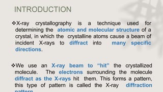
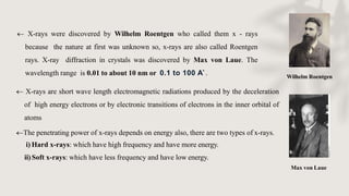
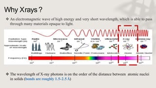
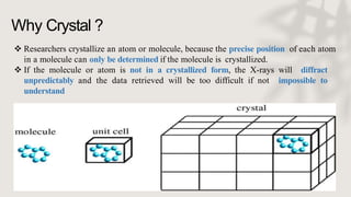
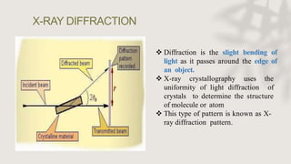
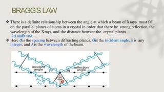
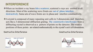
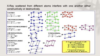
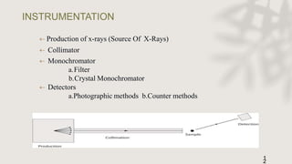
X-RAY CRYSTALLOGRAPHY
PRESENTED BY:
ARMAN DALAL
X-ray crystallography is a technique used for
determining the atomic and molecular structure of a
crystal, in which the crystalline atoms cause a beam of
incident X-rays to diffract into many specific
directions.
We use an X-ray beam to “hit” the crystallized
molecule. The electrons surrounding the molecule
diffract as the X-rays hit them. This forms a pattern,
this type of pattern is called the X-ray diffraction
INTRODUCTION
Wilhelm Roentgen
Max von Laue
 X-rays were discovered by Wilhelm Roentgen who called them x - rays
because the nature at first was unknown so, x-rays are also called Roentgen
rays. X-ray diffraction in crystals was discovered by Max von Laue. The
wavelength range is 0.01 to about 10 nm or 0.1 to 100 A˚.
 X-rays are short wave length electromagnetic radiations produced by the deceleration
of high energy electrons or by electronic transitions of electrons in the inner orbital of
atoms
The penetrating power of x-rays depends on energy also, there are two types of x-rays.
i) Hard x-rays: which have high frequency and have more energy.
ii)Soft x-rays: which have less frequency and have low energy.
Why X-ray ??
Why Crystal ??
Why Xrays ?
 An electromagnetic wave of high energy and very short wavelength, which is able to pass
through many materials opaque to light.
 The wavelength of X-ray photons is on the order of the distance between atomic nuclei
in solids (bonds are roughly 1.5-2.5Å)
Why Crystal ?
 Researchers crystallize an atom or molecule, because the precise position of each atom
in a molecule can only be determined if the molecule is crystallized.
 If the molecule or atom is not in a crystallized form, the X-rays will diffract
unpredictably and the data retrieved will be too difficult if not impossible to
understand
X-RAY DIFFRACTION
 Diffraction is the slight bending of
light as it passes around the edge of
an object.
 X-ray crystallography uses the
uniformity of light diffraction of
crystals to determine the structure
of molecule or atom
 This type of pattern is known as X-
ray diffraction pattern.
BRAGG’SLAW
 There is a definite relationship between the angle at which a beam of Xrays must fall
on the parallel planes of atoms in a crystal in order that there be strong reflection, the
wavelength of the Xrays, and the distance betweenthe crystal planes
2d sinƟ =nλ
 Here dis the spacing between diffracting planes, Ɵis the incident angle, n is any
integer, and λis the wavelength of the beam.
When an incident x-ray beam hits a scatterer, scattered x-rays are emitted in all
directions. Most of the scattering wave fronts are out of phase interfere
destructively. Some sets of wave fronts are in phase and interfere constructively.
A crystal is composed of many repeating unit cells in 3-dimensions, and therefore,
acts like a 3-dimensional diffraction grating. The constructive interference from a
diffracting crystal is observed as a pattern of points on the detector. The relative
positions of these points are related mathematically to the crystal’s unit celldimensions.
Destructive Interference Constructive Interference
INTERFERENCE
X-Ray scattered from different atoms interfere with one another either
constructively or destructively.
 Production of x-rays (Source Of X-Rays)
 Collimator
 Monochromator
a.Filter
b.Crystal Monochromator
 Detectors
a.Photographic methods b.Counter methods
INSTRUMENTATION
1
2
Production of X-Rays
 X-rays are produced whenever a
charged particles are accelerated.
 This Instrument is called as
Coolidge tube or x-ray tube.
 X-rays are generated when high
velocity electrons strike on a
metal target.
 A source of electrons – hot
tungsten filament, a high
accelerating voltage between the
cathode (W) and the anode and a
metal target, Cu, Al, Mo, Mg.
 The anode is a water-cooled
block of Cu containing desired
target metal.
A monochromator is an
optical device that
transmits a
mechanically selectable
narrow band of
wavelengths of light or
other radiation chosen
from a wider range of
wavelengths available at
the input.
Monochromator
Types Of
Monochromators
 Monochromatization can be broadly divided intotwo,
1.Interference Filters
2.Crystal Monochromator
 Crystal Monochromators can be again divided into two
i)Flat crystal Monochromator
ii)Curved crystal Monochromator
Interference Filters
 X-ray beam may be partly monochromatized by
insertion of a suitable filter.
 A filter is a window of material that absorbs
undesirable radiation but allows the radiation of
required wavelength to pass.
 Interference filters contain several optical layers
deposited on a glass substrate or transparent
quartz. The specific performance characteristics
of the filter are determined by the thickness of the
optical layers.
Crystal monochromators are made up of suitable crystalline material positioned in the x-
ray beam so that the angle of reflecting planes satisfied the Bragg’s equation for the
required wavelength the beam is split up into component wavelengths crystals used in
monochromators are made up of materials like NaCl, lithium fluoride , quartz etc.
Pyrolytic graphite can be used for broad band and silicon for narrow band.
Crystal Monochromator
Detectors
X-ray detectors are devices used to measure the flux, spatial distribution,
spectrum, and other properties of X-rays
The x-ray intensities can be measured and
recorded either by
1)Photographic method
2)Counter methods:-
Geiger - Muller tube counter
Scintillation detector
Semi conductor detectors
Photographic method
To record the position and intensity of x-ray
beam a plane or cylindrical film is used.
The film after exposing to x-ray is
developed
Geiger tube is filled with inert gas like
argon
Central wire anodeis maintained at a
positive potential of 800 to 1500V.
The electron is accelerated by the
potential gradient and causes the
ionization of large number of argon
atoms, resulting in the production of
avalanche of electrons that are
travelling towards central anode
Geiger - Muller tube counter
 In a scintillation detector there is large sodium iodide crystal activated with a
small amount of thallium
 When x-ray is incident upon crystal , the pulses ofvisible light are emitted which can be detected
by a photo multiplier tube

Useful for measuring x-ray of short wavelength
Crystals used in scintillation detectors include sodium iodide ,anthracene, napthalene and p-
terphenol

Scintillation Detector
 When x-ray falls on silicon lithium drifted detector an electron
(-e) and a hole (+e)
 Pure silicon made up with thin film of lithium metal plated onto
one end
 Under the influence of voltage electrons moves towards +ve
charge and
holes towards –ve
 Voltage generated is measure of the x-ray intensity falling on
crystal
 Upon arriving at lithium pulse is generated
 Voltage of pulse=q/c; q-total charge collected on electrode, c-
detector capacity.
Semi-conductor
detectors
X-RAY DIFFRACTION METHODS
Single Crystal X-Ray Diffraction
First step to take a crystal. Crystal should be sufficient large (large than 0.1 mm in all
dimensions), pure in composition & regular in structure with no internal defects. Then
crystal placed in intense beam of xrays of single wavelength. Angles & intensities of
diffracted xrays measured. As crystal gradually rotated previous reflection disappear & new
one appears. Intensity of every spot is recorded & multiple data sets collected. Then these
data sets combined computationally with chemical information to produce a model of
arrangement of atoms within crystal
 Single crystal mounted with one axis normal to a monochromatic x-ray beam
 Cylindrical film placed around the sample
 As sample rotates, some sets of planes momentarily satisfy Bragg condition
 When film is laid flat, a series of horizontal lines appears
 Because crystal rotates about a single axis, possible Bragg angles are limited - not every
plane is able to produce a diffracted spot
 Sometimes used to determine unknown crystal structures
Rotating Crystal Method
Fine powder is struck on a hair with a gum ,it is suspended vertically in the axis of a cylindrical
camera
When monochromatic beam is allowed to pass different possibilities may happen
1.There will be some particles out of random orientation of small crystals in the fine powder
2.Another fraction of grains will have another set of planes in the correct positions for the reflections to
occur
3.Reflections are possible in different orders for each set
X-ray powder diffraction (XRD) is a rapid analytical technique primarily used
for phase identification of a crystalline material and can provide information on
unit cell dimensions. The analyzed material is finely ground, homogenized, and
average bulk composition is determined.
Powder Crystal Method
APPLICATIONS
27
1. Structure of crystals
2. Polymer
characterisatio
n
3. State of anneal in
metals
4. Particle size
determination
a) Spot counting method
b) Broadening of
diffraction lines
c) Low-angle scattering
5.Applications of
diffraction
methods to
complexes
a) Determination of
cis- trans
isomerism
b) Determination of
linkage isomerism
6.Miscellaneous
applications
1.STRUCTURE OF CRYSTALS
a-x-ray pattern of salt Nacl
b-x-ray pattern of salt Kcl
c-x-ray pattern of mixture of Nacl &Kcl
d-x-ray pattern of a powder mixed
crystal of Nacl &Kcl
2.POLYMER CHARACTERISATION
Determine degree of crystanillity
Non-crystalline portion scatters x-ray beam to give a
continuous background(amorphous materials)
Crystalline portion causes diffraction lines that are not
continuous.(crystalline materials)
3.State of anneal in metals:
XRD is used to test the metals without removing the part from
its position and without weakening it.
MISCELLANEOUS APPLICATIONS
Soil classification based on crystallinity
Analysis of industrial dusts
Assessment of weathering & degradation of minerals &
polymers
Study of corrosion products
Examination of tooth enamel &dentine
Examination of bone state &tissue state
Structure of DNA& RNA
X-ray diffraction pattern for a single alum crystal.
REFERENCES
1) Instrumental methods of chemical analysis ,B.K.sharma,17th edition 1997-1998,GOEL publishing house.
pageno:329-359
2) Principles of instrumental analysis,5th edition ,by Dougles a.skoog,f.James holles,Timothy A.Niemen.
page no:277-298
3) Instrumental methods of chemical analysis ,Gurudeep R.chatwal, sham k.anand, Himalaya publications page
no:2.303- 2.332
4) http://www.scienceiscool.org/solids/intro.html
5) http://en.wikipedia.org/wiki/X-ray_crystallography
6) https://pt.slideshare.net/praying1/972-b3102005-diffraction-direction/7
7) https://link.springer.com/referenceworkentry/10.1007%2F978-3-540-72816-0_12847
x ray crystallography & diffraction

x ray crystallography & diffraction

  • 1.
  • 2.
    X-ray crystallography isa technique used for determining the atomic and molecular structure of a crystal, in which the crystalline atoms cause a beam of incident X-rays to diffract into many specific directions. We use an X-ray beam to “hit” the crystallized molecule. The electrons surrounding the molecule diffract as the X-rays hit them. This forms a pattern, this type of pattern is called the X-ray diffraction INTRODUCTION
  • 3.
    Wilhelm Roentgen Max vonLaue  X-rays were discovered by Wilhelm Roentgen who called them x - rays because the nature at first was unknown so, x-rays are also called Roentgen rays. X-ray diffraction in crystals was discovered by Max von Laue. The wavelength range is 0.01 to about 10 nm or 0.1 to 100 A˚.  X-rays are short wave length electromagnetic radiations produced by the deceleration of high energy electrons or by electronic transitions of electrons in the inner orbital of atoms The penetrating power of x-rays depends on energy also, there are two types of x-rays. i) Hard x-rays: which have high frequency and have more energy. ii)Soft x-rays: which have less frequency and have low energy.
  • 4.
    Why X-ray ?? WhyCrystal ??
  • 5.
    Why Xrays ? An electromagnetic wave of high energy and very short wavelength, which is able to pass through many materials opaque to light.  The wavelength of X-ray photons is on the order of the distance between atomic nuclei in solids (bonds are roughly 1.5-2.5Å)
  • 6.
    Why Crystal ? Researchers crystallize an atom or molecule, because the precise position of each atom in a molecule can only be determined if the molecule is crystallized.  If the molecule or atom is not in a crystallized form, the X-rays will diffract unpredictably and the data retrieved will be too difficult if not impossible to understand
  • 8.
    X-RAY DIFFRACTION  Diffractionis the slight bending of light as it passes around the edge of an object.  X-ray crystallography uses the uniformity of light diffraction of crystals to determine the structure of molecule or atom  This type of pattern is known as X- ray diffraction pattern.
  • 9.
    BRAGG’SLAW  There isa definite relationship between the angle at which a beam of Xrays must fall on the parallel planes of atoms in a crystal in order that there be strong reflection, the wavelength of the Xrays, and the distance betweenthe crystal planes 2d sinƟ =nλ  Here dis the spacing between diffracting planes, Ɵis the incident angle, n is any integer, and λis the wavelength of the beam.
  • 10.
    When an incidentx-ray beam hits a scatterer, scattered x-rays are emitted in all directions. Most of the scattering wave fronts are out of phase interfere destructively. Some sets of wave fronts are in phase and interfere constructively. A crystal is composed of many repeating unit cells in 3-dimensions, and therefore, acts like a 3-dimensional diffraction grating. The constructive interference from a diffracting crystal is observed as a pattern of points on the detector. The relative positions of these points are related mathematically to the crystal’s unit celldimensions. Destructive Interference Constructive Interference INTERFERENCE
  • 11.
    X-Ray scattered fromdifferent atoms interfere with one another either constructively or destructively.
  • 12.
     Production ofx-rays (Source Of X-Rays)  Collimator  Monochromator a.Filter b.Crystal Monochromator  Detectors a.Photographic methods b.Counter methods INSTRUMENTATION 1 2
  • 13.
    Production of X-Rays X-rays are produced whenever a charged particles are accelerated.  This Instrument is called as Coolidge tube or x-ray tube.  X-rays are generated when high velocity electrons strike on a metal target.  A source of electrons – hot tungsten filament, a high accelerating voltage between the cathode (W) and the anode and a metal target, Cu, Al, Mo, Mg.  The anode is a water-cooled block of Cu containing desired target metal.
  • 14.
    A monochromator isan optical device that transmits a mechanically selectable narrow band of wavelengths of light or other radiation chosen from a wider range of wavelengths available at the input. Monochromator
  • 15.
    Types Of Monochromators  Monochromatizationcan be broadly divided intotwo, 1.Interference Filters 2.Crystal Monochromator  Crystal Monochromators can be again divided into two i)Flat crystal Monochromator ii)Curved crystal Monochromator
  • 16.
    Interference Filters  X-raybeam may be partly monochromatized by insertion of a suitable filter.  A filter is a window of material that absorbs undesirable radiation but allows the radiation of required wavelength to pass.  Interference filters contain several optical layers deposited on a glass substrate or transparent quartz. The specific performance characteristics of the filter are determined by the thickness of the optical layers.
  • 17.
    Crystal monochromators aremade up of suitable crystalline material positioned in the x- ray beam so that the angle of reflecting planes satisfied the Bragg’s equation for the required wavelength the beam is split up into component wavelengths crystals used in monochromators are made up of materials like NaCl, lithium fluoride , quartz etc. Pyrolytic graphite can be used for broad band and silicon for narrow band. Crystal Monochromator
  • 18.
    Detectors X-ray detectors aredevices used to measure the flux, spatial distribution, spectrum, and other properties of X-rays The x-ray intensities can be measured and recorded either by 1)Photographic method 2)Counter methods:- Geiger - Muller tube counter Scintillation detector Semi conductor detectors
  • 19.
    Photographic method To recordthe position and intensity of x-ray beam a plane or cylindrical film is used. The film after exposing to x-ray is developed
  • 20.
    Geiger tube isfilled with inert gas like argon Central wire anodeis maintained at a positive potential of 800 to 1500V. The electron is accelerated by the potential gradient and causes the ionization of large number of argon atoms, resulting in the production of avalanche of electrons that are travelling towards central anode Geiger - Muller tube counter
  • 21.
     In ascintillation detector there is large sodium iodide crystal activated with a small amount of thallium  When x-ray is incident upon crystal , the pulses ofvisible light are emitted which can be detected by a photo multiplier tube  Useful for measuring x-ray of short wavelength Crystals used in scintillation detectors include sodium iodide ,anthracene, napthalene and p- terphenol  Scintillation Detector
  • 22.
     When x-rayfalls on silicon lithium drifted detector an electron (-e) and a hole (+e)  Pure silicon made up with thin film of lithium metal plated onto one end  Under the influence of voltage electrons moves towards +ve charge and holes towards –ve  Voltage generated is measure of the x-ray intensity falling on crystal  Upon arriving at lithium pulse is generated  Voltage of pulse=q/c; q-total charge collected on electrode, c- detector capacity. Semi-conductor detectors
  • 23.
  • 24.
    Single Crystal X-RayDiffraction First step to take a crystal. Crystal should be sufficient large (large than 0.1 mm in all dimensions), pure in composition & regular in structure with no internal defects. Then crystal placed in intense beam of xrays of single wavelength. Angles & intensities of diffracted xrays measured. As crystal gradually rotated previous reflection disappear & new one appears. Intensity of every spot is recorded & multiple data sets collected. Then these data sets combined computationally with chemical information to produce a model of arrangement of atoms within crystal
  • 25.
     Single crystalmounted with one axis normal to a monochromatic x-ray beam  Cylindrical film placed around the sample  As sample rotates, some sets of planes momentarily satisfy Bragg condition  When film is laid flat, a series of horizontal lines appears  Because crystal rotates about a single axis, possible Bragg angles are limited - not every plane is able to produce a diffracted spot  Sometimes used to determine unknown crystal structures Rotating Crystal Method
  • 26.
    Fine powder isstruck on a hair with a gum ,it is suspended vertically in the axis of a cylindrical camera When monochromatic beam is allowed to pass different possibilities may happen 1.There will be some particles out of random orientation of small crystals in the fine powder 2.Another fraction of grains will have another set of planes in the correct positions for the reflections to occur 3.Reflections are possible in different orders for each set X-ray powder diffraction (XRD) is a rapid analytical technique primarily used for phase identification of a crystalline material and can provide information on unit cell dimensions. The analyzed material is finely ground, homogenized, and average bulk composition is determined. Powder Crystal Method
  • 27.
    APPLICATIONS 27 1. Structure ofcrystals 2. Polymer characterisatio n 3. State of anneal in metals 4. Particle size determination a) Spot counting method b) Broadening of diffraction lines c) Low-angle scattering 5.Applications of diffraction methods to complexes a) Determination of cis- trans isomerism b) Determination of linkage isomerism 6.Miscellaneous applications
  • 28.
    1.STRUCTURE OF CRYSTALS a-x-raypattern of salt Nacl b-x-ray pattern of salt Kcl c-x-ray pattern of mixture of Nacl &Kcl d-x-ray pattern of a powder mixed crystal of Nacl &Kcl
  • 29.
    2.POLYMER CHARACTERISATION Determine degreeof crystanillity Non-crystalline portion scatters x-ray beam to give a continuous background(amorphous materials) Crystalline portion causes diffraction lines that are not continuous.(crystalline materials)
  • 30.
    3.State of annealin metals: XRD is used to test the metals without removing the part from its position and without weakening it.
  • 31.
    MISCELLANEOUS APPLICATIONS Soil classificationbased on crystallinity Analysis of industrial dusts Assessment of weathering & degradation of minerals & polymers Study of corrosion products Examination of tooth enamel &dentine Examination of bone state &tissue state Structure of DNA& RNA
  • 32.
    X-ray diffraction patternfor a single alum crystal.
  • 33.
    REFERENCES 1) Instrumental methodsof chemical analysis ,B.K.sharma,17th edition 1997-1998,GOEL publishing house. pageno:329-359 2) Principles of instrumental analysis,5th edition ,by Dougles a.skoog,f.James holles,Timothy A.Niemen. page no:277-298 3) Instrumental methods of chemical analysis ,Gurudeep R.chatwal, sham k.anand, Himalaya publications page no:2.303- 2.332 4) http://www.scienceiscool.org/solids/intro.html 5) http://en.wikipedia.org/wiki/X-ray_crystallography 6) https://pt.slideshare.net/praying1/972-b3102005-diffraction-direction/7 7) https://link.springer.com/referenceworkentry/10.1007%2F978-3-540-72816-0_12847