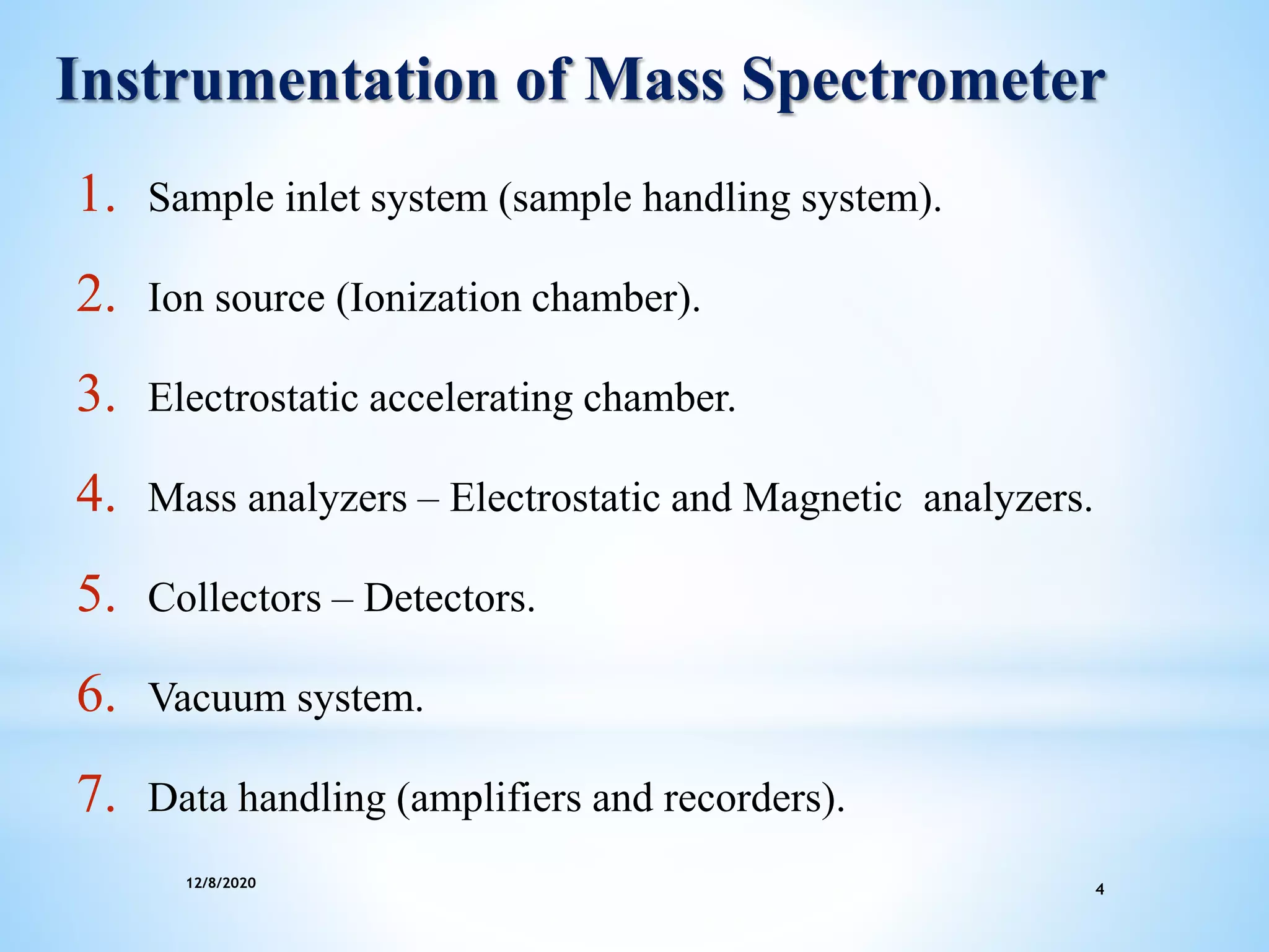
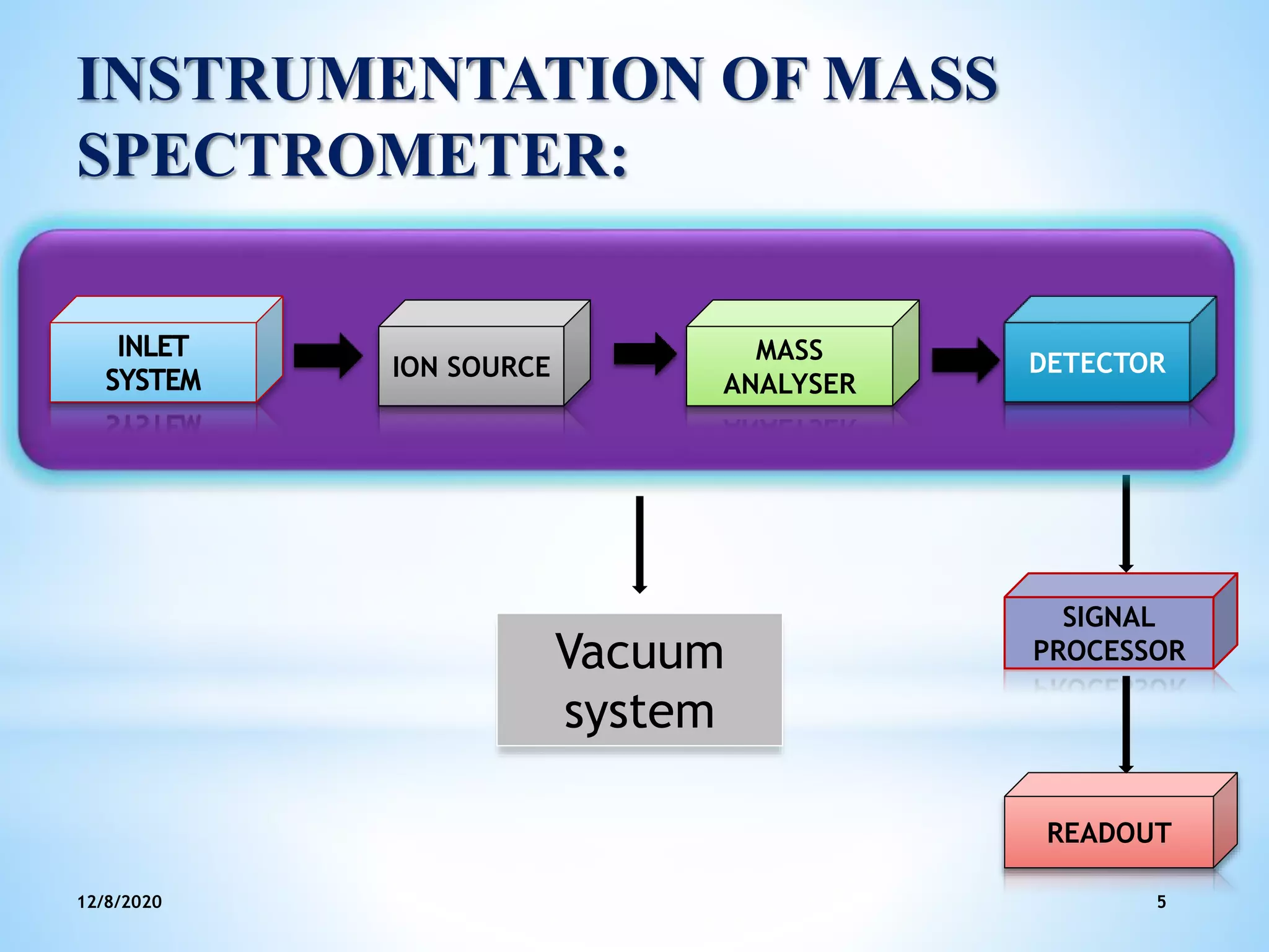
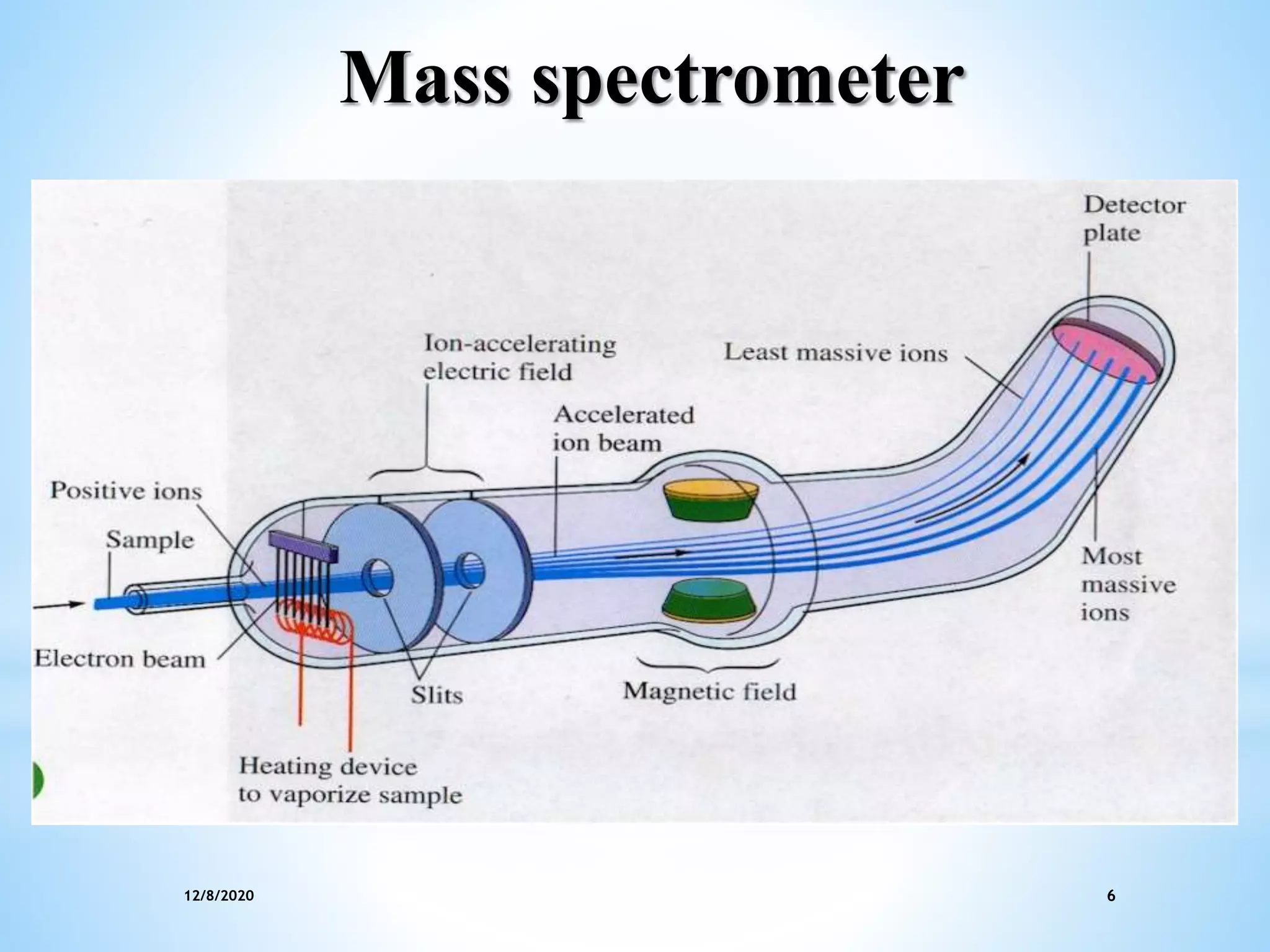
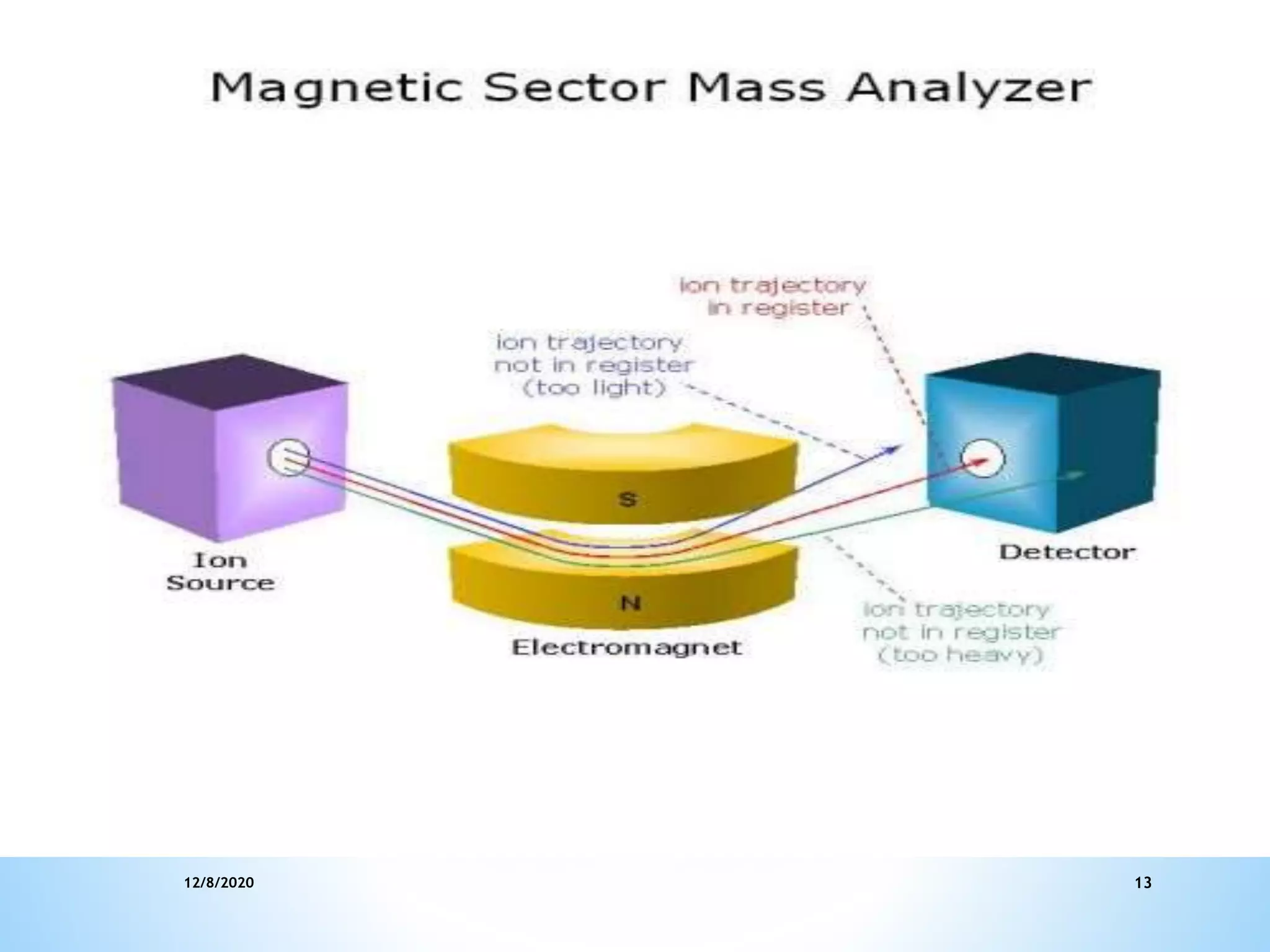
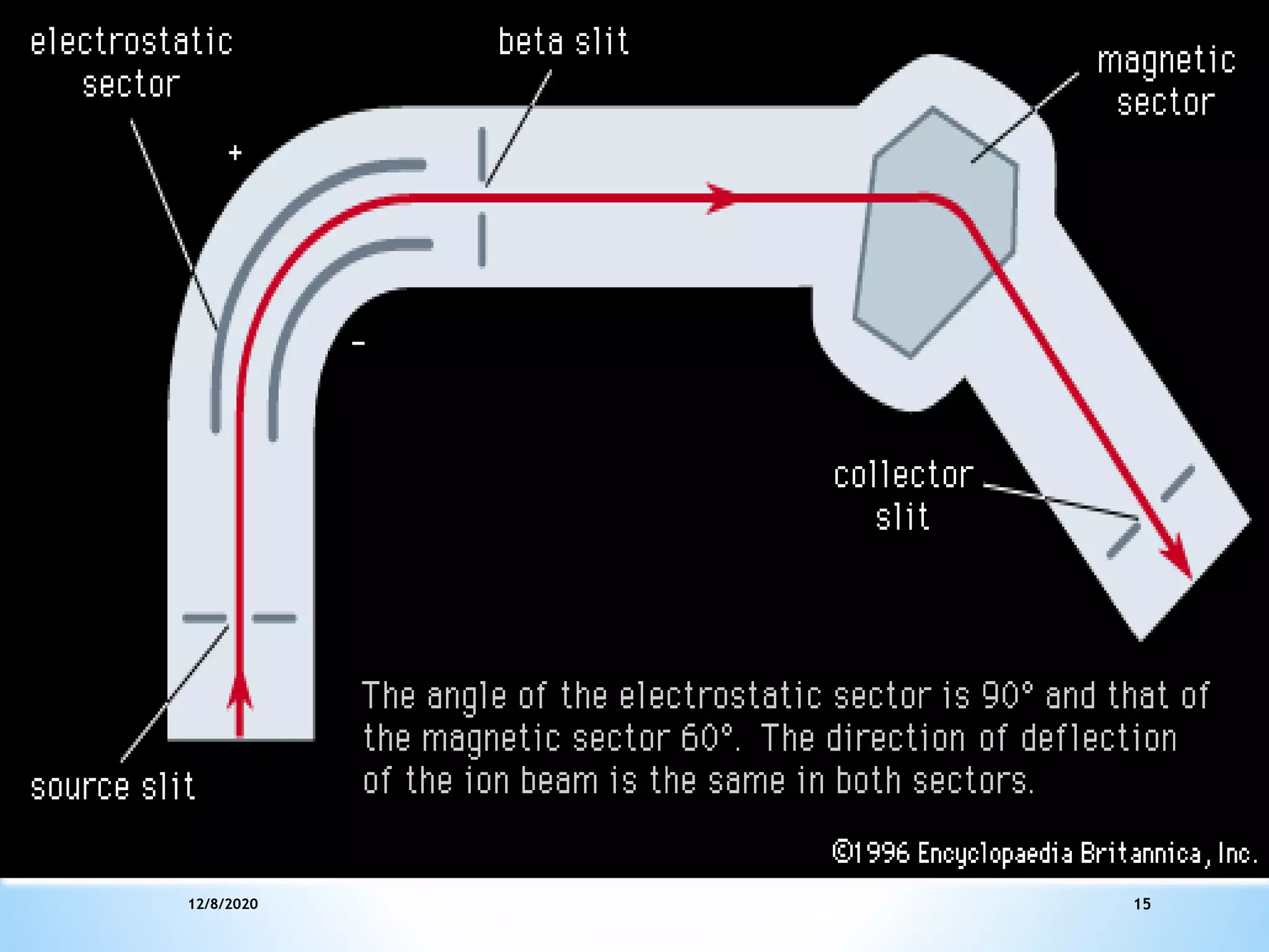
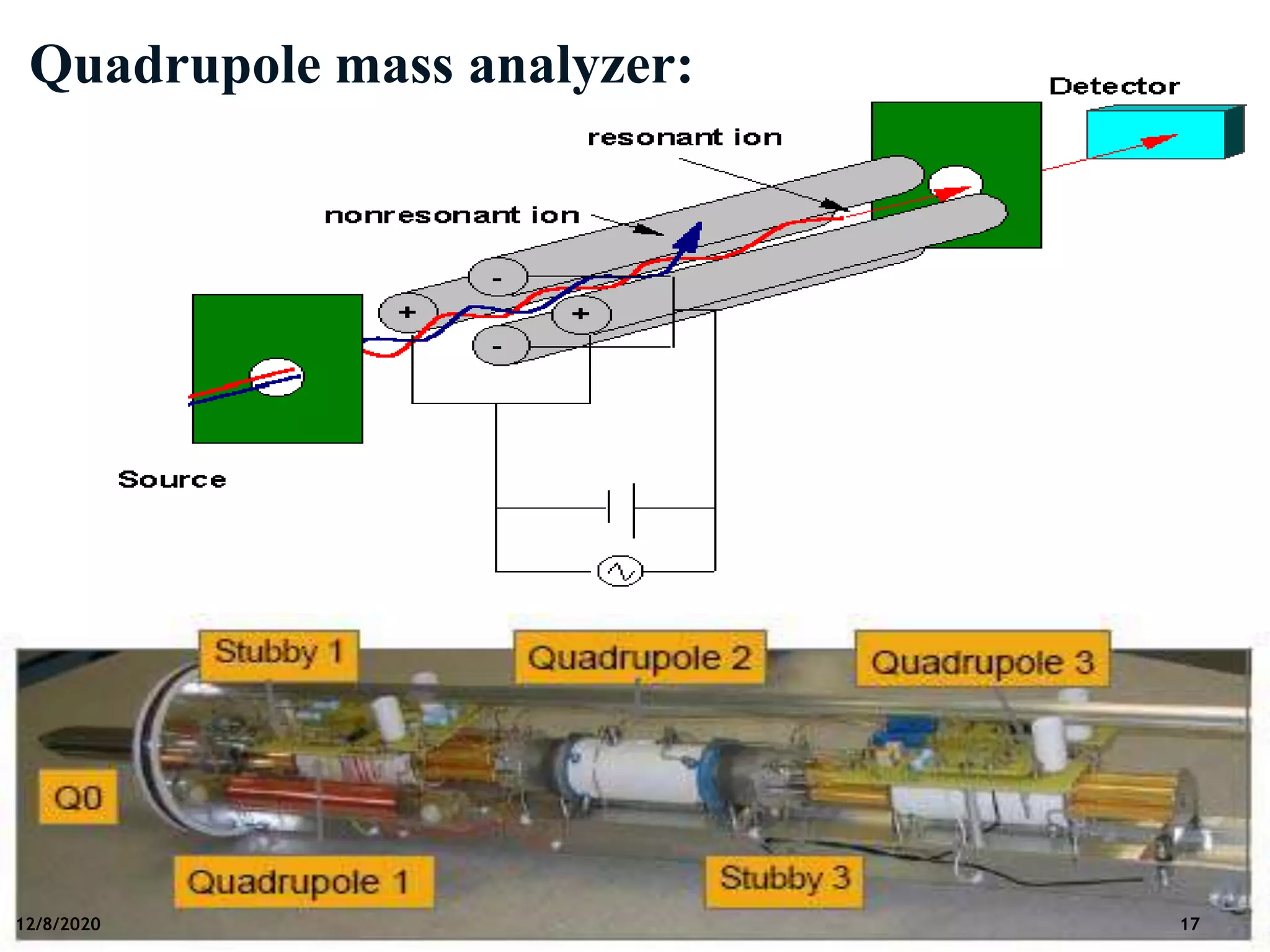
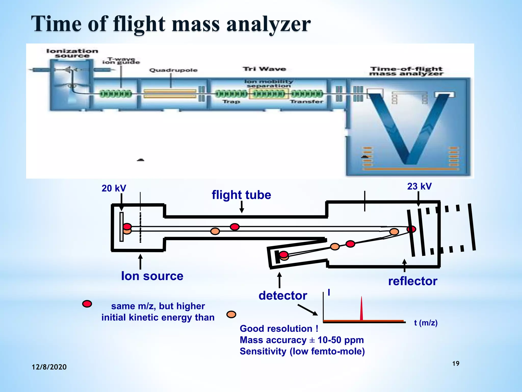
NAVEEN K L
1st M.Pharm
Dept. of Pharmacology
Srinivas College of Pharmacy
Instrumentation and Application
of Mass Spectroscopy
12/8/2020 2
Contents:
Introduction
Instrumentation
Applications
References
12/8/2020 3
Mass Spectroscopy
Mass spectroscopy is a method in which the
compound under investigation is bombarded
with a beam of electrons which produce an
ionic molecule or fragments of the original
species.
The resulting charged particle is separated
according to their masses. The spectrum is
produced, known as mass spectrum.
12/8/2020 4
Instrumentation of Mass Spectrometer
1. Sample inlet system (sample handling system).
2. Ion source (Ionization chamber).
3. Electrostatic accelerating chamber.
4. Mass analyzers – Electrostatic and Magnetic analyzers.
5. Collectors – Detectors.
6. Vacuum system.
7. Data handling (amplifiers and recorders).
INSTRUMENTATION OF MASS
SPECTROMETER:
12/8/2020 5
SIGNAL
PROCESSOR
READOUT
ION SOURCE
MASS
ANALYSER
DETECTOR
Vacuum
system
12/8/2020 6
Mass spectrometer
12/8/2020 7
1. Sample inlet systems
Permits introduction of sample in to the ion source.
Mass spectrometer should have a vapour sample and
it should enter the ionisation chamber at constant rate.
The sample is converted into gaseous state in the
inlet system.
12/8/2020 8
The rate at which sample is introduced into ionization
chamber must remain constant so that to achieve relative
abundance of different species in mass spectrum can be
determined.
A sample size of about 1μ is required.
Only about 0.1% is ionized in the chamber.
Gas samples:
Transfer of sample from small containers like glass bulb, of
known volume( about 3ml) coupled to a mercury manometer.
12/8/2020 9
Liquid samples:
Hypodermic needle injection through a silicone
rubber dam.
The low pressure in reservoir draws in the liquid and
vaporizes it instantly.
Solids or Non-volatile liquids:
 Directly introduced on silica or platinum probe and
then volatilized by gentle heating .
12/8/2020 10
2. Ion Sources:
Chemical ionization:
In this method ionization results from ion molecule chemical
interaction.
The interaction process involves large amount of reagent gas
and a small amount of sample.
The reagent ion and sample molecule may react via any of the
several modes like proton exchange.
Reagent gas : Argon, helium and nitrogen show fragmentation
patterns with increased sensitivity.
12/8/2020 11
3. Mass analyzers and ion separator:
After passing through the electrostatic slits, ions enter in to the
analyzers.
Separate the ions according to their mass by charge ratio.
Different types of mass analyzers are:
1.Magnetic deflection or sector mass analyzer .
2.Double focus mass analyzer .
3.Quadrupole mass analyzer .
4.Time of flight mass analyzer.
12/8/2020 12
1. Magnetic deflection or sector mass analyzer:
 In this type, the homogeneous beam of ions from the slit can
be focus by a magnetic field in a shape of a sector.
 Gas phase molecules are ionized by a beam of high energy
electrons.
 Electrons may be ejected from molecules (ionization) or
bonds in molecules may rapture (fragmentation)
 Ions are then accelerated in a field (sector) at a voltage V.
 Basis of separation by m/z
12/8/2020 13
12/8/2020 14
2. Double focus mass analyzer :
 Here the ions are analyzed according to their
momentum and translational energies.
 There is an electrostatic deflection field between the
ion source and the mass analyzer.
 As the ions are passed through the electrostatic
sector, they are deflected according to their
translational energy.
 Finally the magnetic sector disperses the ions
according to their momentum.
12/8/2020 15
12/8/2020 16
3.Quadrupole mass analyzer:
The Quadrupole consists of four parallel rods. The opposing
rods have the same polarity while adjacent rods have opposite
polarity.
Each rod is applied with a DC and RF (Oscillating radio
frequency) voltage.Ions are scanned by varying the DC/RF
quadrupole voltages.
Only ions with the selected mass to charge ratio will have the
correct oscillatory pathway in the RF field.
12/8/202017
12/8/2020 17
Quadrupole mass analyzer:
12/8/2020 18
4. Time of flight mass analyzer:
As the name indicates the mass spectrum is obtained depending upon
the time it took for the ion to reach the detector from accelerating
chamber.
The ions are accelerated and accelerated particles entering the tube have
same kinetic energies .
 The lighter particles arrives at the collector earlier than heavier
particles.
t = L/v ------- time of flight equation.
t - time taken for the ion to pass through a drift tube.
v - velocity of the charged particle.
L - length of the tube.
19
12/8/2020
Time of flight mass analyzer
Ion source reflector
flight tube
detector
same m/z, but higher
initial kinetic energy than
20 kV 23 kV
t (m/z)
Good resolution !
Mass accuracy ± 10-50 ppm
Sensitivity (low femto-mole)
I
12/8/2020 20
4. Detectors:
Ions are amplified and recorded.
Measure one m/z value at a time (single channel
detectors)
 Multiple detectors are used for multiple ion
detection
High resolution magnetic sector instruments use
multiple detectors (called Multicollectors)
12/8/2020 21
Various detectors used are-
I. Faraday cup collector.
II. Electron multiplier.
III.Ion sensitive photographic plates.
IV.Array detector.
12/8/2020 22
I. Faraday cup collector:
Simple and effective means of monitoring ion current.
When sufficient amount of sample is available, the
measurement can be done by collecting them in single Faraday
cup and scanning for different mass by charge values.
•Advantages:
Current as low as 10-5A may be detected.
•Disadvantage:
It needs high impedance amplifier , which limits the speed at
which spectrum can be scanned.
12/8/2020 23
Faraday cup collector
24
II. Electron multiplier:
 When the amount of sample available is small, the ion current
produced is such a low current, which cannot be measured
directly with a Faraday cup and further amplification is carried
out by the secondary electron multiplier.
12/8/2020
12/8/2020 25
III. Ion sensitive photographic plates:
Here the ions directly exposed to a photographic film.
This type of detectors used in mass analyzers. Which disperse
ions according to their mass to charge ratio.
The ion sensitive photographic plate integrate the ions and
record them.
This detector provides greater resolution and highest
sensitivity.
12/8/2020 26
IV. Array Detectors:
Used in Time of Flight Mass instruments
Employs a focal plane camera (FPC) consisting of an array of
31 Faraday Cup
Up to 15 m/z values can be measured simultaneously
 Exhibits improved precision compared with single channel
detectors.
12/8/2020 27
5. The vacuum system:
Whole instrument is kept under high vacuum conditions
10-6 to 10-7 torr.
Types of pump systems are used -
1. Oil diffusion pumps
2. Mercury diffusion pump
12/8/2020 28
6. Data handling:
Data are digitalized and collected on magnetic tape or stored in
a memory of computer.
7. Recorders:
Multichannel ultraviolet recorder.
Pens, tape recorders, oscillators other data collecting systems
are used as recording devices.
12/8/2020 29
Applications:
It is used in environmental analysis.
Identification of unknown compounds.
Used in forensic analysis.
Also used in clinical research.
In generation of Physico-chemical data.
To measure the relative molecular masses (Molecular
weight) with very high accuracy; from this can be
deduced exact molecular formula.
12/8/2020 30
Cont…
It is also used to find out the places within molecule
at which it prefers to fragment.
Mass spectrometry is being used to identify the
mixtures of complicated samples such as urine,
lymph, blood, and it is used with high sensitivity
methods to measure low doses and long time point
data.
Determination of isotopic composition of different
elements.
12/8/2020 31
References:
1. YR Sharma, S Chand. Elementary Organic Spectroscopy
Principle and Chemical application; P.No:292-95.
2. Gurudeep R. Chatwal, Sham K. Anand, ‘Instrumental methods
of chemical analysis; P.No: 2.278-2.283.
3. Skoog Holler, Crouch. Instrumemal Analysis. Ind six ed;
P.No:606-08.
4. Douglas A Skoog, Donald M West, F James Holler, Stanley R
Crouch. Fundamentals of Analytical Chemistry. Ninth ed;
P.No:806-16
SlideShare Presentation on Mass spectrophotometer

SlideShare Presentation on Mass spectrophotometer

  • 1.
    NAVEEN K L 1stM.Pharm Dept. of Pharmacology Srinivas College of Pharmacy Instrumentation and Application of Mass Spectroscopy
  • 2.
  • 3.
    12/8/2020 3 Mass Spectroscopy Massspectroscopy is a method in which the compound under investigation is bombarded with a beam of electrons which produce an ionic molecule or fragments of the original species. The resulting charged particle is separated according to their masses. The spectrum is produced, known as mass spectrum.
  • 4.
    12/8/2020 4 Instrumentation ofMass Spectrometer 1. Sample inlet system (sample handling system). 2. Ion source (Ionization chamber). 3. Electrostatic accelerating chamber. 4. Mass analyzers – Electrostatic and Magnetic analyzers. 5. Collectors – Detectors. 6. Vacuum system. 7. Data handling (amplifiers and recorders).
  • 5.
    INSTRUMENTATION OF MASS SPECTROMETER: 12/8/20205 SIGNAL PROCESSOR READOUT ION SOURCE MASS ANALYSER DETECTOR Vacuum system
  • 6.
  • 7.
    12/8/2020 7 1. Sampleinlet systems Permits introduction of sample in to the ion source. Mass spectrometer should have a vapour sample and it should enter the ionisation chamber at constant rate. The sample is converted into gaseous state in the inlet system.
  • 8.
    12/8/2020 8 The rateat which sample is introduced into ionization chamber must remain constant so that to achieve relative abundance of different species in mass spectrum can be determined. A sample size of about 1μ is required. Only about 0.1% is ionized in the chamber. Gas samples: Transfer of sample from small containers like glass bulb, of known volume( about 3ml) coupled to a mercury manometer.
  • 9.
    12/8/2020 9 Liquid samples: Hypodermicneedle injection through a silicone rubber dam. The low pressure in reservoir draws in the liquid and vaporizes it instantly. Solids or Non-volatile liquids:  Directly introduced on silica or platinum probe and then volatilized by gentle heating .
  • 10.
    12/8/2020 10 2. IonSources: Chemical ionization: In this method ionization results from ion molecule chemical interaction. The interaction process involves large amount of reagent gas and a small amount of sample. The reagent ion and sample molecule may react via any of the several modes like proton exchange. Reagent gas : Argon, helium and nitrogen show fragmentation patterns with increased sensitivity.
  • 11.
    12/8/2020 11 3. Massanalyzers and ion separator: After passing through the electrostatic slits, ions enter in to the analyzers. Separate the ions according to their mass by charge ratio. Different types of mass analyzers are: 1.Magnetic deflection or sector mass analyzer . 2.Double focus mass analyzer . 3.Quadrupole mass analyzer . 4.Time of flight mass analyzer.
  • 12.
    12/8/2020 12 1. Magneticdeflection or sector mass analyzer:  In this type, the homogeneous beam of ions from the slit can be focus by a magnetic field in a shape of a sector.  Gas phase molecules are ionized by a beam of high energy electrons.  Electrons may be ejected from molecules (ionization) or bonds in molecules may rapture (fragmentation)  Ions are then accelerated in a field (sector) at a voltage V.  Basis of separation by m/z
  • 13.
  • 14.
    12/8/2020 14 2. Doublefocus mass analyzer :  Here the ions are analyzed according to their momentum and translational energies.  There is an electrostatic deflection field between the ion source and the mass analyzer.  As the ions are passed through the electrostatic sector, they are deflected according to their translational energy.  Finally the magnetic sector disperses the ions according to their momentum.
  • 15.
  • 16.
    12/8/2020 16 3.Quadrupole massanalyzer: The Quadrupole consists of four parallel rods. The opposing rods have the same polarity while adjacent rods have opposite polarity. Each rod is applied with a DC and RF (Oscillating radio frequency) voltage.Ions are scanned by varying the DC/RF quadrupole voltages. Only ions with the selected mass to charge ratio will have the correct oscillatory pathway in the RF field.
  • 17.
  • 18.
    12/8/2020 18 4. Timeof flight mass analyzer: As the name indicates the mass spectrum is obtained depending upon the time it took for the ion to reach the detector from accelerating chamber. The ions are accelerated and accelerated particles entering the tube have same kinetic energies .  The lighter particles arrives at the collector earlier than heavier particles. t = L/v ------- time of flight equation. t - time taken for the ion to pass through a drift tube. v - velocity of the charged particle. L - length of the tube.
  • 19.
    19 12/8/2020 Time of flightmass analyzer Ion source reflector flight tube detector same m/z, but higher initial kinetic energy than 20 kV 23 kV t (m/z) Good resolution ! Mass accuracy ± 10-50 ppm Sensitivity (low femto-mole) I
  • 20.
    12/8/2020 20 4. Detectors: Ionsare amplified and recorded. Measure one m/z value at a time (single channel detectors)  Multiple detectors are used for multiple ion detection High resolution magnetic sector instruments use multiple detectors (called Multicollectors)
  • 21.
    12/8/2020 21 Various detectorsused are- I. Faraday cup collector. II. Electron multiplier. III.Ion sensitive photographic plates. IV.Array detector.
  • 22.
    12/8/2020 22 I. Faradaycup collector: Simple and effective means of monitoring ion current. When sufficient amount of sample is available, the measurement can be done by collecting them in single Faraday cup and scanning for different mass by charge values. •Advantages: Current as low as 10-5A may be detected. •Disadvantage: It needs high impedance amplifier , which limits the speed at which spectrum can be scanned.
  • 23.
  • 24.
    24 II. Electron multiplier: When the amount of sample available is small, the ion current produced is such a low current, which cannot be measured directly with a Faraday cup and further amplification is carried out by the secondary electron multiplier. 12/8/2020
  • 25.
    12/8/2020 25 III. Ionsensitive photographic plates: Here the ions directly exposed to a photographic film. This type of detectors used in mass analyzers. Which disperse ions according to their mass to charge ratio. The ion sensitive photographic plate integrate the ions and record them. This detector provides greater resolution and highest sensitivity.
  • 26.
    12/8/2020 26 IV. ArrayDetectors: Used in Time of Flight Mass instruments Employs a focal plane camera (FPC) consisting of an array of 31 Faraday Cup Up to 15 m/z values can be measured simultaneously  Exhibits improved precision compared with single channel detectors.
  • 27.
    12/8/2020 27 5. Thevacuum system: Whole instrument is kept under high vacuum conditions 10-6 to 10-7 torr. Types of pump systems are used - 1. Oil diffusion pumps 2. Mercury diffusion pump
  • 28.
    12/8/2020 28 6. Datahandling: Data are digitalized and collected on magnetic tape or stored in a memory of computer. 7. Recorders: Multichannel ultraviolet recorder. Pens, tape recorders, oscillators other data collecting systems are used as recording devices.
  • 29.
    12/8/2020 29 Applications: It isused in environmental analysis. Identification of unknown compounds. Used in forensic analysis. Also used in clinical research. In generation of Physico-chemical data. To measure the relative molecular masses (Molecular weight) with very high accuracy; from this can be deduced exact molecular formula.
  • 30.
    12/8/2020 30 Cont… It isalso used to find out the places within molecule at which it prefers to fragment. Mass spectrometry is being used to identify the mixtures of complicated samples such as urine, lymph, blood, and it is used with high sensitivity methods to measure low doses and long time point data. Determination of isotopic composition of different elements.
  • 31.
    12/8/2020 31 References: 1. YRSharma, S Chand. Elementary Organic Spectroscopy Principle and Chemical application; P.No:292-95. 2. Gurudeep R. Chatwal, Sham K. Anand, ‘Instrumental methods of chemical analysis; P.No: 2.278-2.283. 3. Skoog Holler, Crouch. Instrumemal Analysis. Ind six ed; P.No:606-08. 4. Douglas A Skoog, Donald M West, F James Holler, Stanley R Crouch. Fundamentals of Analytical Chemistry. Ninth ed; P.No:806-16

Editor's Notes

  • #8 3. usually this can be achieved by the aid of heat. So if the samples are less volatile, it may be heated in flask before introducing into the ionisation chamber.
  • #9 Mercury monometer-to measure the pressure of gasses
  • #11 From the inlet system sample is introduced to ionisation chamber. Where it Gets ionised due to chemical ionisation.
  • #19 3. Here we can calculate the time taken by the ions to reach the detector from accelerating chamber using following equation
  • #21 here
  • #23 A-Ampere
  • #30 Orgaic pollutants, water quality, food contamination Forensic-Determination of drug abuse. To predict solubility ionization isoelectric point ewtc
  • #31 Number and abundance of the isotopes of the element.