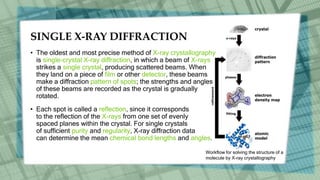
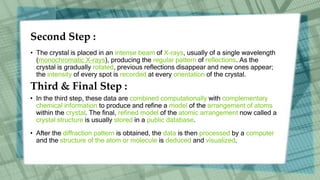
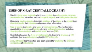
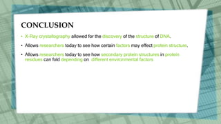
This is the short description about x ray crystallography.
simplest and easy to understand.
Procedure of X ray Diffraction.
Pros and Cons of X ray Crystallography
X-ray Crystallography is a scientific method used to determine the arrangement of atoms of a crystalline solid in three dimension. It is based on x ray diffraction. Reveals structure of a crystal at atomic level.
X-Ray Crystallography is a technique used to determine the atomic and molecular structure of a crystal, in which the crystalline atoms cause a beam of incident X-rays to diffract into many specific directions.
X-ray crystallography is a technique used for determining the atomic and molecular structure of a crystal, in which the crystalline atoms cause a beam of incident X-rays to diffract into many specific directions.
X ray crystallography and X ray DiffractionFaisal Hussain
This is the short description about x ray crystallography.
simplest and easy to understand.
Procedure of X ray Diffraction.
Advantages and Disadvantages of X ray Crystallography
X ray, invisible, highly penetrating electromagnetic radiation of much shorter wavelength (higher frequency) than visible light. The wavelength range for X rays is from about 10-8 m to about 10-11 m, the corresponding frequency range is from about 3 × 10¹⁶ Hz to about 3 × 10¹⁹ Hz.
It would be use full to All Needy People. It involve information about NMR Spectroscopy ( a spectroscopic techniques), factors influencing , proton NMR and their applications of NMR as well as Nuclear magnetic imaging.
X-ray crystallography is the experimental science determining the atomic and molecular structure of a crystal, in which the crystalline structure causes a beam of incident X-rays to diffract into many specific directions. By measuring the angles and intensities of these diffracted beams, a crystallographer can produce a three-dimensional picture of the density of electrons within the crystal. From this electron density, the mean positions of the atoms in the crystal can be determined, as well as their chemical bonds, their crystallographic disorder, and various other information.
X ray crystallography to visualize protein structure.Ritam38
This ppt discusses in detail the process of X ray Crystallography.
Made by the following 3rd year Bs-Ms students of IISER Kolkata:
B Sri Sindhu
Rasiwala Hassan Shabbir
Ritam Samanta
Himanshu Gupta
Sakshi Ajay Shrisath
Aditya Borkar
Diana Denzil Fernandez
Neha Kumari
.Sowmya
Anjali Mohan
Debanjana Mondal
Aanandita Gope
Shruti Santosh Sail
X-ray diffraction (XRD) is a versatile non-destructive analytical technique used to analyze physical properties such as phase composition, crystal structure and orientation of powder, solid and liquid samples. Many materials are made up of tiny crystallites. The chemical composition and structural type of these crystals is called their 'phase'. Materials can be single phase or multiphase mixtures and may contain crystalline and non-crystalline components. In an X-ray diffractometer, different crystalline phases give different diffraction patterns. Phase identification can be performed by comparing X-ray diffraction patterns obtained from unknown samples to patterns in reference databases.
principles:
X-Ray Diffraction is the result of constructive interference between X-rays and a crystalline sample. The wavelength of the X-rays used is of the same order of magnitude of the distance between the atoms in a crystalline lattice. This gives rise to a diffraction pattern that can be analysed in a number of ways, the most popular being applying the famous Bragg’s Law (nλ=2d sin θ) which is used in the measurement of crystals and their phases.
Applictions:
Many researchers, in industrial as well as in scientific laboratories, rely on X-ray diffraction (XRD) as a tool to develop new materials or to improve production efficiency. Innovations in X-ray diffraction closely follow the research on new materials, such as in semiconductor technologies or pharmaceutical investigations. Industrial research is directed toward the ever-increasing speed and efficiency of production processes. Fully automated X-ray diffraction analyses in mining and building materials production sites result in more cost-effective solutions for production control.
The main uses of X-ray diffraction are:
Qualitative and quantitative phase analysis of pure substances and mixtures. The most common method for phase analysis is often called 'X-ray powder diffraction' (XRPD).
X-ray crystallography is a powerful technique used in determining the three-dimensional structure of molecules at atomic resolution. It involves the use of X-rays to probe the arrangement of atoms in a crystal lattice. The information obtained from X-ray crystallography can be used to understand the function of biomolecules such as proteins, DNA, and RNA.
Memory organization in computer architectureFaisal Hussain
Memory organization in computer architecture
Volatile Memory
Non-Volatile Memory
Memory Hierarchy
Memory Access Methods
Random Access
Sequential Access
Direct Access
Main Memory
DRAM
SRAM
NVRAM
RAM: Random Access Memory
ROM: Read Only Memory
Auxiliary Memory
Cache Memory
Hit Ratio
Associative Memory
Numerous emerging and reemerging infectious microorganisms can pose a serious threat to public health and food security.
Pakistan needs to identify these threats, taking into consideration local, regional, and global health security issues. Irrespective of region, country, and race, these infections are serious threats.
its a complete procedure of software testing.
Software Testing Research Paper.
step by step procedure of Software testing.
Software testing Techniques in this research paper.
introduction and Procedure software testing.
Professional air quality monitoring systems provide immediate, on-site data for analysis, compliance, and decision-making.
Monitor common gases, weather parameters, particulates.
(May 29th, 2024) Advancements in Intravital Microscopy- Insights for Preclini...Scintica Instrumentation
Intravital microscopy (IVM) is a powerful tool utilized to study cellular behavior over time and space in vivo. Much of our understanding of cell biology has been accomplished using various in vitro and ex vivo methods; however, these studies do not necessarily reflect the natural dynamics of biological processes. Unlike traditional cell culture or fixed tissue imaging, IVM allows for the ultra-fast high-resolution imaging of cellular processes over time and space and were studied in its natural environment. Real-time visualization of biological processes in the context of an intact organism helps maintain physiological relevance and provide insights into the progression of disease, response to treatments or developmental processes.
In this webinar we give an overview of advanced applications of the IVM system in preclinical research. IVIM technology is a provider of all-in-one intravital microscopy systems and solutions optimized for in vivo imaging of live animal models at sub-micron resolution. The system’s unique features and user-friendly software enables researchers to probe fast dynamic biological processes such as immune cell tracking, cell-cell interaction as well as vascularization and tumor metastasis with exceptional detail. This webinar will also give an overview of IVM being utilized in drug development, offering a view into the intricate interaction between drugs/nanoparticles and tissues in vivo and allows for the evaluation of therapeutic intervention in a variety of tissues and organs. This interdisciplinary collaboration continues to drive the advancements of novel therapeutic strategies.
Slide 1: Title Slide
Extrachromosomal Inheritance
Slide 2: Introduction to Extrachromosomal Inheritance
Definition: Extrachromosomal inheritance refers to the transmission of genetic material that is not found within the nucleus.
Key Components: Involves genes located in mitochondria, chloroplasts, and plasmids.
Slide 3: Mitochondrial Inheritance
Mitochondria: Organelles responsible for energy production.
Mitochondrial DNA (mtDNA): Circular DNA molecule found in mitochondria.
Inheritance Pattern: Maternally inherited, meaning it is passed from mothers to all their offspring.
Diseases: Examples include Leber’s hereditary optic neuropathy (LHON) and mitochondrial myopathy.
Slide 4: Chloroplast Inheritance
Chloroplasts: Organelles responsible for photosynthesis in plants.
Chloroplast DNA (cpDNA): Circular DNA molecule found in chloroplasts.
Inheritance Pattern: Often maternally inherited in most plants, but can vary in some species.
Examples: Variegation in plants, where leaf color patterns are determined by chloroplast DNA.
Slide 5: Plasmid Inheritance
Plasmids: Small, circular DNA molecules found in bacteria and some eukaryotes.
Features: Can carry antibiotic resistance genes and can be transferred between cells through processes like conjugation.
Significance: Important in biotechnology for gene cloning and genetic engineering.
Slide 6: Mechanisms of Extrachromosomal Inheritance
Non-Mendelian Patterns: Do not follow Mendel’s laws of inheritance.
Cytoplasmic Segregation: During cell division, organelles like mitochondria and chloroplasts are randomly distributed to daughter cells.
Heteroplasmy: Presence of more than one type of organellar genome within a cell, leading to variation in expression.
Slide 7: Examples of Extrachromosomal Inheritance
Four O’clock Plant (Mirabilis jalapa): Shows variegated leaves due to different cpDNA in leaf cells.
Petite Mutants in Yeast: Result from mutations in mitochondrial DNA affecting respiration.
Slide 8: Importance of Extrachromosomal Inheritance
Evolution: Provides insight into the evolution of eukaryotic cells.
Medicine: Understanding mitochondrial inheritance helps in diagnosing and treating mitochondrial diseases.
Agriculture: Chloroplast inheritance can be used in plant breeding and genetic modification.
Slide 9: Recent Research and Advances
Gene Editing: Techniques like CRISPR-Cas9 are being used to edit mitochondrial and chloroplast DNA.
Therapies: Development of mitochondrial replacement therapy (MRT) for preventing mitochondrial diseases.
Slide 10: Conclusion
Summary: Extrachromosomal inheritance involves the transmission of genetic material outside the nucleus and plays a crucial role in genetics, medicine, and biotechnology.
Future Directions: Continued research and technological advancements hold promise for new treatments and applications.
Slide 11: Questions and Discussion
Invite Audience: Open the floor for any questions or further discussion on the topic.
Richard's entangled aventures in wonderlandRichard Gill
Since the loophole-free Bell experiments of 2020 and the Nobel prizes in physics of 2022, critics of Bell's work have retreated to the fortress of super-determinism. Now, super-determinism is a derogatory word - it just means "determinism". Palmer, Hance and Hossenfelder argue that quantum mechanics and determinism are not incompatible, using a sophisticated mathematical construction based on a subtle thinning of allowed states and measurements in quantum mechanics, such that what is left appears to make Bell's argument fail, without altering the empirical predictions of quantum mechanics. I think however that it is a smoke screen, and the slogan "lost in math" comes to my mind. I will discuss some other recent disproofs of Bell's theorem using the language of causality based on causal graphs. Causal thinking is also central to law and justice. I will mention surprising connections to my work on serial killer nurse cases, in particular the Dutch case of Lucia de Berk and the current UK case of Lucy Letby.
2. INTRODUCTION
• X-Ray Crystallography is a technique used for identifying the atomic and
molecular structure of a crystal, in which the crystalline atoms cause a beam of
incident X-rays to diffract into many specific directions. By measuring the angles
and intensities of these diffracted beams, a crystallographer can produce a three-
dimensional picture of the density of electrons within the crystal. From
this electron density, we can be determined:
(i)- Positions of atoms within Crystal (ii)- Chemical bonds
(iii)- Disorder (iv)- And various other Information's
• The method also revealed the structure and function of many biological
molecules, including vitamins, drugs, proteins and nucleic acids such as DNA.
3. HISTORY
• The English physicist Sir William Henry Bragg pioneered the determination of
crustal structure by X-ray diffraction methods.
• X-ray crystallography is a complex field that has been associated with several of
science’s major breakthroughs in the 20th century
• Using X-ray crystal data, Dr. James Watson and Dr. Francis Crick were able to
determine the helix structure of DNA in 1953.
• In 1998 Dr. Peter Kim, a scientist, was able to determine the structure of a key
protein responsible for the HIV infection process.
4. X-RAY DIFFRACTION
• X-Ray Crystallography uses the uniformity of light diffraction of crystals to
determine the structure of a molecule or atom.
• Then they use an X-ray beam to “hit” the crystallized molecule. The electrons
surrounding the molecule diffract as the X-rays hit them. This forms a pattern, this
type of pattern is called the X-ray diffraction pattern.
• This is an X-ray diffraction pattern formed when X-rays are
focused on a crystalline material, in this case a protein. Each
dot, called a reflection, forms from the coherent interference of
scattered X-rays passing through the crystal.
5. SINGLE X-RAY DIFFRACTION
• The oldest and most precise method of X-ray crystallography
is single-crystal X-ray diffraction, in which a beam of X-rays
strikes a single crystal, producing scattered beams. When
they land on a piece of film or other detector, these beams
make a diffraction pattern of spots; the strengths and angles
of these beams are recorded as the crystal is gradually
rotated.
• Each spot is called a reflection, since it corresponds
to the reflection of the X-rays from one set of evenly
spaced planes within the crystal. For single crystals
of sufficient purity and regularity, X-ray diffraction data
can determine the mean chemical bond lengths and angles.
Workflow for solving the structure of a
molecule by X-ray crystallography
7. INSTRUMENT COMPONENTS OF
X-RAY DIFFRACTION
Generally a typical x-ray diffraction contain
below parts:
• Detector
• X-ray source
• Crystal on the end of mounting needle
• Liquid nitrogen steam to keep crystal cold
• Movable mount to rotate crystal
8. PROCEDURE
First Step :
• The first-and often most difficult-step is to obtain an adequate crystal of the
material under study. The crystal should be sufficiently large (typically larger than
0.1 mm in all dimensions), pure in composition and regular in structure, with no
significant internal imperfections such as cracks or twinning.
• Researchers crystallize an atom or molecule, because the precise position of
each atom in a molecule can only be determined if the molecule is crystallized. If
the molecule or atom is not in a crystallized form, the X-rays will diffract
unpredictably and the data retrieved will be too difficult if not impossible to
understand.
9. Second Step :
• The crystal is placed in an intense beam of X-rays, usually of a single wavelength
(monochromatic X-rays), producing the regular pattern of reflections. As the
crystal is gradually rotated, previous reflections disappear and new ones appear;
the intensity of every spot is recorded at every orientation of the crystal.
Third & Final Step :
• In the third step, these data are combined computationally with complementary
chemical information to produce and refine a model of the arrangement of atoms
within the crystal. The final, refined model of the atomic arrangement now called a
crystal structure is usually stored in a public database.
• After the diffraction pattern is obtained, the data is then processed by a computer
and the structure of the atom or molecule is deduced and visualized.
10. USES OF X-RAY CRSYTALLOGRAPHY
• Used to study many materials which form crystals like salts, metals, minerals,
semiconductors, as well as various inorganic, organic and biological molecules.
• Determine electron density, the mean positions of the atoms in the crystal their
chemical bonds, their disorder and various other information.
• Size of atoms, the lengths and types of chemical bonds. The method also
revealed the structure and function of many biological molecules, including
vitamins, drugs, proteins and nucleic acids such as DNA.
• Scientists also uses the X-ray crystallography to determine structure of HIV
protease, that is a viral enzyme critical in HIV’s life cycle.
• X-ray diffraction technique has also been applied for analyzing the chemical
composition of milk stones.
12. CONCLUSION
• X-Ray crystallography allowed for the discovery of the structure of DNA.
• Allows researchers today to see how certain factors may effect protein structure.
• Allows researchers today to see how secondary protein structures in protein
residues can fold depending on different environmental factors