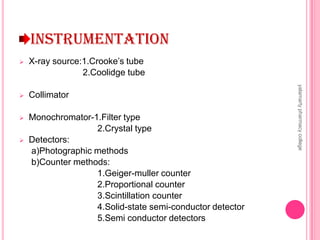
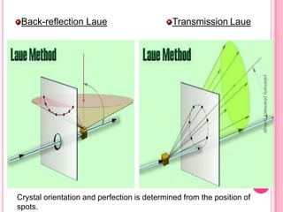
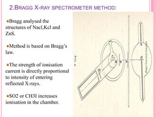
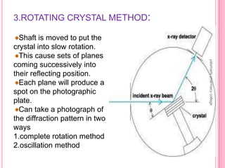
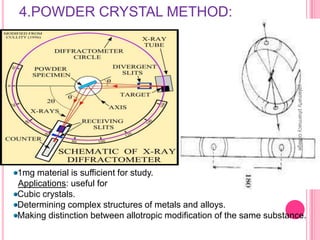
X-Ray Diffraction
Presented by…
J.N.V.Indira devi
M.Pharmacy 1st year
Pharmaceutical analysis and
quality assurance
Yalamarty pharmacy college
Andhra university
Visakhapatnam
yalamartypharmacycollege
CONTENTS
INTRODUCTION
BRAGG EQUATION
INSTRUMENTATION
X-RAY DIFFRACTION METHODS
APPLICATIONS
yalamartypharmacycollege
ELECTROMAGNETIC SPECTRUM
yalamartypharmacycollege
INTRODUCTION
X-Rays :
 X-rays are short wave length electromagnetic
radiations produced by the deceleration of high energy
electrons or by electronic transitions of electrons in the
inner orbital of atoms
 X-ray region 0.1to100 A˚
 Analytical purpose 0.7 to 2 A˚
 More energetic (i.e. can penetrate deeper into a
material).
 Emitted in a continuous band of white radiation as well
as a series of discrete lines that are characteristic of
the target material.
yalamartypharmacycollege
Characteristic Radiation:
 The characteristic lines in an atom's emission spectra are called K, L, M,
... and correspond to the n = 1, 2, 3, ... quantum levels of the electron
energy states, respectively.
 α lines (n = 2 to n = 1, or n = 3 to n = 2).
 β lines (n = 3 to n = 1 or n = 4 to n = 2).
 Moseley found that :1/λ = K2 [Z - σ]2.
Electronic energy levels of an atom
yalamartypharmacycollege
GENERATION OF X-RAYS
 Generated by bombarding a metal target with an energetic beam of
electrons.
 Synchrotrons:
Very expensive to build and maintain.
 By use of radioactive source like 55Fe.
 A new method of generating X rays that is not yet commercially
available uses an electron-impact beam impinging on a stream of
liquid gallium.
 X rays may also be produced while unrolling adhesive tape from a
tape dispenser.
yalamartypharmacycollege
X-RAY TECHNIQUES
X-ray absorption methods
 Fraction of X-ray photons absorbed is considered.
 Used in elemental analysis and thickness measurements.
X-ray fluorescence methods
 Wavelength and intensity of generated X-rays are measured
for qualitative and quantitative analysis.
 Non-destructive and requires little sample preparation.
X-ray diffraction methods
 Scattering of X-rays by crystals.
 Determines crystalline structure.
yalamartypharmacycollege
X-RAY DIFFRACTOMETER
The Bruker-D8
diffractometer
yalamartypharmacycollege
d
 The path difference between ray 1 and ray 2 = 2d Sin
 For constructive interference: n = 2d Sin
Ray 1
Ray 2
Deviation = 2
Constructive interference of the reflected beams emerging from two different planes
will take place if the difference in path lengths of two rays is equal to whole number of
wavelengths.
BRAGG’s EQUATION
yalamartypharmacycollege
INSTRUMENTATION
 X-ray source:1.Crooke’s tube
2.Coolidge tube
 Collimator
 Monochromator-1.Filter type
2.Crystal type
 Detectors:
a)Photographic methods
b)Counter methods:
1.Geiger-muller counter
2.Proportional counter
3.Scintillation counter
4.Solid-state semi-conductor detector
5.Semi conductor detectors
yalamartypharmacycollege
INSTRUMENTATION OF XRD
yalamartypharmacycollege
X-RAY SOURCE
Crookes tube
Called as cold cathode tube.
Electrons are generated by ionization
of the residual air in the tube, instead of
heated filament.
An aluminum cathode plate at one end
of the tube created a beam of electrons,
which struck a platinum anode target at
the center generating X-rays.
Advantage
Point source X-rays, which resulted in
sharper images.
Disadvantage :Unreliable
Crookes tube
yalamartypharmacycollege
Coolidge tube
Called as hot cathode tube.
Works with a very good quality vacuum (about 10-4 Pa,
The electrons are produced by thermionic effect from a tungsten filament
heated by an electric current.
There are two designs:
1.End-window tubes: Have thin "transmission target" to allow X-rays to
pass through the target
2.side-window tubes:An Electrostatic Lens to focus the beam onto a very
small spot on the anode.
• A window designed for escape of the generated X-ray photons.
• Power 0.1 to 18 kW.
yalamartypharmacycollege
COLLIMATOR
 Inserted in the diffracted-beam to
get a narrow x-ray beam.
 It consists two sets of closely
packed metal plates seperated by a
gap.
 The left end of the collimator
shown is mounted on the X-ray tube.
 The yellow-colored region at the
left end determines the the size of
the beam.
 The green region at the right end
removes parasitic radiation.
yalamartypharmacycollege
MATERIALS USED:NACL,LIF,QUARTZ ETC,.
Filter type
A window that absorbs undesirable radiation
and allows required wavelength to pass.
Eg:Zr absorbs x-rays emitted by Mo.
Crystal type
Positioned in the x-ray beam so that the angle
of the reflecting planes satisfied the Bragg’s
equation for the required wave length.
Characteristics of a crystal:
Mechanically strong and stable
The mosaicity and resolution of the crystal,
should be small.
MONOCHROMATOR
yalamartypharmacycollege
DETECTORS
Photographic methods
Contains photographic plate
Blackening of developed film is expressed in terms of density ,D
D=Log Io/I.
Photostimulable phosphors
An increasingly common method is the use of photo stimulated luminescence
Photostimulable phosphor plate (PSP plate) is used in place of the photographic
plate.
After the plate is X-rayed, excited electrons in the phosphor material remain ‘trapped'
in'colour centres' in the crystal lattice until stimulated by a laser beam passed over the
plate surface.
The light given off during laser stimulation is collected by a photomultiplier tube
ADVANTAGE:The PSP plate can be reused
yalamartypharmacycollege
Counter methods
1.Geiger-muller counter:
Filled with an inert gas like argon.
Measures ionizing radiation.
Detect the emission of nuclear
radiation: alpha particles, beta
particles or gamma rays
Advantages:
a)Trouble free
b)Inexpensive
Disadvantages:
a)Cannot be used to measure
energy of ionizing radiation.
b)Used for low counting rates
c)Efficiency falls off below 1A
yalamartypharmacycollege
2.Proportional counter:
Filled with heavier gas like xenon
or krypton as it is easily ionized.
Output pulse is dependent on
intensity of X-rays falling on
counter.
Count the particles of ionizing
radiation and measures their
energy.
Advantages:
a)Count high rates with out
significant error.
Disadvantages:
a)Associated electronic circuit is
complex.
b)Expensive.
yalamartypharmacycollege
3.Scintillation counter:
Measures X-rays of shorter
wavelengths.
The sensor, called a scintillator,
consists of a transparent crystal,
usually phosphor, plastic (usually
containing anthracene), or organic
liquid that fluoresces when struck
by ionizing radiation.
The PMT is attached to an
electronic amplifier to count and
possibly quantify the amplitude of
the signals.
Advantages:
a)Count high rates.
yalamartypharmacycollege
4.Solid state semi-conductor detector:
 The electrons produced by X-ray beam are promoted into
conduction bands and the current which flows is directly
proportional to the incident X-ray energy.
Disadvantage:
 Maintainted at very low Temp to minimise the noise and
prevent deterioration of the detector.
5.Semi-conductor detectors:
 Silicon-lithium drifted detector.
 The principle is similar to gas
ionization detector.
 Voltage of pulse=Q/C
Application: In neutron activation
analysis
Semi-conductor detector
yalamartypharmacycollege
X-RAY DIFFRACTION METHODS
1.LAUE PHOTOGRAPHIC METHOD:
The Laue method is mainly used to determine the orientation of large
single crystals. White radiation is reflected from, or transmitted through, a
fixed crystal.
 Back-reflection Laue
In the back-reflection
method, the film is Placed
between X-ray source and
crystal.
The beams which are
diffracted in a backward
direction are recorded.
 Transmission Laue
The film is placed behind the
crystal to record beams which
are transmitted through the
crystal.
Disadvantage: Big crystals are required
yalamartypharmacycollege
Back-reflection Laue Transmission Laue
Crystal orientation and perfection is determined from the position of
spots.
yalamartypharmacycollege
2.BRAGG X-RAY SPECTROMETER METHOD:
Bragg analysed the
structures of Nacl,Kcl and
ZnS.
Method is based on Bragg’s
law.
The strength of ionisation
current is directly proportional
to intensity of entering
reflected X-rays.
SO2 or CH3I increases
ionisation in the chamber.
yalamartypharmacycollege
3.ROTATING CRYSTAL METHOD:
Shaft is moved to put the
crystal into slow rotation.
This cause sets of planes
coming successively into
their reflecting position.
Each plane will produce a
spot on the photographic
plate.
Can take a photograph of
the diffraction pattern in two
ways
1.complete rotation method
2.oscillation method
yalamartypharmacycollege
4.POWDER CRYSTAL METHOD:
1mg material is sufficient for study.
Applications: useful for
Cubic crystals.
Determining complex structures of metals and alloys.
Making distinction between allotropic modification of the same substance.
yalamartypharmacycollege
APPLICATIONS
Structure of crystals
Polymer characterization
State of anneal in metals
Particle size determination
Spot counting method
v=V.δθ.cosθ/2n
Broadening of diffraction lines
Low-angle scattering
Applications of diffraction methods to complexes
Determination of cis-trans isomerism
Eg:Bis(pyridine-2-carboxamido)nickel(II) chloride
Determination of linkage isomerism
Eg:Biuret+copper(II)=pottassium bis(biureto)cuprate(II)
tetrahydrate
yalamartypharmacycollege
MISCELLANEOUS APPLICATIONS
 Soil classification based on crystallinity.
 Analysis of industrial dusts.
 Assessment of weathering & degradation of
minerals & polymers.
 Study of corrosion products.
 Examination of tooth enamel & dentine.
 Examination of bone state & tissue state.
 Structure of DNA&RNA.
 X-ray Diffraction on aerosol drugs -
Measurements on small quantities of
pharmaceutical samples using the X'Celerator
detector.
yalamartypharmacycollege
REFERENCES
1)Instrumental methods of chemical analysis ,B.K.sharma,17th
edition 1997-1998,GOEL publishing house.page no:329-359
2)Principles of instrumental analysis,5th edition ,by Dougles
a.skoog,f.James holles,Timothy A.Niemen.page no:277-298
3)Instrumental methods of chemical analysis ,Gurudeep
R.chatwal,sham k.anand,Himalaya publications page no:2.303-
2.332
4) Instrumental Methods Of Chemical Analysis –
H. Kaur pg.no:727-729,737
5) http://www.scienceiscool.org/solids/intro.html
6) http://en.wikipedia.org/wiki/X-ray_crystallography
yalamartypharmacycollege

X ray diffraction

  • 1.
    X-Ray Diffraction Presented by… J.N.V.Indiradevi M.Pharmacy 1st year Pharmaceutical analysis and quality assurance Yalamarty pharmacy college Andhra university Visakhapatnam yalamartypharmacycollege
  • 2.
  • 3.
  • 4.
    INTRODUCTION X-Rays :  X-raysare short wave length electromagnetic radiations produced by the deceleration of high energy electrons or by electronic transitions of electrons in the inner orbital of atoms  X-ray region 0.1to100 A˚  Analytical purpose 0.7 to 2 A˚  More energetic (i.e. can penetrate deeper into a material).  Emitted in a continuous band of white radiation as well as a series of discrete lines that are characteristic of the target material. yalamartypharmacycollege
  • 5.
    Characteristic Radiation:  Thecharacteristic lines in an atom's emission spectra are called K, L, M, ... and correspond to the n = 1, 2, 3, ... quantum levels of the electron energy states, respectively.  α lines (n = 2 to n = 1, or n = 3 to n = 2).  β lines (n = 3 to n = 1 or n = 4 to n = 2).  Moseley found that :1/λ = K2 [Z - σ]2. Electronic energy levels of an atom yalamartypharmacycollege
  • 6.
    GENERATION OF X-RAYS Generated by bombarding a metal target with an energetic beam of electrons.  Synchrotrons: Very expensive to build and maintain.  By use of radioactive source like 55Fe.  A new method of generating X rays that is not yet commercially available uses an electron-impact beam impinging on a stream of liquid gallium.  X rays may also be produced while unrolling adhesive tape from a tape dispenser. yalamartypharmacycollege
  • 7.
    X-RAY TECHNIQUES X-ray absorptionmethods  Fraction of X-ray photons absorbed is considered.  Used in elemental analysis and thickness measurements. X-ray fluorescence methods  Wavelength and intensity of generated X-rays are measured for qualitative and quantitative analysis.  Non-destructive and requires little sample preparation. X-ray diffraction methods  Scattering of X-rays by crystals.  Determines crystalline structure. yalamartypharmacycollege
  • 8.
  • 9.
    d  The pathdifference between ray 1 and ray 2 = 2d Sin  For constructive interference: n = 2d Sin Ray 1 Ray 2 Deviation = 2 Constructive interference of the reflected beams emerging from two different planes will take place if the difference in path lengths of two rays is equal to whole number of wavelengths. BRAGG’s EQUATION yalamartypharmacycollege
  • 10.
    INSTRUMENTATION  X-ray source:1.Crooke’stube 2.Coolidge tube  Collimator  Monochromator-1.Filter type 2.Crystal type  Detectors: a)Photographic methods b)Counter methods: 1.Geiger-muller counter 2.Proportional counter 3.Scintillation counter 4.Solid-state semi-conductor detector 5.Semi conductor detectors yalamartypharmacycollege
  • 11.
  • 12.
    X-RAY SOURCE Crookes tube Calledas cold cathode tube. Electrons are generated by ionization of the residual air in the tube, instead of heated filament. An aluminum cathode plate at one end of the tube created a beam of electrons, which struck a platinum anode target at the center generating X-rays. Advantage Point source X-rays, which resulted in sharper images. Disadvantage :Unreliable Crookes tube yalamartypharmacycollege
  • 13.
    Coolidge tube Called ashot cathode tube. Works with a very good quality vacuum (about 10-4 Pa, The electrons are produced by thermionic effect from a tungsten filament heated by an electric current. There are two designs: 1.End-window tubes: Have thin "transmission target" to allow X-rays to pass through the target 2.side-window tubes:An Electrostatic Lens to focus the beam onto a very small spot on the anode. • A window designed for escape of the generated X-ray photons. • Power 0.1 to 18 kW. yalamartypharmacycollege
  • 14.
    COLLIMATOR  Inserted inthe diffracted-beam to get a narrow x-ray beam.  It consists two sets of closely packed metal plates seperated by a gap.  The left end of the collimator shown is mounted on the X-ray tube.  The yellow-colored region at the left end determines the the size of the beam.  The green region at the right end removes parasitic radiation. yalamartypharmacycollege
  • 15.
    MATERIALS USED:NACL,LIF,QUARTZ ETC,. Filtertype A window that absorbs undesirable radiation and allows required wavelength to pass. Eg:Zr absorbs x-rays emitted by Mo. Crystal type Positioned in the x-ray beam so that the angle of the reflecting planes satisfied the Bragg’s equation for the required wave length. Characteristics of a crystal: Mechanically strong and stable The mosaicity and resolution of the crystal, should be small. MONOCHROMATOR yalamartypharmacycollege
  • 16.
    DETECTORS Photographic methods Contains photographicplate Blackening of developed film is expressed in terms of density ,D D=Log Io/I. Photostimulable phosphors An increasingly common method is the use of photo stimulated luminescence Photostimulable phosphor plate (PSP plate) is used in place of the photographic plate. After the plate is X-rayed, excited electrons in the phosphor material remain ‘trapped' in'colour centres' in the crystal lattice until stimulated by a laser beam passed over the plate surface. The light given off during laser stimulation is collected by a photomultiplier tube ADVANTAGE:The PSP plate can be reused yalamartypharmacycollege
  • 17.
    Counter methods 1.Geiger-muller counter: Filledwith an inert gas like argon. Measures ionizing radiation. Detect the emission of nuclear radiation: alpha particles, beta particles or gamma rays Advantages: a)Trouble free b)Inexpensive Disadvantages: a)Cannot be used to measure energy of ionizing radiation. b)Used for low counting rates c)Efficiency falls off below 1A yalamartypharmacycollege
  • 18.
    2.Proportional counter: Filled withheavier gas like xenon or krypton as it is easily ionized. Output pulse is dependent on intensity of X-rays falling on counter. Count the particles of ionizing radiation and measures their energy. Advantages: a)Count high rates with out significant error. Disadvantages: a)Associated electronic circuit is complex. b)Expensive. yalamartypharmacycollege
  • 19.
    3.Scintillation counter: Measures X-raysof shorter wavelengths. The sensor, called a scintillator, consists of a transparent crystal, usually phosphor, plastic (usually containing anthracene), or organic liquid that fluoresces when struck by ionizing radiation. The PMT is attached to an electronic amplifier to count and possibly quantify the amplitude of the signals. Advantages: a)Count high rates. yalamartypharmacycollege
  • 20.
    4.Solid state semi-conductordetector:  The electrons produced by X-ray beam are promoted into conduction bands and the current which flows is directly proportional to the incident X-ray energy. Disadvantage:  Maintainted at very low Temp to minimise the noise and prevent deterioration of the detector. 5.Semi-conductor detectors:  Silicon-lithium drifted detector.  The principle is similar to gas ionization detector.  Voltage of pulse=Q/C Application: In neutron activation analysis Semi-conductor detector yalamartypharmacycollege
  • 21.
    X-RAY DIFFRACTION METHODS 1.LAUEPHOTOGRAPHIC METHOD: The Laue method is mainly used to determine the orientation of large single crystals. White radiation is reflected from, or transmitted through, a fixed crystal.  Back-reflection Laue In the back-reflection method, the film is Placed between X-ray source and crystal. The beams which are diffracted in a backward direction are recorded.  Transmission Laue The film is placed behind the crystal to record beams which are transmitted through the crystal. Disadvantage: Big crystals are required yalamartypharmacycollege
  • 22.
    Back-reflection Laue TransmissionLaue Crystal orientation and perfection is determined from the position of spots. yalamartypharmacycollege
  • 23.
    2.BRAGG X-RAY SPECTROMETERMETHOD: Bragg analysed the structures of Nacl,Kcl and ZnS. Method is based on Bragg’s law. The strength of ionisation current is directly proportional to intensity of entering reflected X-rays. SO2 or CH3I increases ionisation in the chamber. yalamartypharmacycollege
  • 24.
    3.ROTATING CRYSTAL METHOD: Shaftis moved to put the crystal into slow rotation. This cause sets of planes coming successively into their reflecting position. Each plane will produce a spot on the photographic plate. Can take a photograph of the diffraction pattern in two ways 1.complete rotation method 2.oscillation method yalamartypharmacycollege
  • 25.
    4.POWDER CRYSTAL METHOD: 1mgmaterial is sufficient for study. Applications: useful for Cubic crystals. Determining complex structures of metals and alloys. Making distinction between allotropic modification of the same substance. yalamartypharmacycollege
  • 26.
    APPLICATIONS Structure of crystals Polymercharacterization State of anneal in metals Particle size determination Spot counting method v=V.δθ.cosθ/2n Broadening of diffraction lines Low-angle scattering Applications of diffraction methods to complexes Determination of cis-trans isomerism Eg:Bis(pyridine-2-carboxamido)nickel(II) chloride Determination of linkage isomerism Eg:Biuret+copper(II)=pottassium bis(biureto)cuprate(II) tetrahydrate yalamartypharmacycollege
  • 27.
    MISCELLANEOUS APPLICATIONS  Soilclassification based on crystallinity.  Analysis of industrial dusts.  Assessment of weathering & degradation of minerals & polymers.  Study of corrosion products.  Examination of tooth enamel & dentine.  Examination of bone state & tissue state.  Structure of DNA&RNA.  X-ray Diffraction on aerosol drugs - Measurements on small quantities of pharmaceutical samples using the X'Celerator detector. yalamartypharmacycollege
  • 28.
    REFERENCES 1)Instrumental methods ofchemical analysis ,B.K.sharma,17th edition 1997-1998,GOEL publishing house.page no:329-359 2)Principles of instrumental analysis,5th edition ,by Dougles a.skoog,f.James holles,Timothy A.Niemen.page no:277-298 3)Instrumental methods of chemical analysis ,Gurudeep R.chatwal,sham k.anand,Himalaya publications page no:2.303- 2.332 4) Instrumental Methods Of Chemical Analysis – H. Kaur pg.no:727-729,737 5) http://www.scienceiscool.org/solids/intro.html 6) http://en.wikipedia.org/wiki/X-ray_crystallography yalamartypharmacycollege
  • 29.