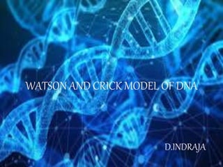
The document summarizes the Watson and Crick model of DNA, which proposed in 1953 that DNA consists of two helical strands coiled around a common axis to form a double helix. Each strand contains alternating sugar and phosphate groups, with nitrogenous base pairs connecting the strands via hydrogen bonds. Adenine always pairs with thymine and guanine pairs with cytosine. The structure explained DNA's ability to replicate itself accurately during cell division. Rosalind Franklin's X-ray diffraction images provided key evidence for the double helix structure, though she did not receive full credit at the time due to sexism in the field.
RNA- A polymer of ribonucleotides, is a single stranded structure. There are three major types of RNA- m RNA,t RNA and r RNA. Besides that there are small nuclear,micro RNAs, small interfering and heterogeneous RNAs. Each of them has a specific structure and performs a specific function.
RNA- A polymer of ribonucleotides, is a single stranded structure. There are three major types of RNA- m RNA,t RNA and r RNA. Besides that there are small nuclear,micro RNAs, small interfering and heterogeneous RNAs. Each of them has a specific structure and performs a specific function.
DNA is tightly packed in the nucleus of every cell. DNA wraps around special proteins called histones, which form loops of DNA called nucleosomes. These nucleosomes coil and stack together to form fibers called chromatin. Chromatin in turn forms larger loops and coils to form chromosomes.
DNA packaging is crucial because it makes sure that those excessive DNA are able to fit nicely in a cell that is many times smaller.
The DNA in bacterial cells are either circular or linear. To accommodate the size of bacterial cell, supercoiled DNA are folded into loops with each loop resembles shape of bead-like packets containing small basic proteins that is analogous to histone found in Eukaryotes.
One of the first plausible models to account for the preceding observations was
formulated by Robin Holliday.
The key features of the Holliday model are the formation of heteroduplex DNA; the
creation of a cross bridge; its migration along the two heteroduplex strands,
termed branch migration; the occurrence of mismatch repair; and the
subsequent resolution, or splicing, of the intermediate structure to yield different
typesof recombinant molecules.
Nucleic Acids
DNA
Eukaryotic Chromosomes
The Histones
Deoxynucleic acid ( DNA )
Importance of Nucleotides
Base pairing
Denaturation and Renaturation
Determination GC content
Prokaryotic DNA synthesis
Prokaryotic DNA Replication
Transcription
Coding Strand and Template Strand
Steps of RNA synthesize
A detailed explanation of cloning strategies which involves isolation of DNA fragments from the sample and introduction in to a vector with restriction enzymes and introduced in to host by different methods and finally screening of the host cells with the recombinants based on protein,nucleicacid and antibiotic assays
control of gene expression by sigma factor and post transcriptional controlIndrajaDoradla
explanation of control of gene expression by sigma factor and decription of sigma factor and detailed explation of post transcriptional control by antisense technology and rna i
description of transgenic animals and production with desired traits using different methods and their applications and their advantages and disadvantages
Cancer cell metabolism: special Reference to Lactate PathwayAADYARAJPANDEY1
Normal Cell Metabolism:
Cellular respiration describes the series of steps that cells use to break down sugar and other chemicals to get the energy we need to function.
Energy is stored in the bonds of glucose and when glucose is broken down, much of that energy is released.
Cell utilize energy in the form of ATP.
The first step of respiration is called glycolysis. In a series of steps, glycolysis breaks glucose into two smaller molecules - a chemical called pyruvate. A small amount of ATP is formed during this process.
Most healthy cells continue the breakdown in a second process, called the Kreb's cycle. The Kreb's cycle allows cells to “burn” the pyruvates made in glycolysis to get more ATP.
The last step in the breakdown of glucose is called oxidative phosphorylation (Ox-Phos).
It takes place in specialized cell structures called mitochondria. This process produces a large amount of ATP. Importantly, cells need oxygen to complete oxidative phosphorylation.
If a cell completes only glycolysis, only 2 molecules of ATP are made per glucose. However, if the cell completes the entire respiration process (glycolysis - Kreb's - oxidative phosphorylation), about 36 molecules of ATP are created, giving it much more energy to use.
IN CANCER CELL:
Unlike healthy cells that "burn" the entire molecule of sugar to capture a large amount of energy as ATP, cancer cells are wasteful.
Cancer cells only partially break down sugar molecules. They overuse the first step of respiration, glycolysis. They frequently do not complete the second step, oxidative phosphorylation.
This results in only 2 molecules of ATP per each glucose molecule instead of the 36 or so ATPs healthy cells gain. As a result, cancer cells need to use a lot more sugar molecules to get enough energy to survive.
introduction to WARBERG PHENOMENA:
WARBURG EFFECT Usually, cancer cells are highly glycolytic (glucose addiction) and take up more glucose than do normal cells from outside.
Otto Heinrich Warburg (; 8 October 1883 – 1 August 1970) In 1931 was awarded the Nobel Prize in Physiology for his "discovery of the nature and mode of action of the respiratory enzyme.
WARNBURG EFFECT : cancer cells under aerobic (well-oxygenated) conditions to metabolize glucose to lactate (aerobic glycolysis) is known as the Warburg effect. Warburg made the observation that tumor slices consume glucose and secrete lactate at a higher rate than normal tissues.
Slide 1: Title Slide
Extrachromosomal Inheritance
Slide 2: Introduction to Extrachromosomal Inheritance
Definition: Extrachromosomal inheritance refers to the transmission of genetic material that is not found within the nucleus.
Key Components: Involves genes located in mitochondria, chloroplasts, and plasmids.
Slide 3: Mitochondrial Inheritance
Mitochondria: Organelles responsible for energy production.
Mitochondrial DNA (mtDNA): Circular DNA molecule found in mitochondria.
Inheritance Pattern: Maternally inherited, meaning it is passed from mothers to all their offspring.
Diseases: Examples include Leber’s hereditary optic neuropathy (LHON) and mitochondrial myopathy.
Slide 4: Chloroplast Inheritance
Chloroplasts: Organelles responsible for photosynthesis in plants.
Chloroplast DNA (cpDNA): Circular DNA molecule found in chloroplasts.
Inheritance Pattern: Often maternally inherited in most plants, but can vary in some species.
Examples: Variegation in plants, where leaf color patterns are determined by chloroplast DNA.
Slide 5: Plasmid Inheritance
Plasmids: Small, circular DNA molecules found in bacteria and some eukaryotes.
Features: Can carry antibiotic resistance genes and can be transferred between cells through processes like conjugation.
Significance: Important in biotechnology for gene cloning and genetic engineering.
Slide 6: Mechanisms of Extrachromosomal Inheritance
Non-Mendelian Patterns: Do not follow Mendel’s laws of inheritance.
Cytoplasmic Segregation: During cell division, organelles like mitochondria and chloroplasts are randomly distributed to daughter cells.
Heteroplasmy: Presence of more than one type of organellar genome within a cell, leading to variation in expression.
Slide 7: Examples of Extrachromosomal Inheritance
Four O’clock Plant (Mirabilis jalapa): Shows variegated leaves due to different cpDNA in leaf cells.
Petite Mutants in Yeast: Result from mutations in mitochondrial DNA affecting respiration.
Slide 8: Importance of Extrachromosomal Inheritance
Evolution: Provides insight into the evolution of eukaryotic cells.
Medicine: Understanding mitochondrial inheritance helps in diagnosing and treating mitochondrial diseases.
Agriculture: Chloroplast inheritance can be used in plant breeding and genetic modification.
Slide 9: Recent Research and Advances
Gene Editing: Techniques like CRISPR-Cas9 are being used to edit mitochondrial and chloroplast DNA.
Therapies: Development of mitochondrial replacement therapy (MRT) for preventing mitochondrial diseases.
Slide 10: Conclusion
Summary: Extrachromosomal inheritance involves the transmission of genetic material outside the nucleus and plays a crucial role in genetics, medicine, and biotechnology.
Future Directions: Continued research and technological advancements hold promise for new treatments and applications.
Slide 11: Questions and Discussion
Invite Audience: Open the floor for any questions or further discussion on the topic.
Observation of Io’s Resurfacing via Plume Deposition Using Ground-based Adapt...Sérgio Sacani
Since volcanic activity was first discovered on Io from Voyager images in 1979, changes
on Io’s surface have been monitored from both spacecraft and ground-based telescopes.
Here, we present the highest spatial resolution images of Io ever obtained from a groundbased telescope. These images, acquired by the SHARK-VIS instrument on the Large
Binocular Telescope, show evidence of a major resurfacing event on Io’s trailing hemisphere. When compared to the most recent spacecraft images, the SHARK-VIS images
show that a plume deposit from a powerful eruption at Pillan Patera has covered part
of the long-lived Pele plume deposit. Although this type of resurfacing event may be common on Io, few have been detected due to the rarity of spacecraft visits and the previously low spatial resolution available from Earth-based telescopes. The SHARK-VIS instrument ushers in a new era of high resolution imaging of Io’s surface using adaptive
optics at visible wavelengths.
Richard's entangled aventures in wonderlandRichard Gill
Since the loophole-free Bell experiments of 2020 and the Nobel prizes in physics of 2022, critics of Bell's work have retreated to the fortress of super-determinism. Now, super-determinism is a derogatory word - it just means "determinism". Palmer, Hance and Hossenfelder argue that quantum mechanics and determinism are not incompatible, using a sophisticated mathematical construction based on a subtle thinning of allowed states and measurements in quantum mechanics, such that what is left appears to make Bell's argument fail, without altering the empirical predictions of quantum mechanics. I think however that it is a smoke screen, and the slogan "lost in math" comes to my mind. I will discuss some other recent disproofs of Bell's theorem using the language of causality based on causal graphs. Causal thinking is also central to law and justice. I will mention surprising connections to my work on serial killer nurse cases, in particular the Dutch case of Lucia de Berk and the current UK case of Lucy Letby.
A brief information about the SCOP protein database used in bioinformatics.
The Structural Classification of Proteins (SCOP) database is a comprehensive and authoritative resource for the structural and evolutionary relationships of proteins. It provides a detailed and curated classification of protein structures, grouping them into families, superfamilies, and folds based on their structural and sequence similarities.
The increased availability of biomedical data, particularly in the public domain, offers the opportunity to better understand human health and to develop effective therapeutics for a wide range of unmet medical needs. However, data scientists remain stymied by the fact that data remain hard to find and to productively reuse because data and their metadata i) are wholly inaccessible, ii) are in non-standard or incompatible representations, iii) do not conform to community standards, and iv) have unclear or highly restricted terms and conditions that preclude legitimate reuse. These limitations require a rethink on data can be made machine and AI-ready - the key motivation behind the FAIR Guiding Principles. Concurrently, while recent efforts have explored the use of deep learning to fuse disparate data into predictive models for a wide range of biomedical applications, these models often fail even when the correct answer is already known, and fail to explain individual predictions in terms that data scientists can appreciate. These limitations suggest that new methods to produce practical artificial intelligence are still needed.
In this talk, I will discuss our work in (1) building an integrative knowledge infrastructure to prepare FAIR and "AI-ready" data and services along with (2) neurosymbolic AI methods to improve the quality of predictions and to generate plausible explanations. Attention is given to standards, platforms, and methods to wrangle knowledge into simple, but effective semantic and latent representations, and to make these available into standards-compliant and discoverable interfaces that can be used in model building, validation, and explanation. Our work, and those of others in the field, creates a baseline for building trustworthy and easy to deploy AI models in biomedicine.
Bio
Dr. Michel Dumontier is the Distinguished Professor of Data Science at Maastricht University, founder and executive director of the Institute of Data Science, and co-founder of the FAIR (Findable, Accessible, Interoperable and Reusable) data principles. His research explores socio-technological approaches for responsible discovery science, which includes collaborative multi-modal knowledge graphs, privacy-preserving distributed data mining, and AI methods for drug discovery and personalized medicine. His work is supported through the Dutch National Research Agenda, the Netherlands Organisation for Scientific Research, Horizon Europe, the European Open Science Cloud, the US National Institutes of Health, and a Marie-Curie Innovative Training Network. He is the editor-in-chief for the journal Data Science and is internationally recognized for his contributions in bioinformatics, biomedical informatics, and semantic technologies including ontologies and linked data.
(May 29th, 2024) Advancements in Intravital Microscopy- Insights for Preclini...Scintica Instrumentation
Intravital microscopy (IVM) is a powerful tool utilized to study cellular behavior over time and space in vivo. Much of our understanding of cell biology has been accomplished using various in vitro and ex vivo methods; however, these studies do not necessarily reflect the natural dynamics of biological processes. Unlike traditional cell culture or fixed tissue imaging, IVM allows for the ultra-fast high-resolution imaging of cellular processes over time and space and were studied in its natural environment. Real-time visualization of biological processes in the context of an intact organism helps maintain physiological relevance and provide insights into the progression of disease, response to treatments or developmental processes.
In this webinar we give an overview of advanced applications of the IVM system in preclinical research. IVIM technology is a provider of all-in-one intravital microscopy systems and solutions optimized for in vivo imaging of live animal models at sub-micron resolution. The system’s unique features and user-friendly software enables researchers to probe fast dynamic biological processes such as immune cell tracking, cell-cell interaction as well as vascularization and tumor metastasis with exceptional detail. This webinar will also give an overview of IVM being utilized in drug development, offering a view into the intricate interaction between drugs/nanoparticles and tissues in vivo and allows for the evaluation of therapeutic intervention in a variety of tissues and organs. This interdisciplinary collaboration continues to drive the advancements of novel therapeutic strategies.
2. DNA MODEL
• The three-dimensional structure of DNA, first proposed by
James D. Watson and Francis H. C. Crick in 1953, consists of
two long helical strands that are coiled around a common axis
to form a double helix.
• Each DNA molecule is comprised of two biopolymer strands
coiling around each other.
• Each strand has a 5′end (with a phosphate group) and a 3′end
(with a hydroxyl group).
• The strands are antiparallel, meaning that one strand runs in a
5′to 3′direction, while the other strand runs in a 3′to
5′direction.
• The diameter of the double helix is 2nm and the double helical
structure repeats at an interval of 3.4nm(34A) which
corresponds to ten base pairs which is none other than helix
measure
3. • The distance between two succesive base pairs is 0.34nm
(3.4A)
• The two strands are held together by hydrogen bonds and
are complementary to each other.
• The two DNA strands are called polynucleotides, as they
are made of simpler monomer units called nucleotides.
Basically, the DNA is composed of deoxyribonucleotides.
• The deoxyribonucleotides are linked together by 3′-
5′phosphodiester bonds.
• The nitrogenous bases that compose the
deoxyribonucleotides include adenine, cytosine, thymine,
and guanine.
• The structure of DNA -DNA is a double helix structure
because it looks like a twisted ladder.
4. • The sides of the ladder are made of alternating sugar
(deoxyribose) and phosphate molecules while the steps of the
ladder are made up of a pair of nitrogen bases.
• As a result of the double helical nature of DNA, the molecule
has two asymmetric grooves. One groove is smaller than the
other.
• The larger groove is called the major groove, occurs when the
backbones are far apart; while the smaller one is called
the minor groove, and occurs when they are close together.
• A always pairs with T with 2 hydrogen bonds and G pairs with
C with 3 hydrogen bonds hence two strands are
complimentary to each other
• Because of specificity in base pairing the amount of purines is
equal to the amount of pyramidines this is called as chargaffs
rule of base equivalence.
6. COMPONENTS OF DNA
• Nucleoside is a compound formed by the combination
of a pentose sugar and nitrogen base
• Nucleotide is compound formed by the combination of
nucleoside and phosphate group
• Nucleotides are the building blocks of nucleic acids
• Nucleotide have three charecteristic components
• Nitrogen base
• Deoxyribose sugar
• Phosphate
9. DEOXYRIBOSE SUGAR
• Deoxyribose, also known as D-Deoxyribose and 2-
deoxyribose, is a pentose sugar (monosaccharide
containing five carbon atoms) that is a key
component of the nucleic acid deoxyribonucleic acid
(DNA).
• It is derived from the pentose sugar ribose.
Deoxyribose has the chemical formula C5H10O4.
• Deoxyribose is the sugar component of DNA
• Alternating with phosphate bases, deoxyribose forms
the backbone of the DNA, binding to the nitrogenous
bases adenine, thymine, guanine, and cytosine.
10. • The sugar-phosphate backbone forms the structural
framework of nucleic acids, including DNA.
• This backbone is composed of alternating sugar and
phosphate groups and defines directionality of the
molecule.
PHOSPHATE GROUP
11. • DNA are composed of nucleotides that are linked to one
another in a chain by chemical bonds, called ester bonds
• These bonds are called phosphodiester bonds
• In double-stranded DNA, the molecular double-helix
shape is formed by two linear sugar-phosphate backbones
that run opposite each other and twist together in a helical
shape.
• The sugar-phosphate backbone is negatively charged and
hydrophilic, which allows the DNA backbone to form
bonds with water.
12.
13. Secret behind the discovery
• At King's College in London, Rosalind Franklin and
Maurice Wilkins were studying DNA. Wilkins and
Franklin used X-ray diffraction as their main tool --
beaming X-rays through the molecule yielded a shadow
picture of the molecule's structure, by how the X-rays
bounced off its component parts.
Franklin, a shy and inward young woman, suffered from
patronizing attitudes and sexism that forced her to do
much of her work alone. And her senior partner, Wilkins,
showed some of Franklin's findings to Watson in January
1953 without her knowledge.
14. • Shortly after, Watson and Crick made a crucial advance when
they proposed that the DNA molecule was made up of two
chains of nucleotides paired in such a way to form a double
helix, like a spiral staircase. This structure, announced in their
famous paper in the April 1953 issue of Nature, explained how
the DNA molecule could replicate itself during cell division,
enabling organisms to reproduce themselves with amazing
accuracy except for occasional mutations.
For their work, Watson, Crick, and Wilkins received the Nobel
Prize in 1962. Despite her contribution to the discovery of
DNA's helical structure, Rosalind Franklin was not named a
prize winner: She had died of cancer four years earlier, at the
age of 37.