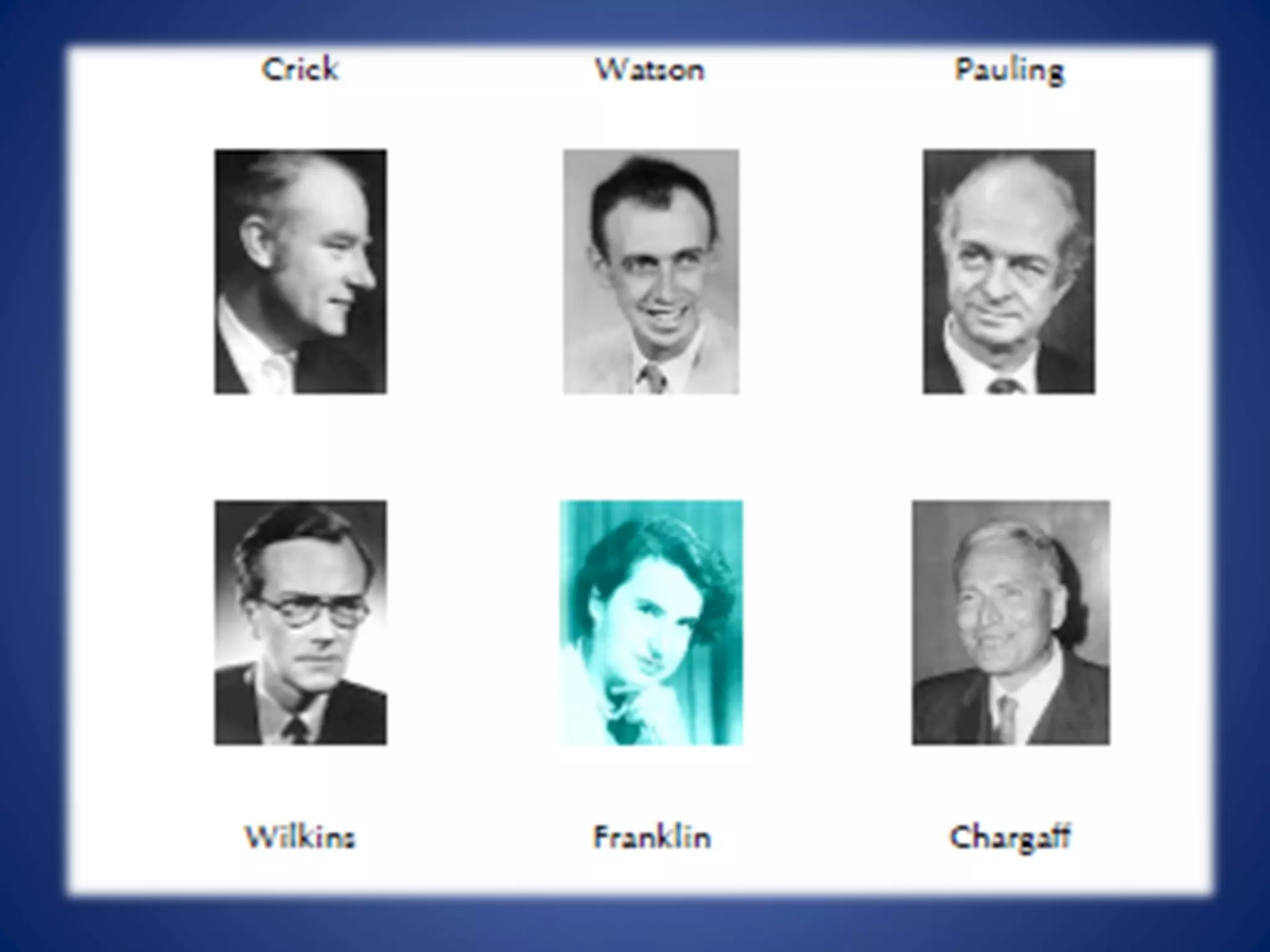
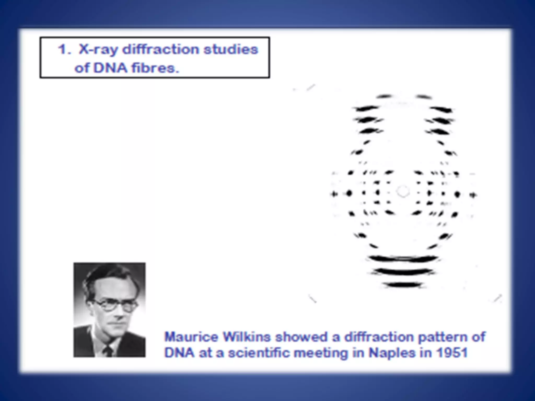
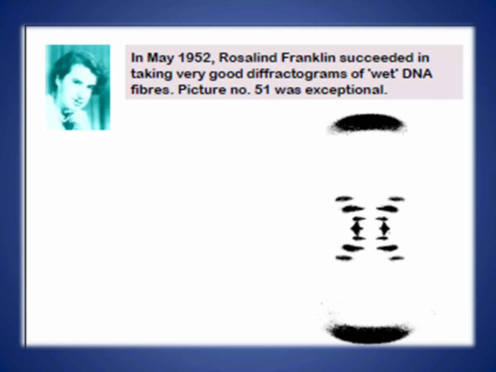
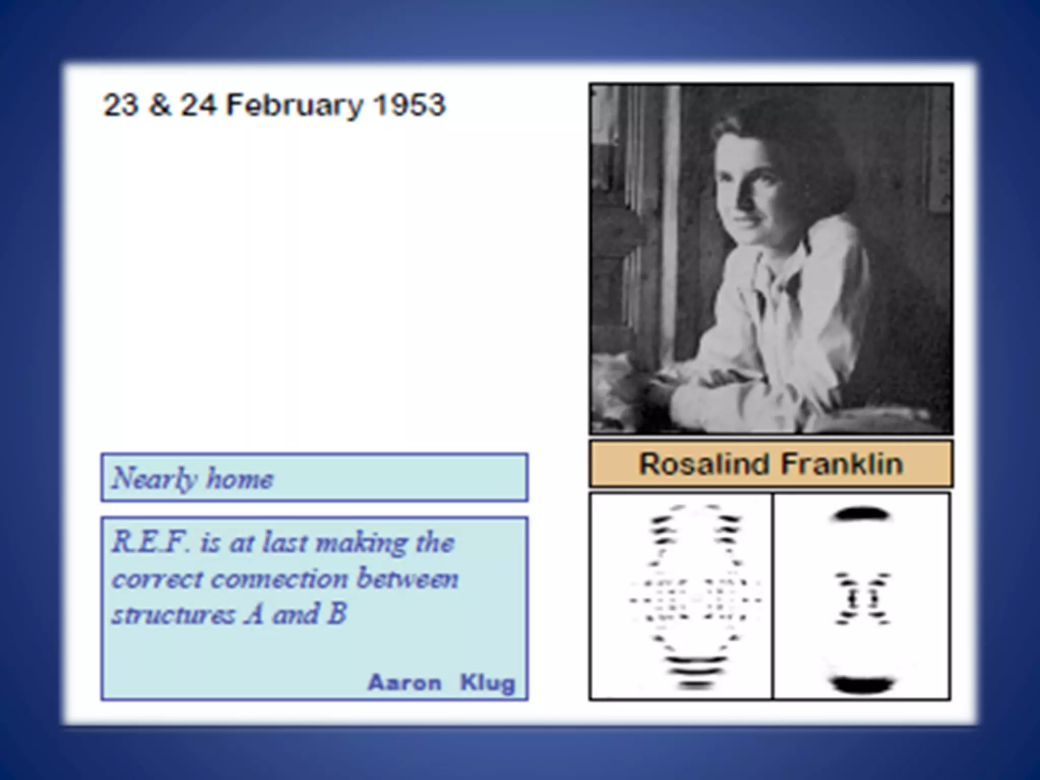
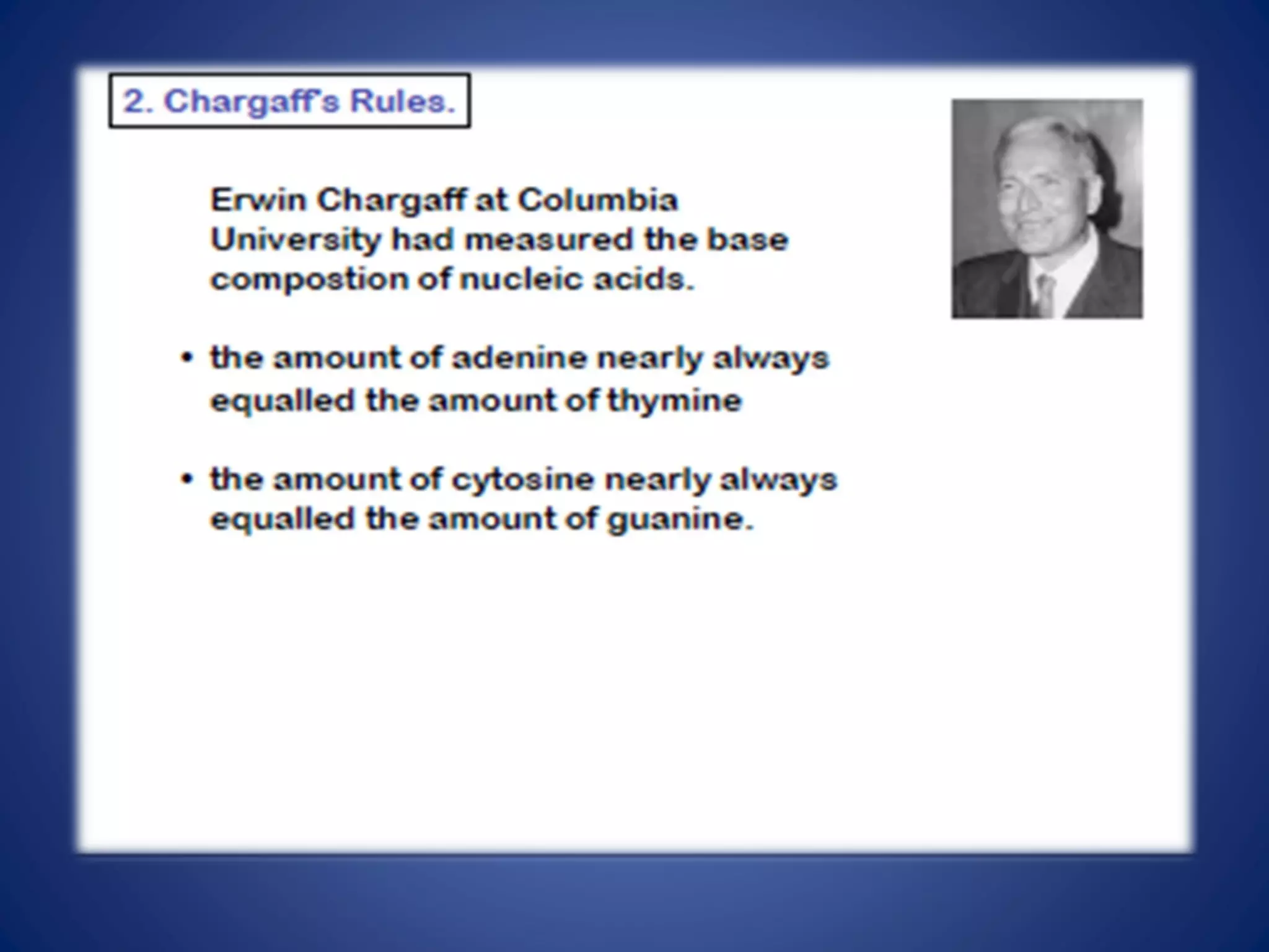
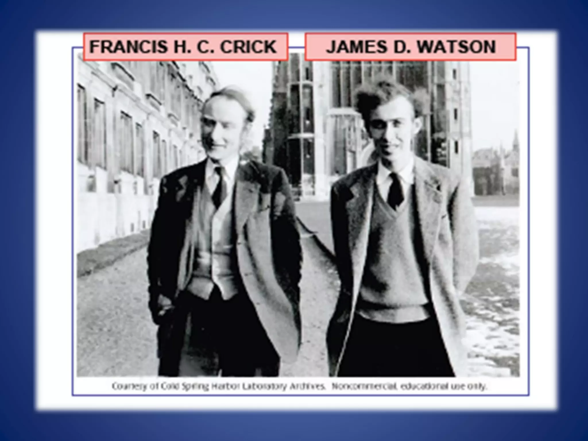
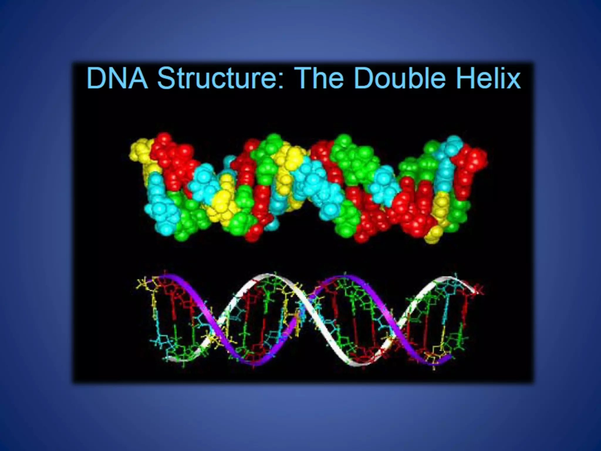
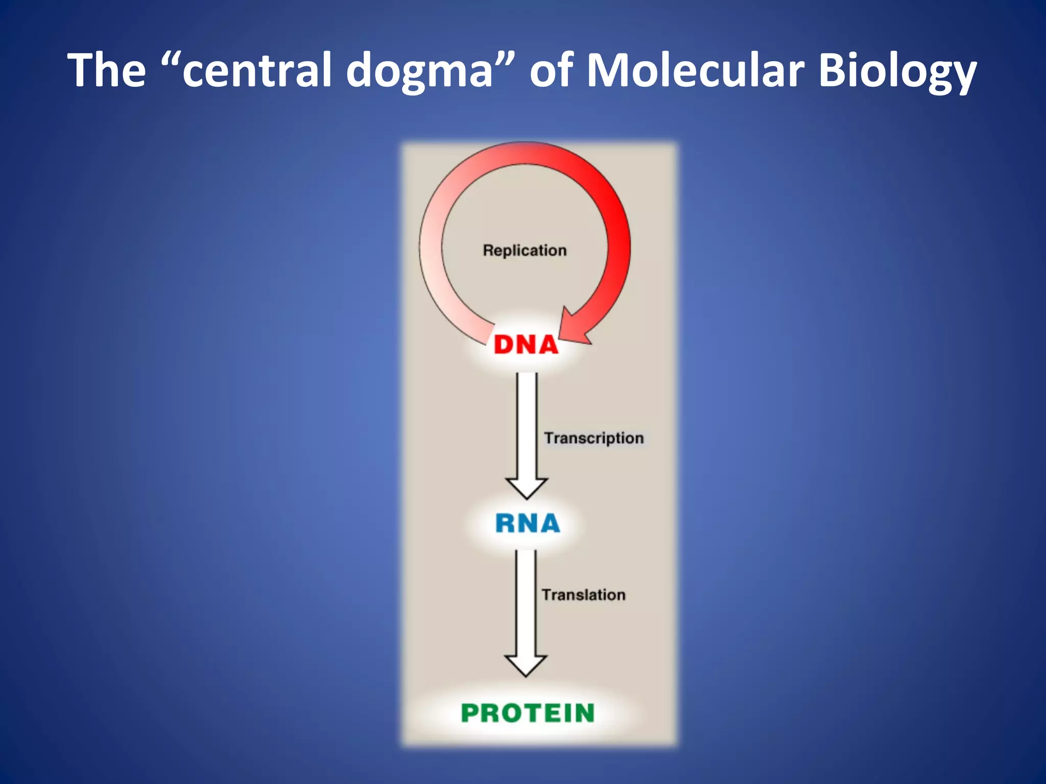
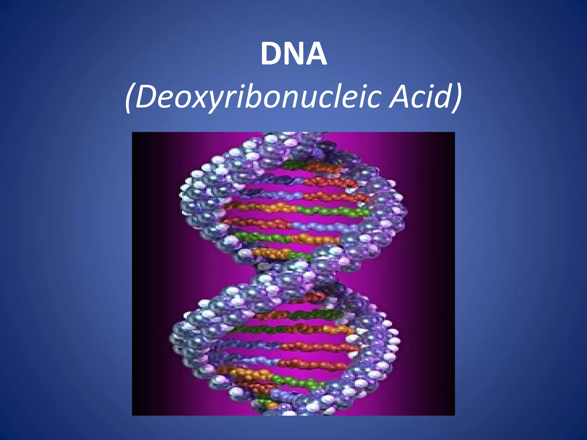
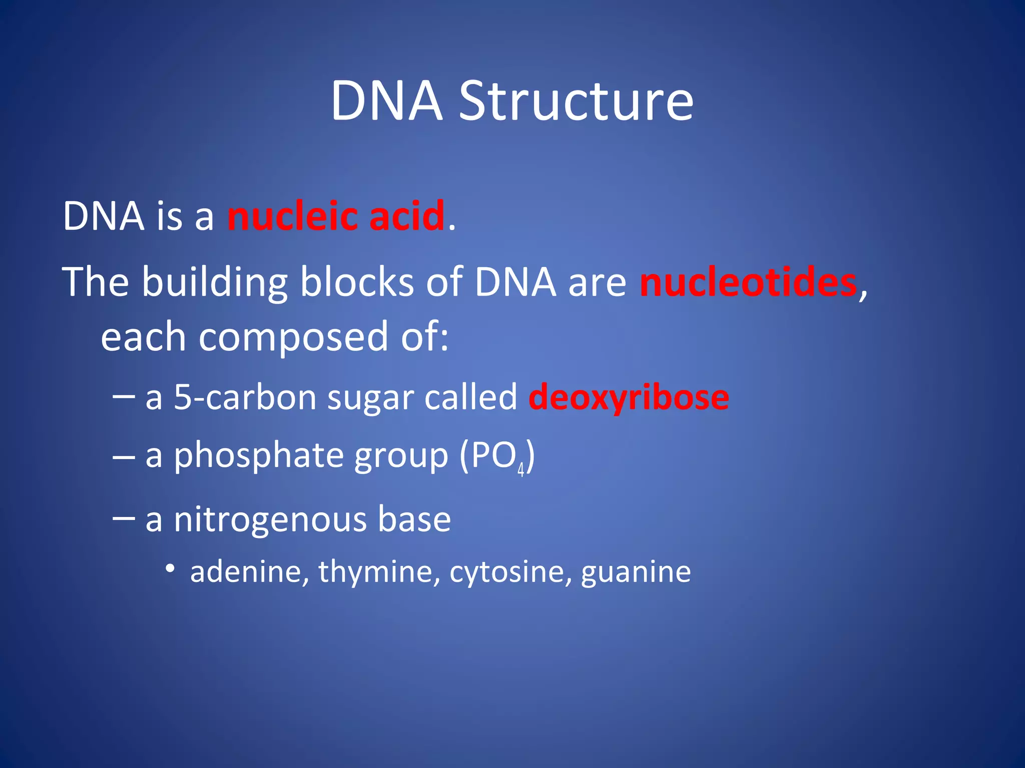
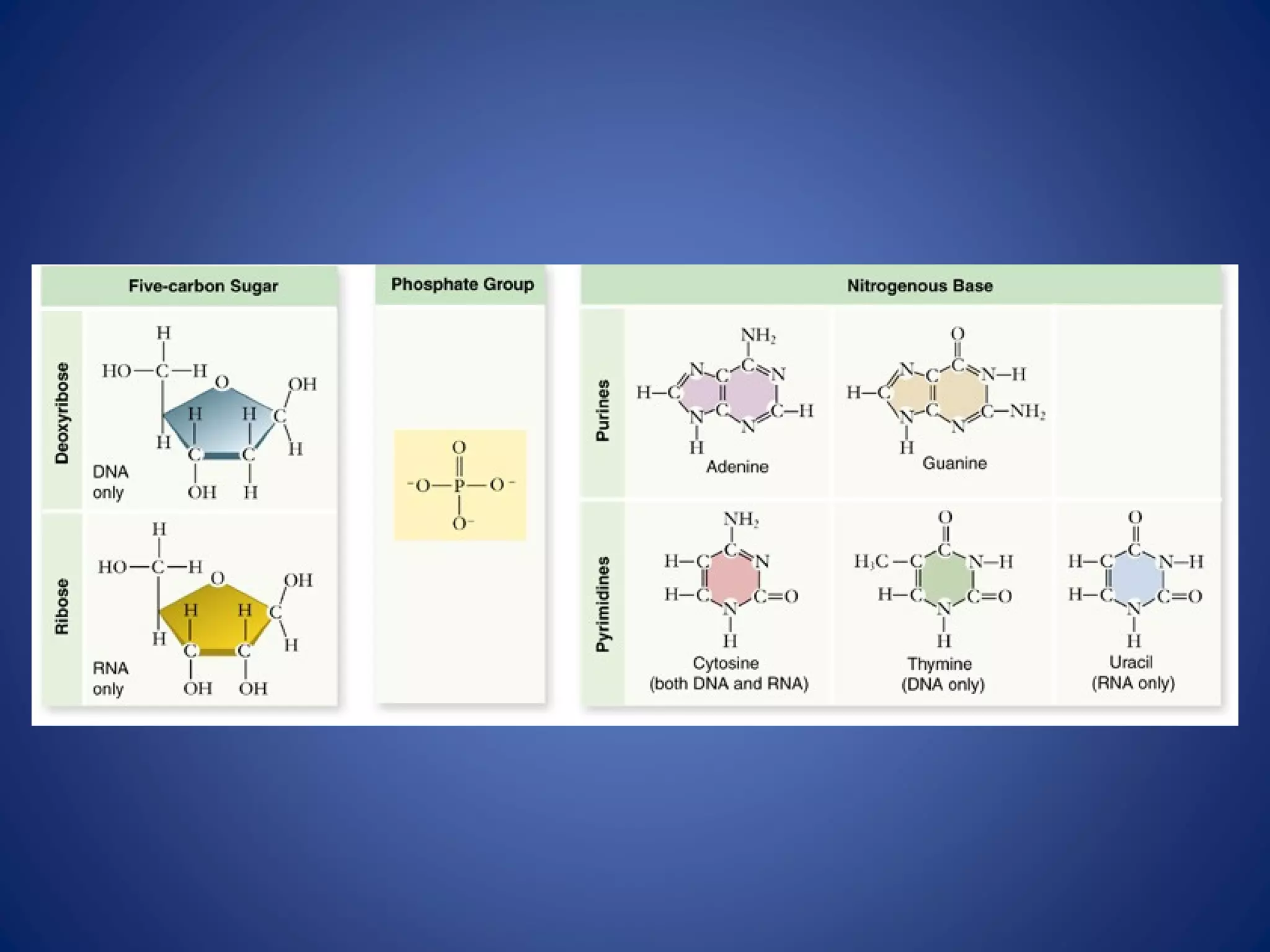
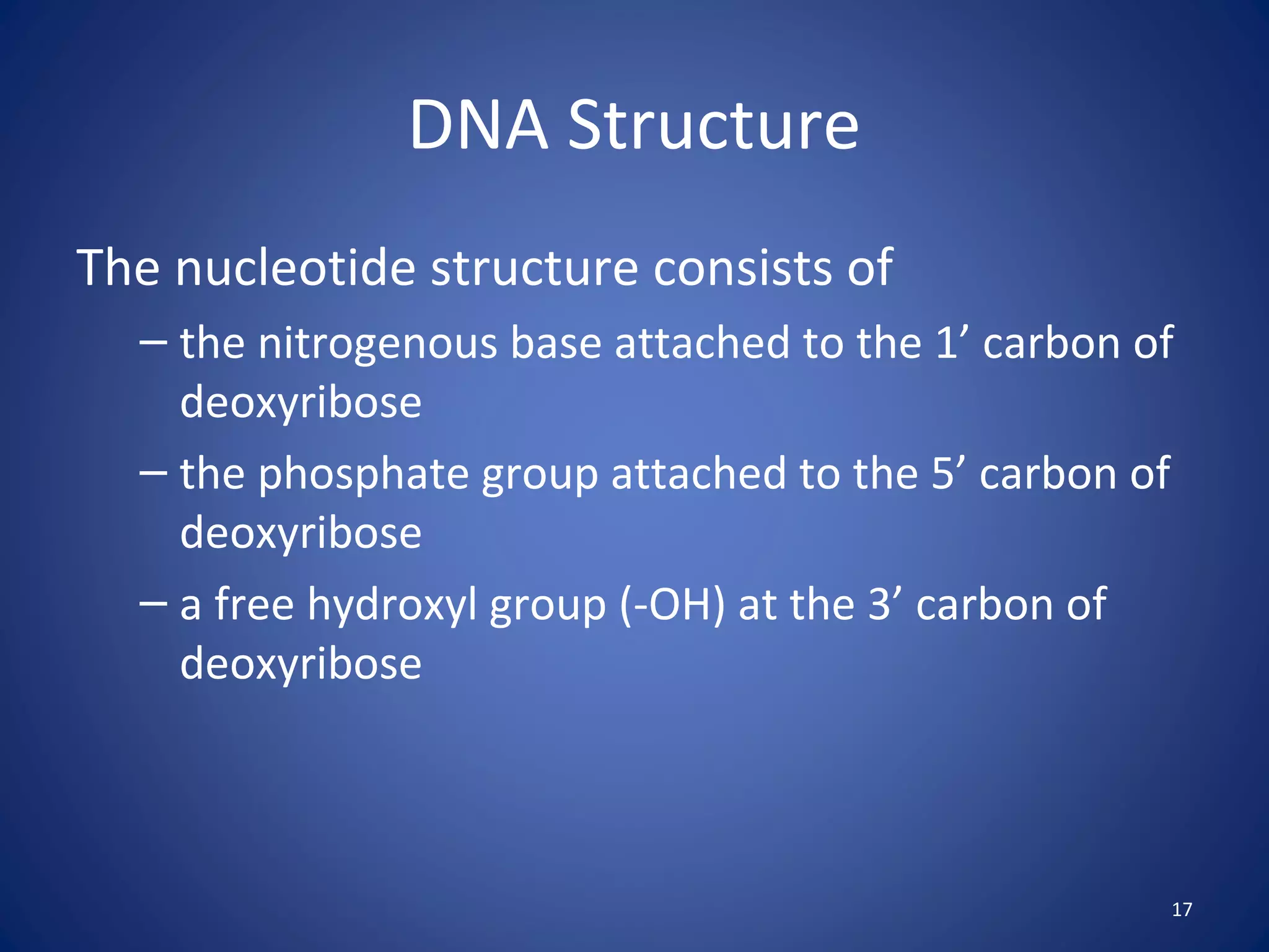
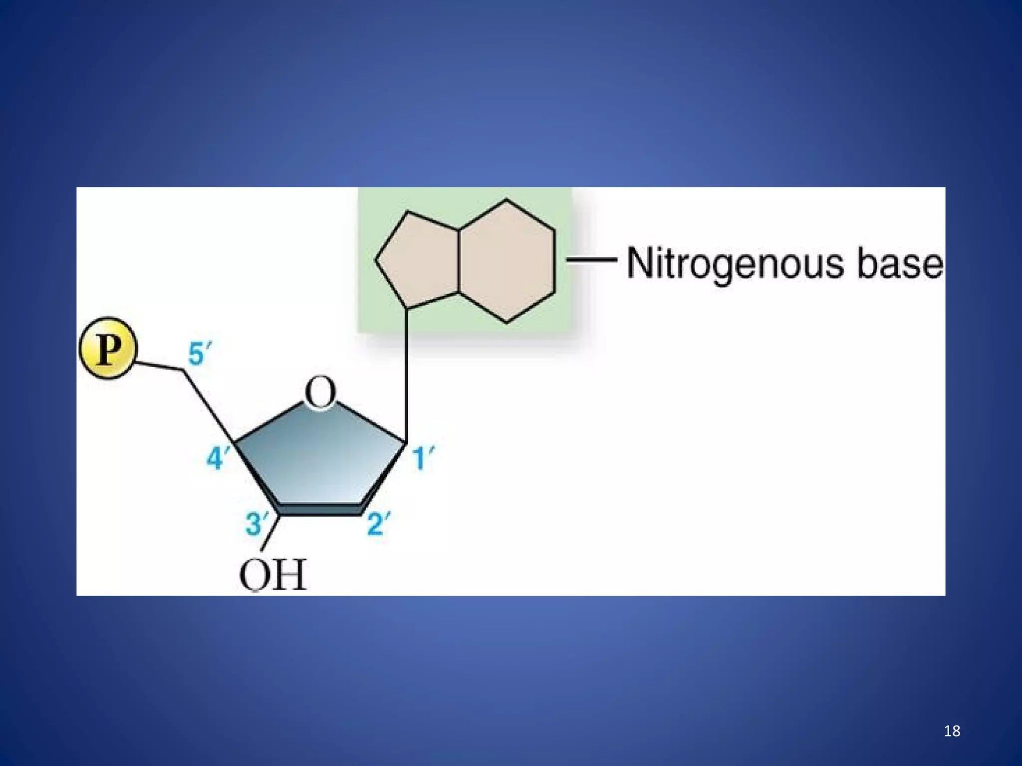
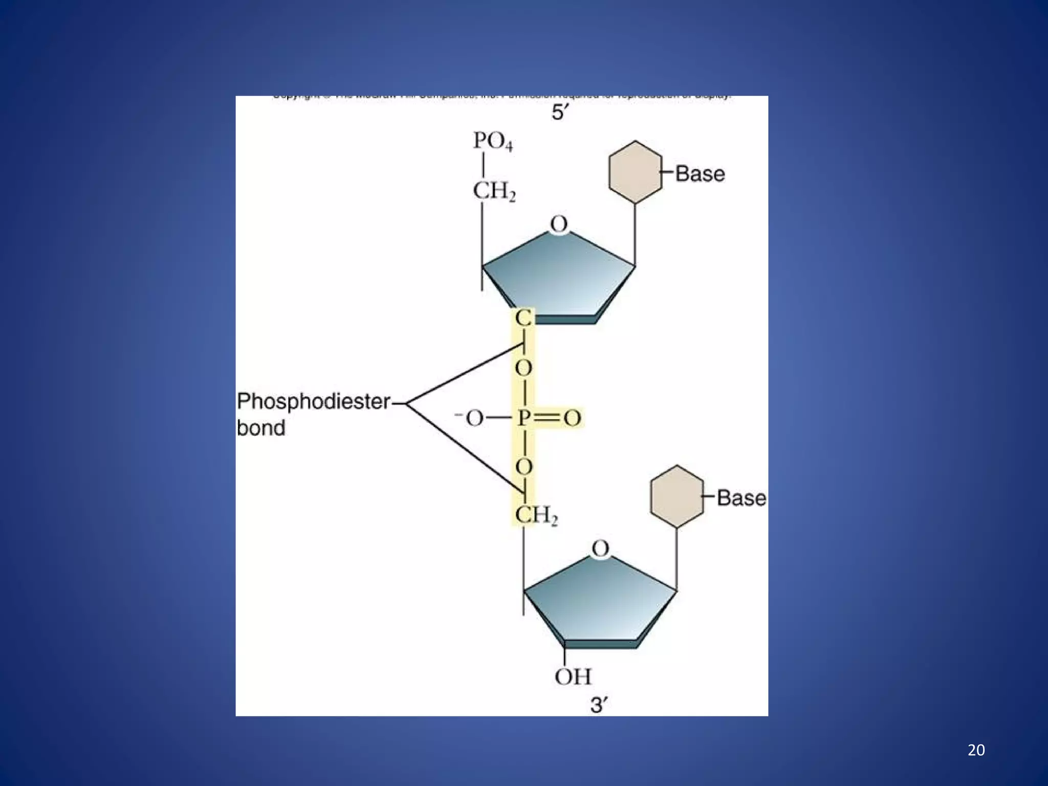
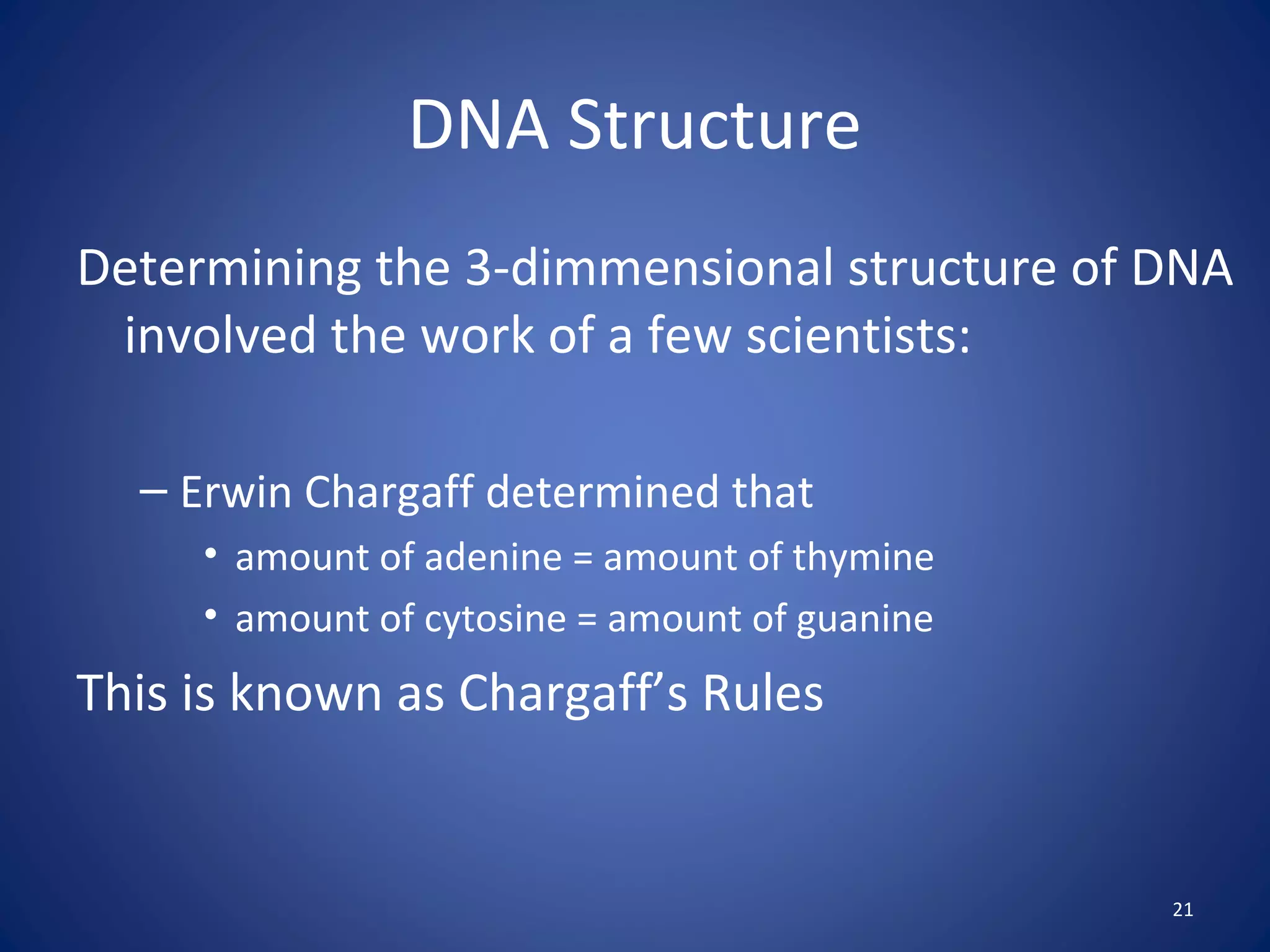
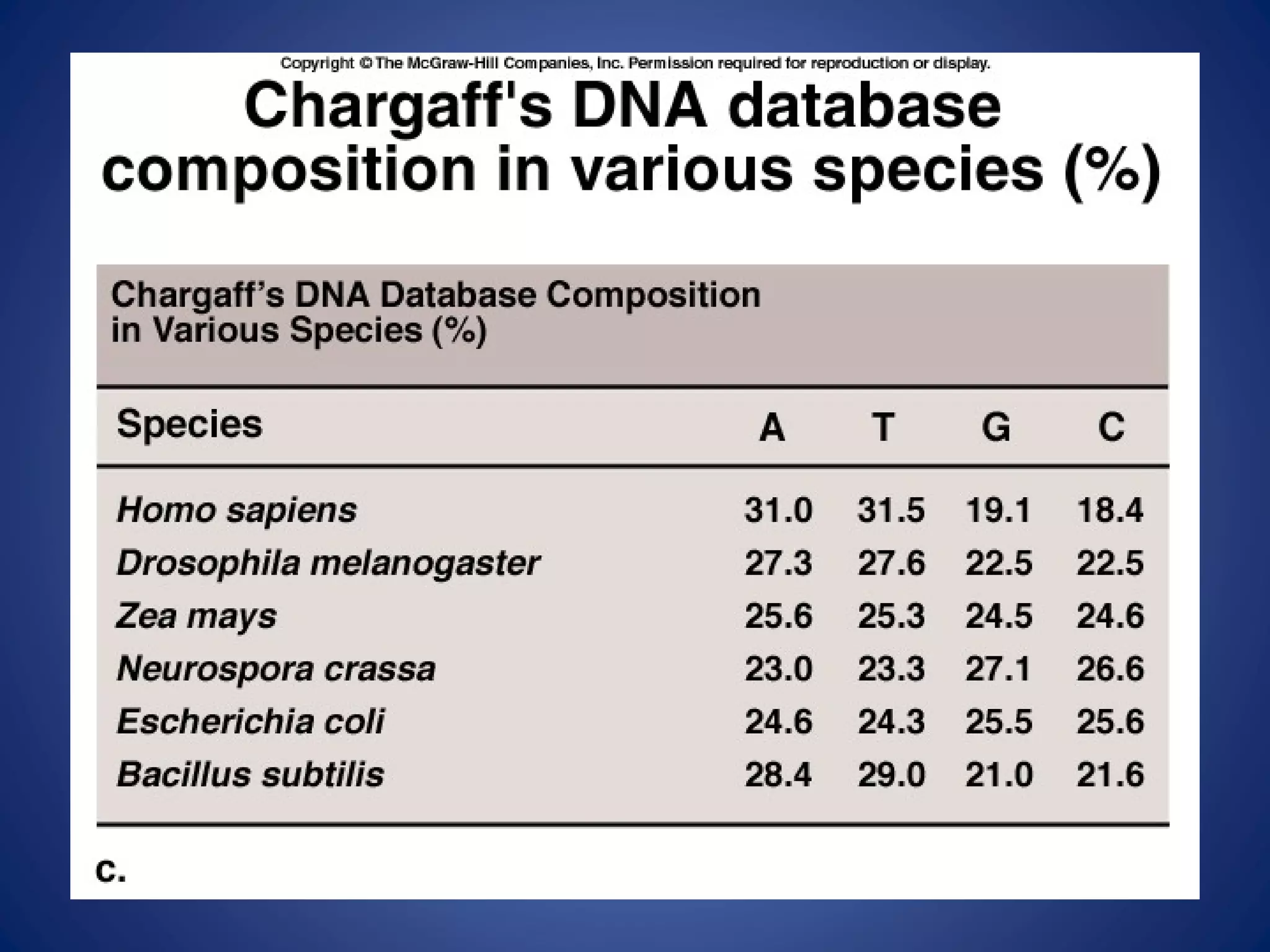
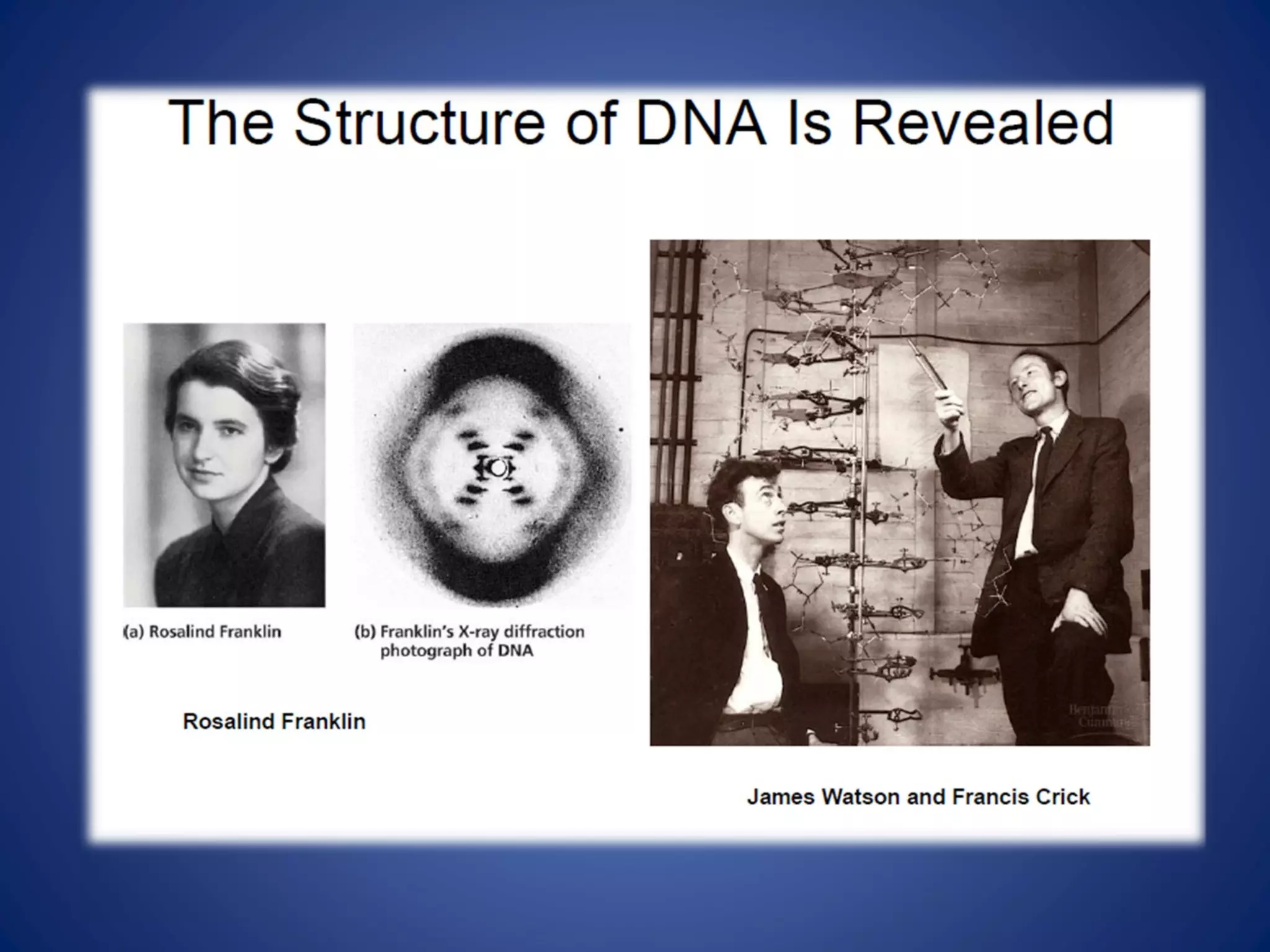
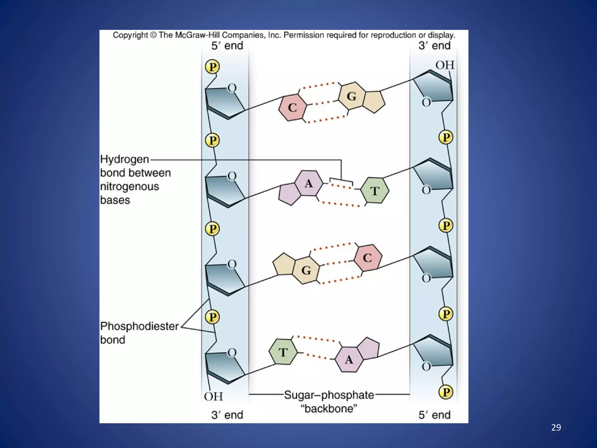
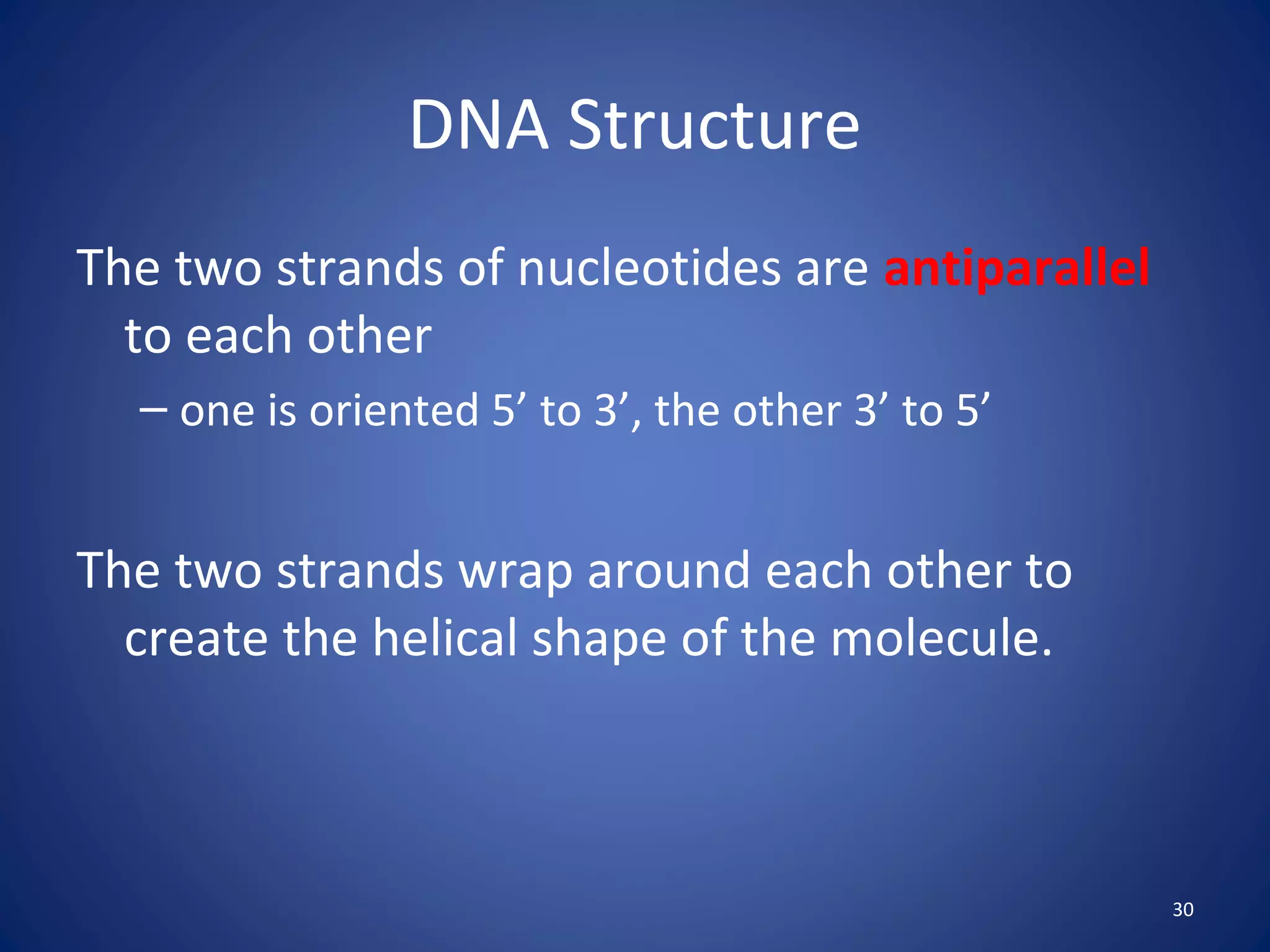
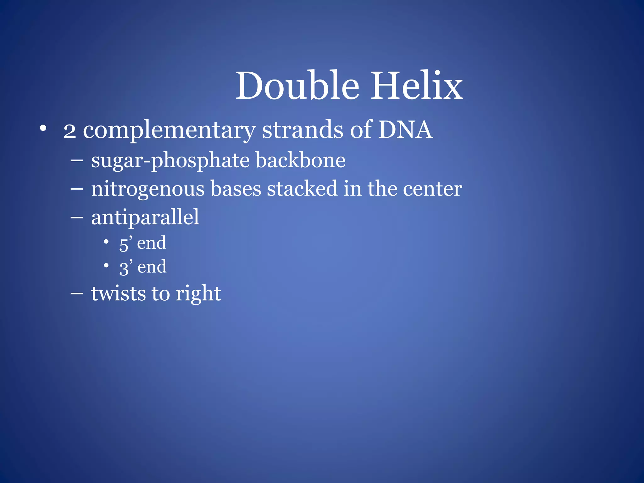
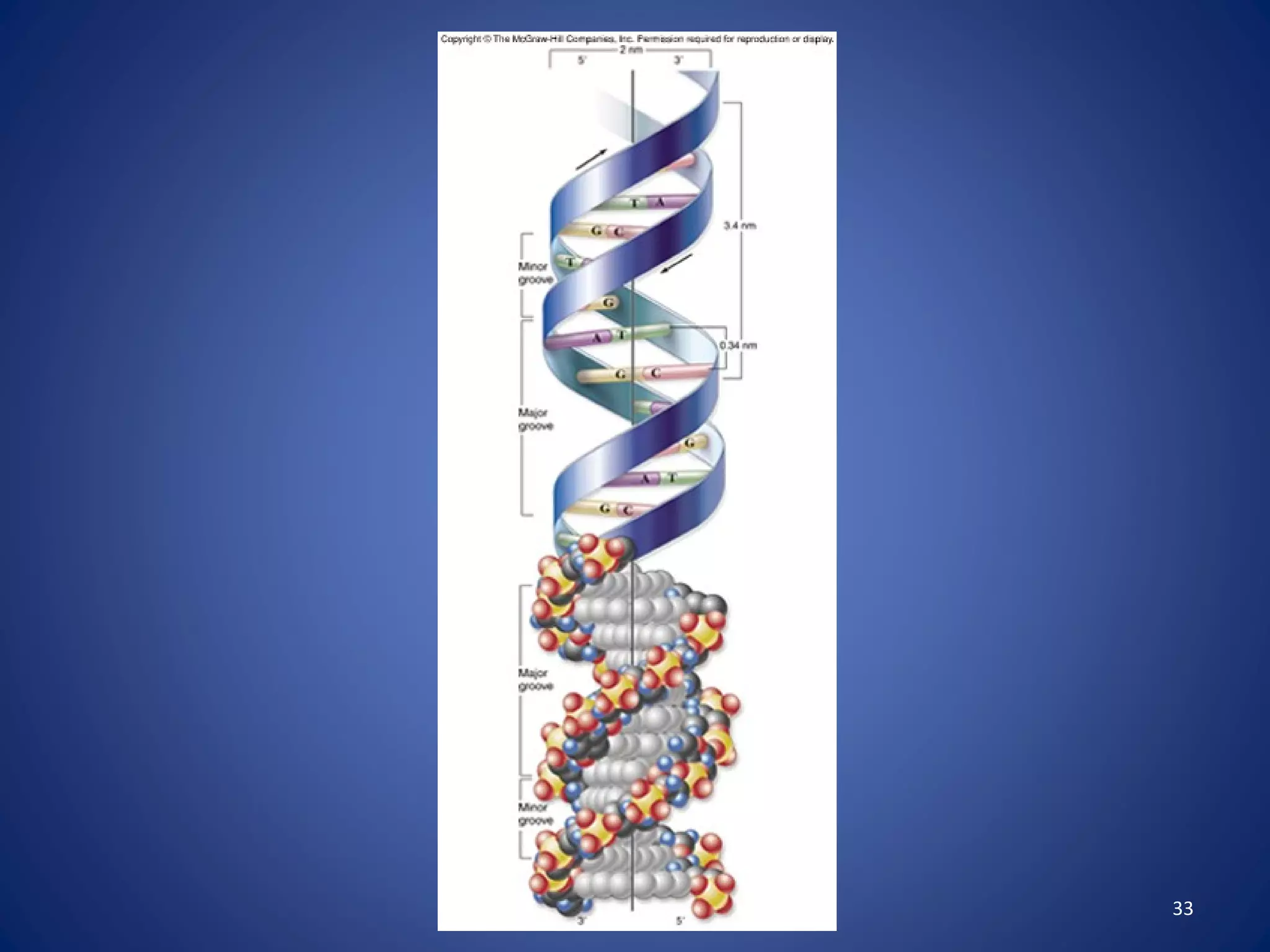
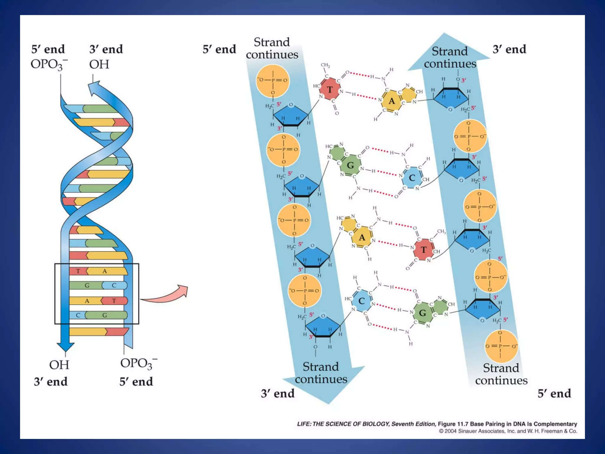
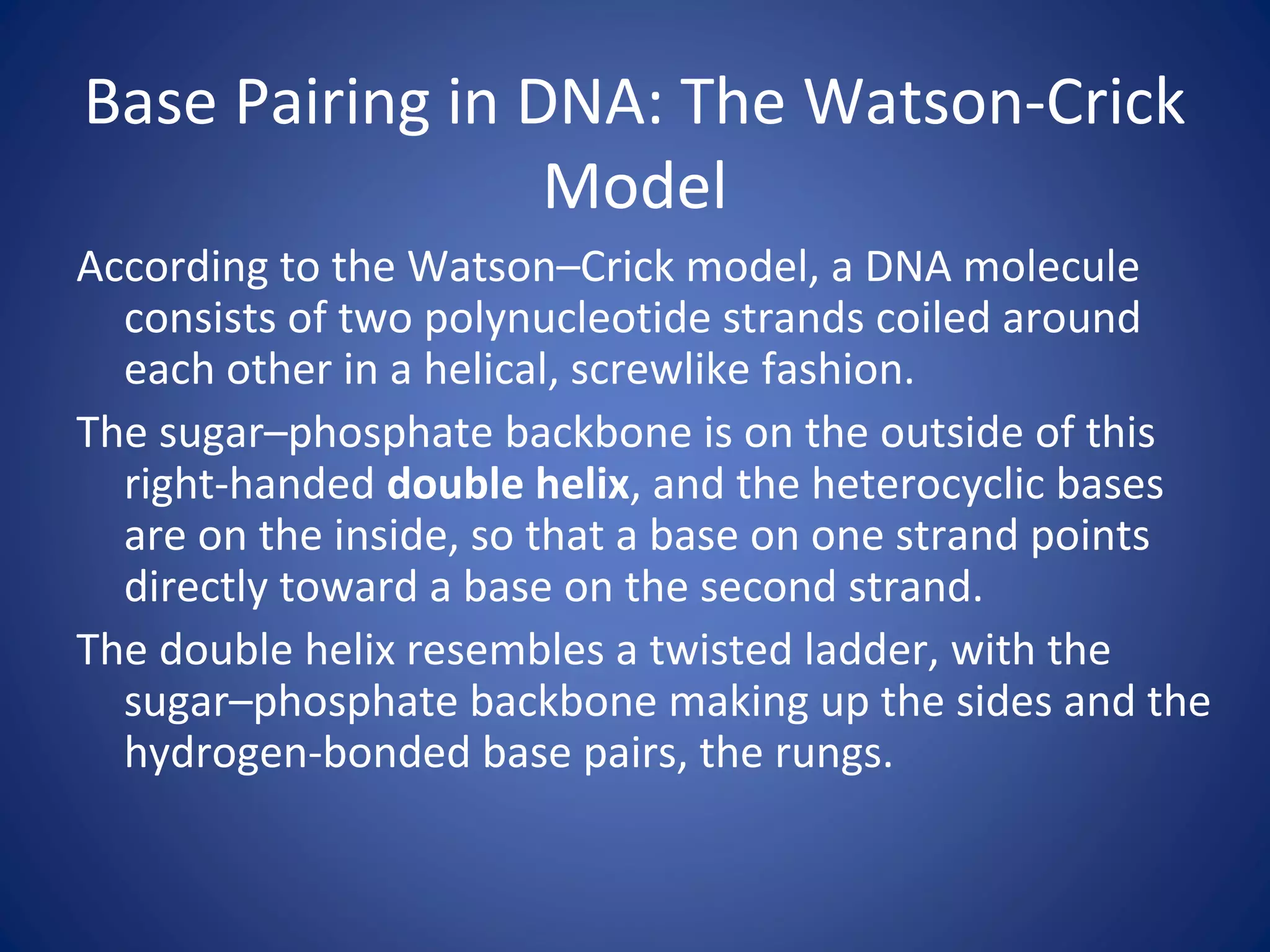
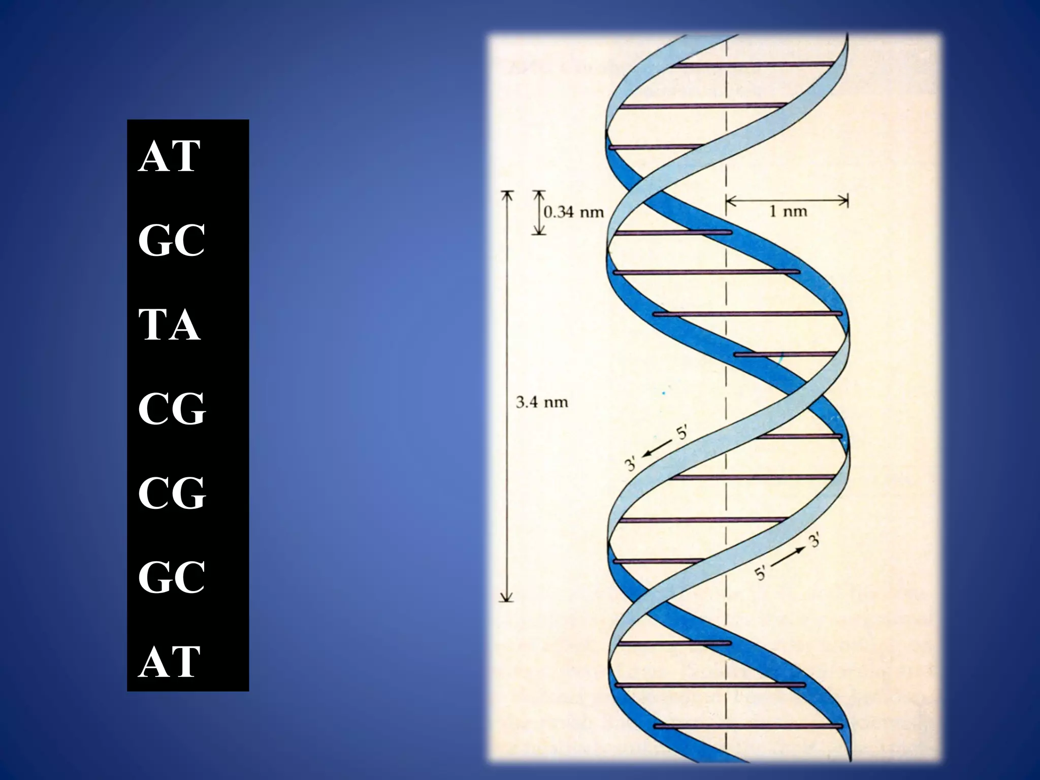
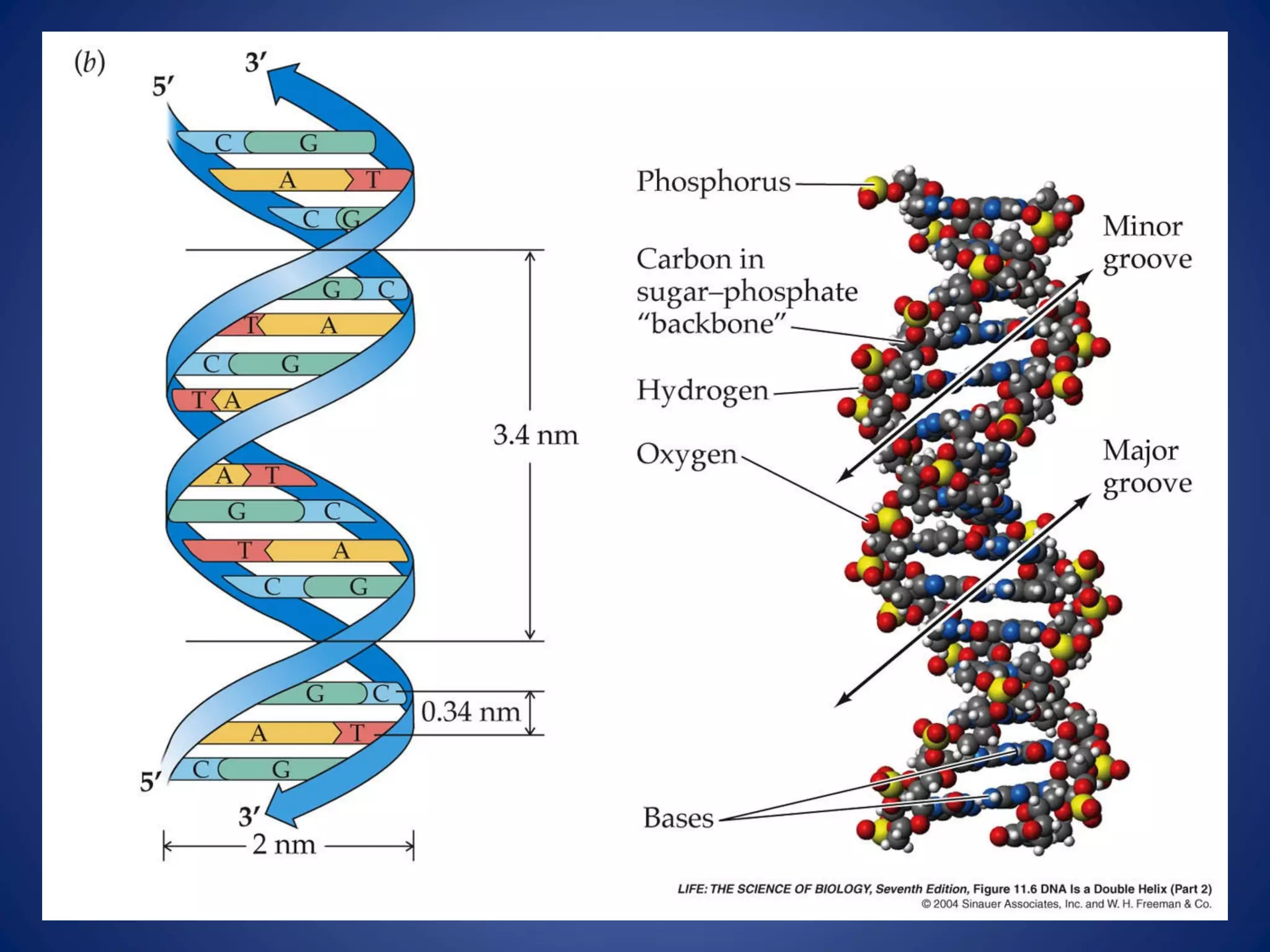
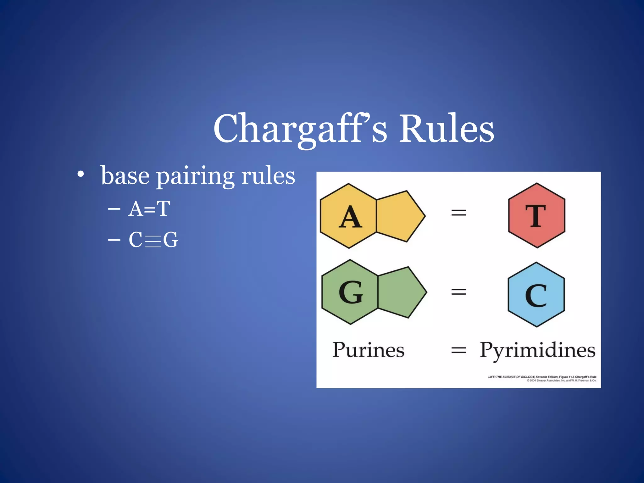
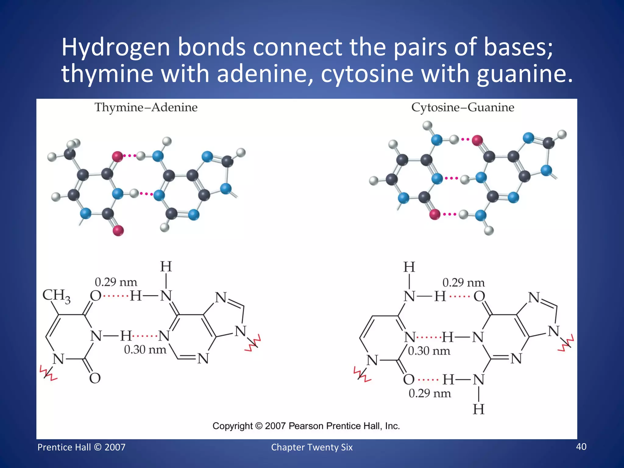
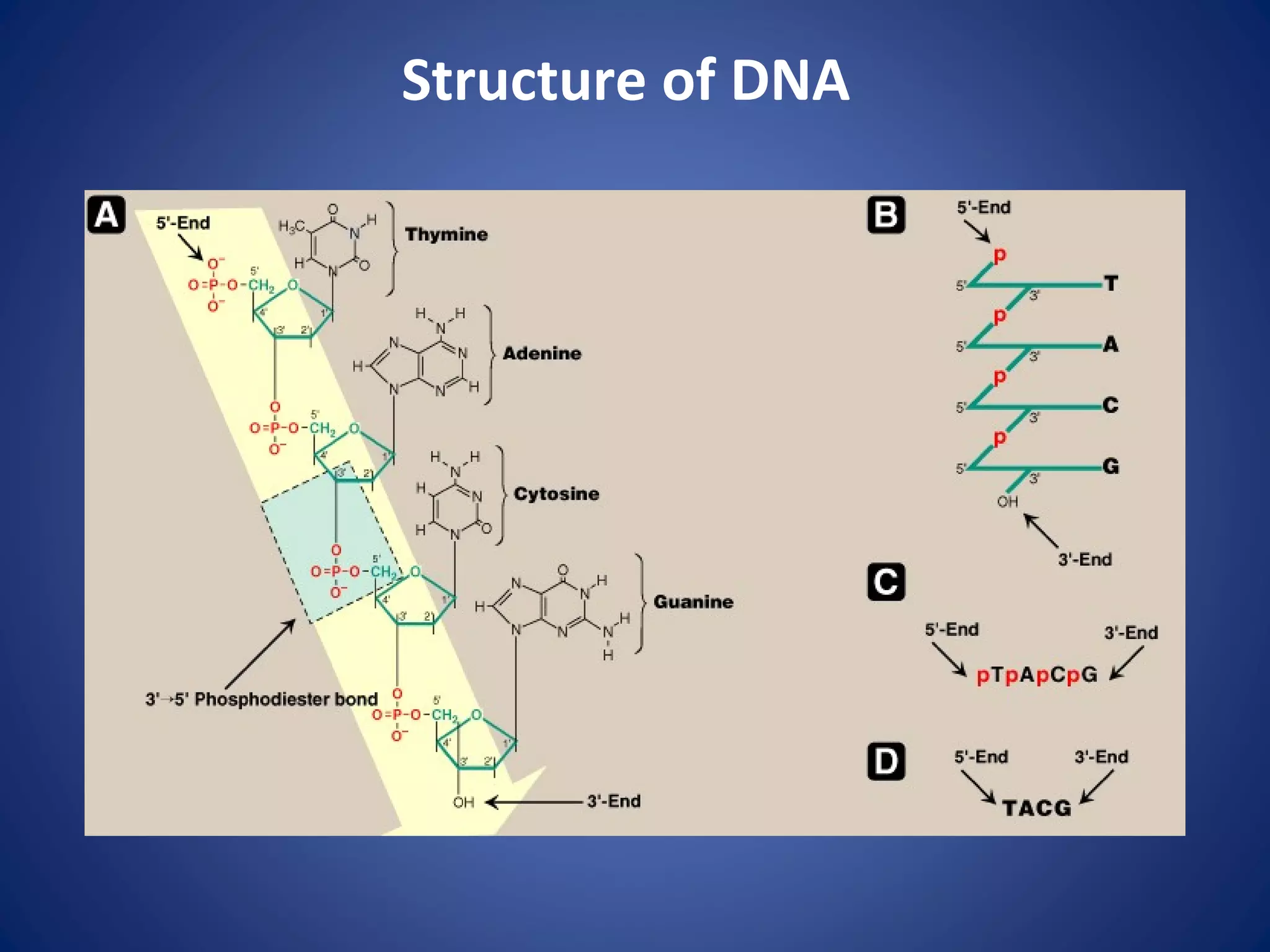
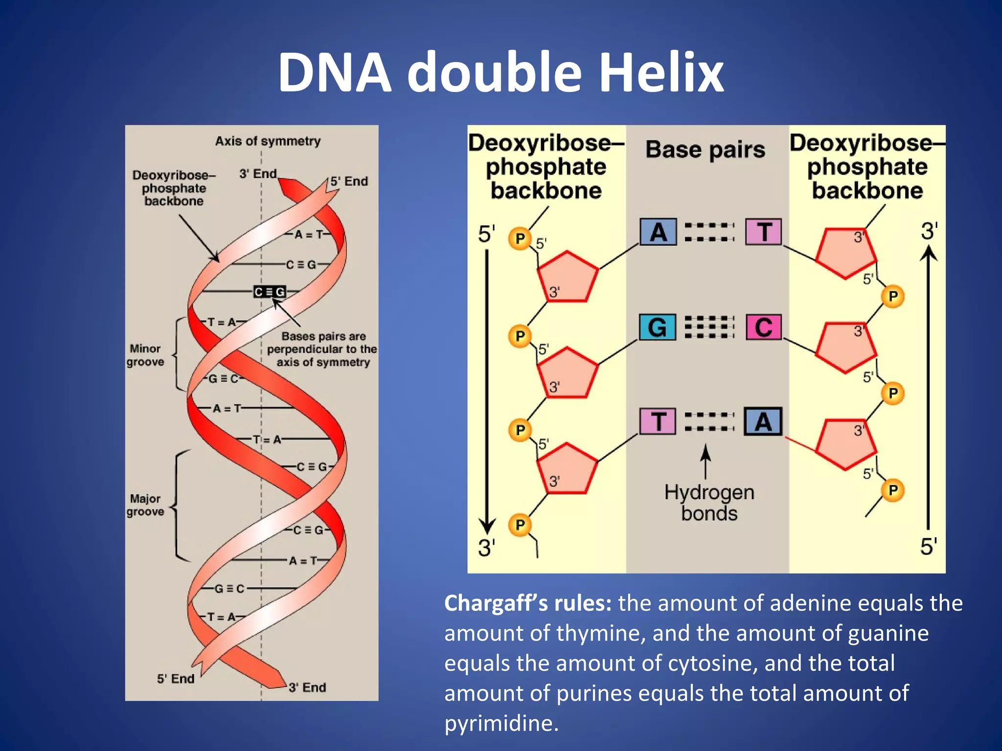
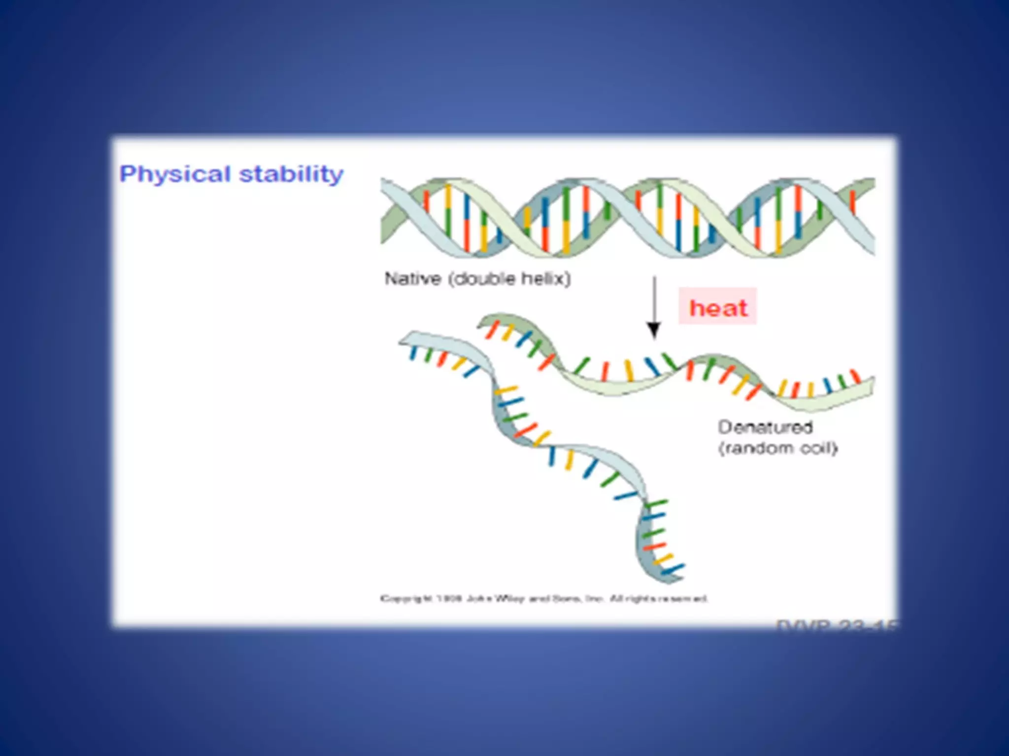
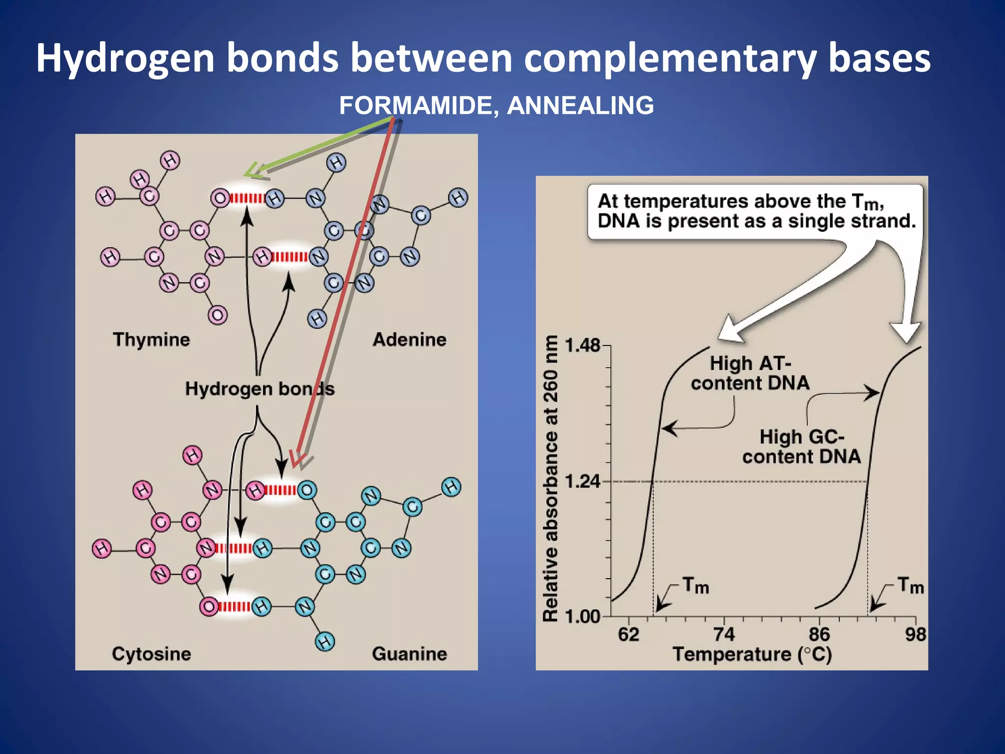
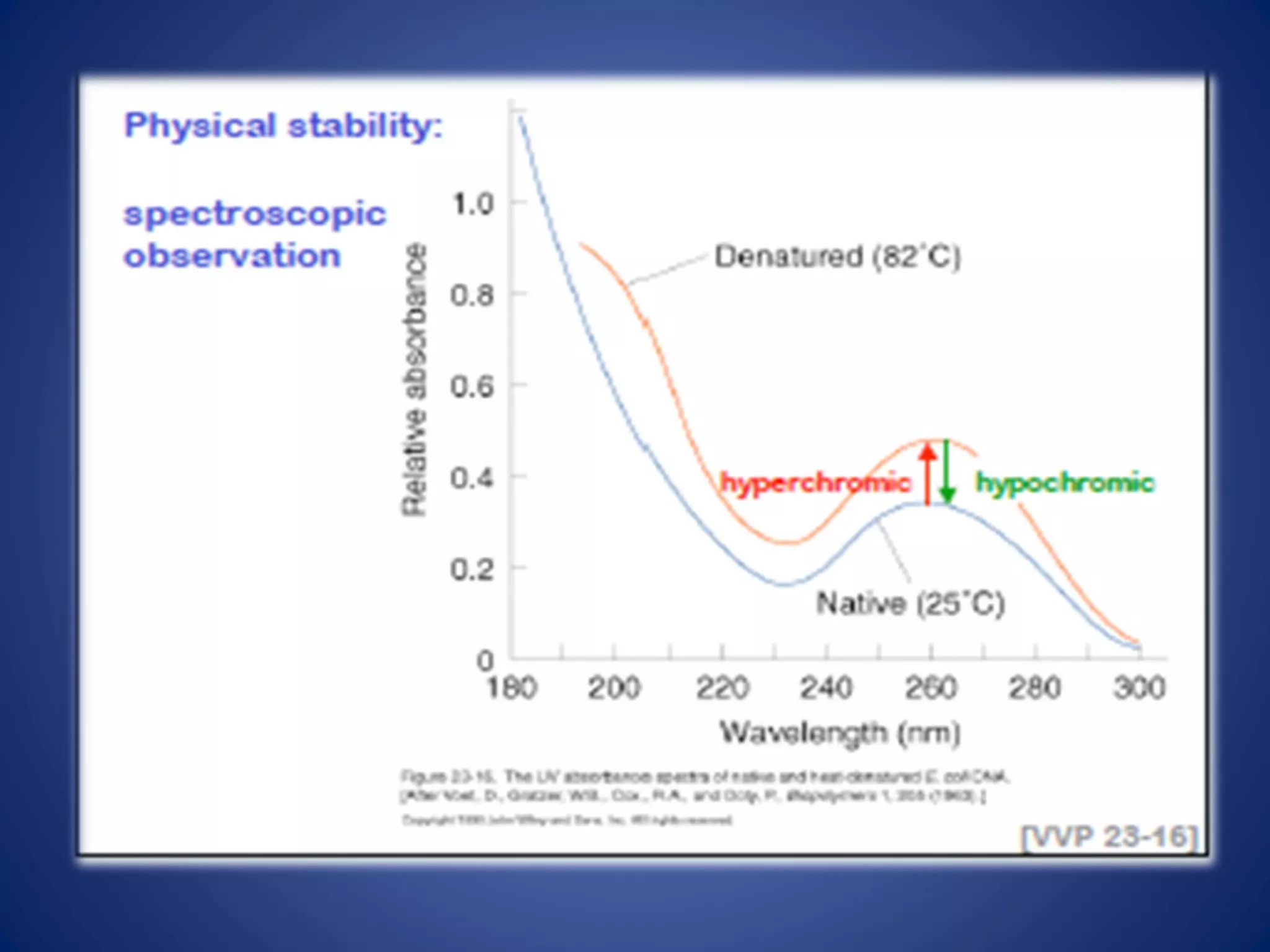
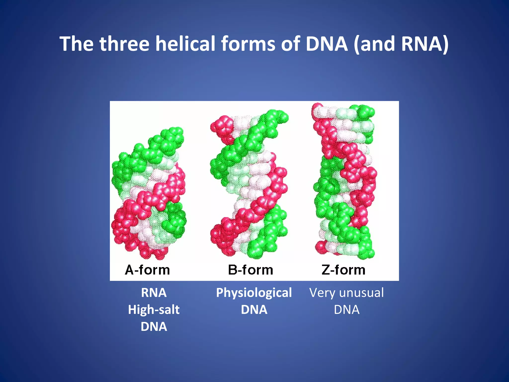
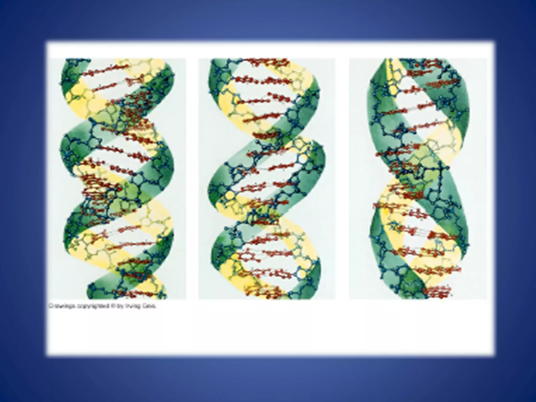
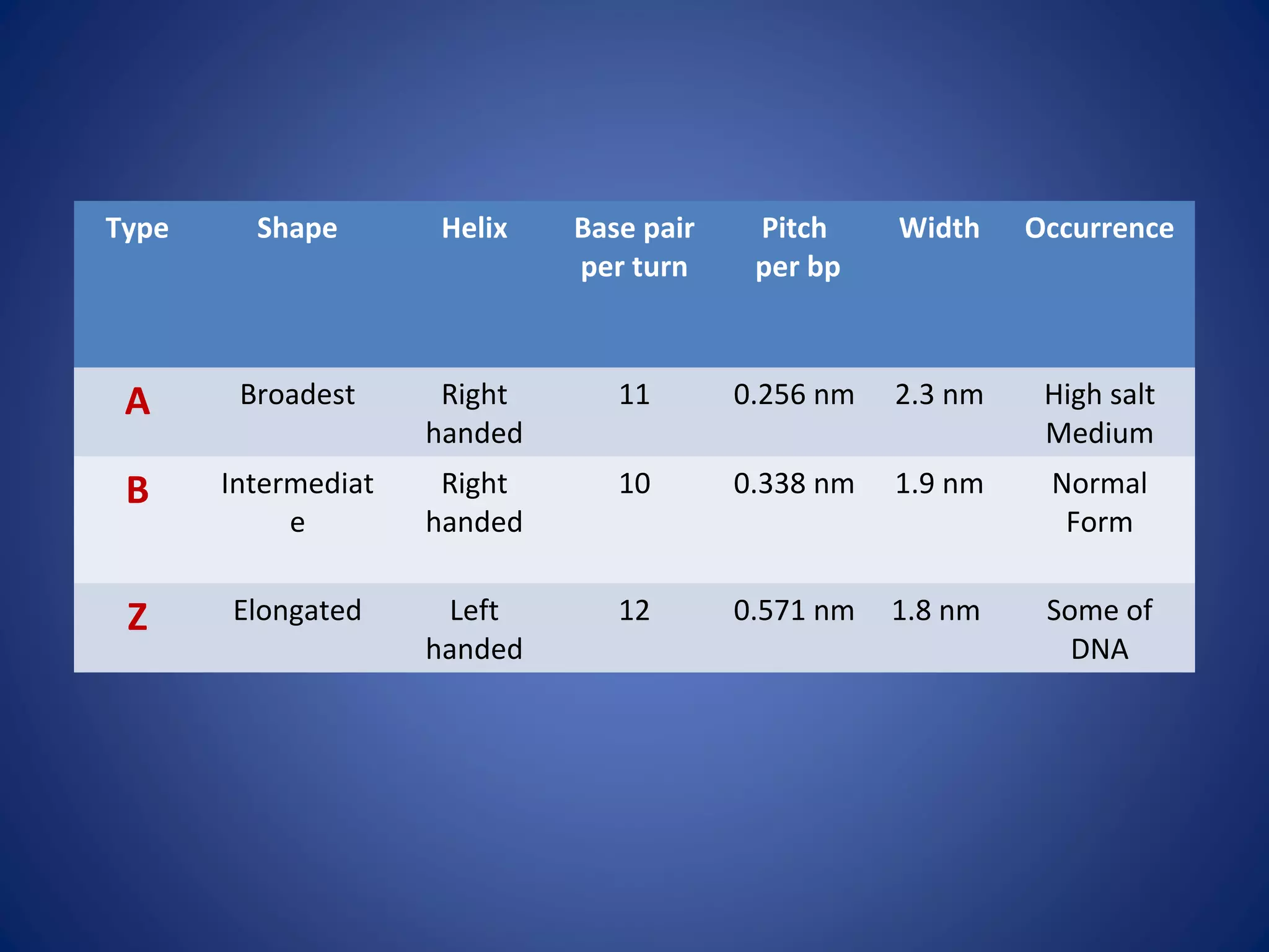
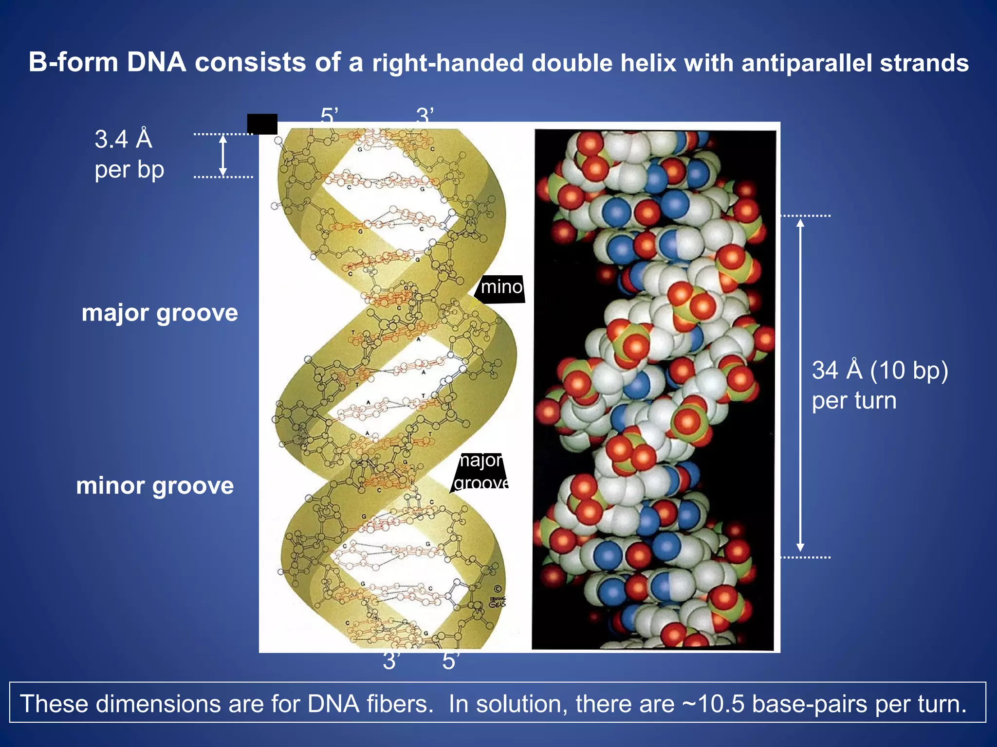
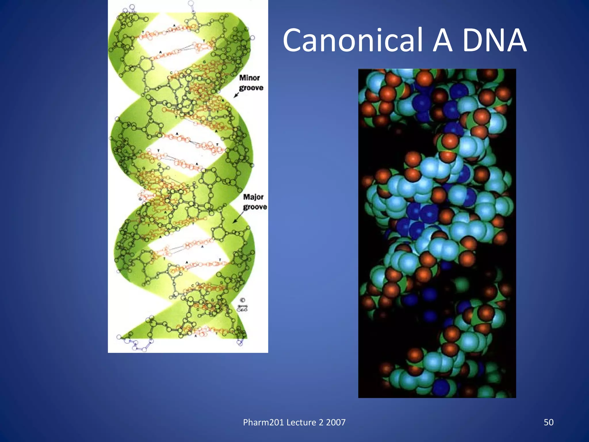
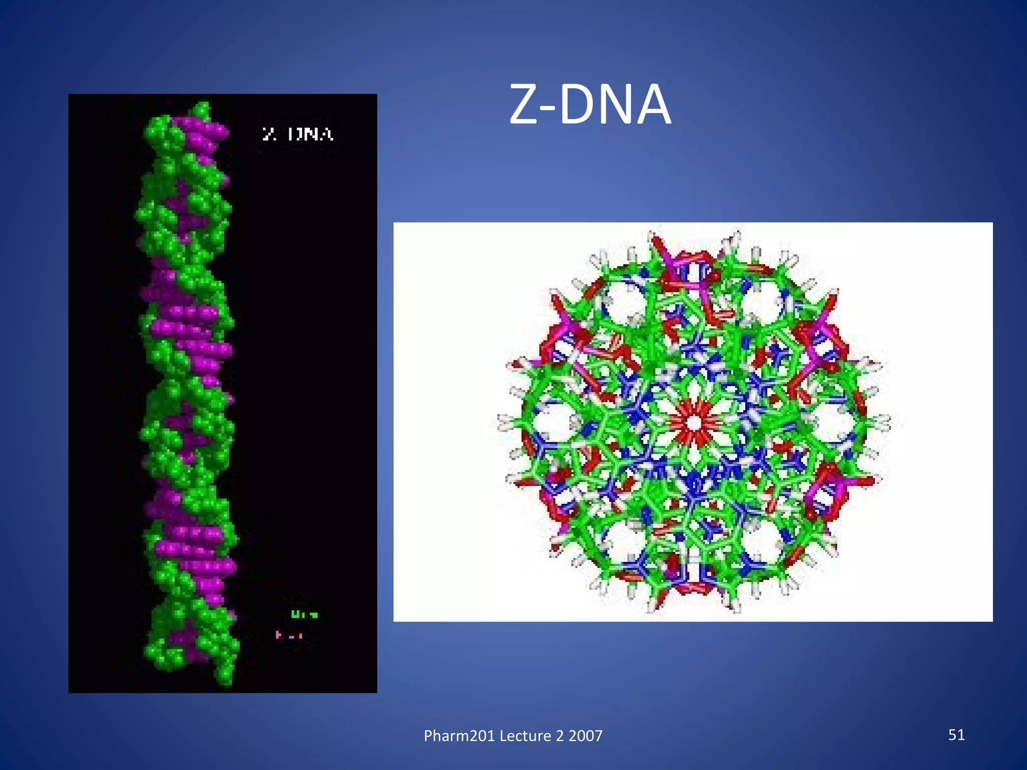
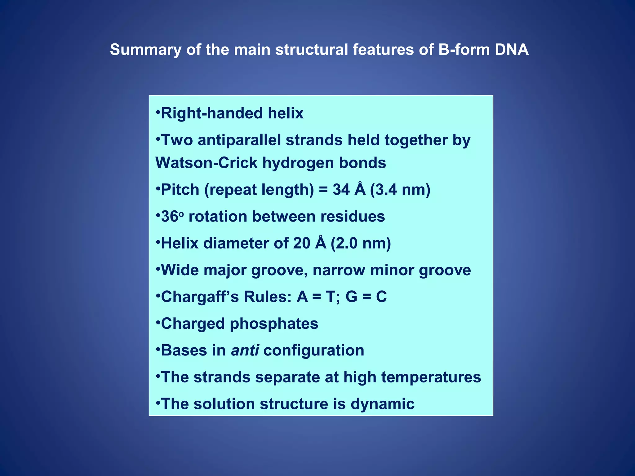
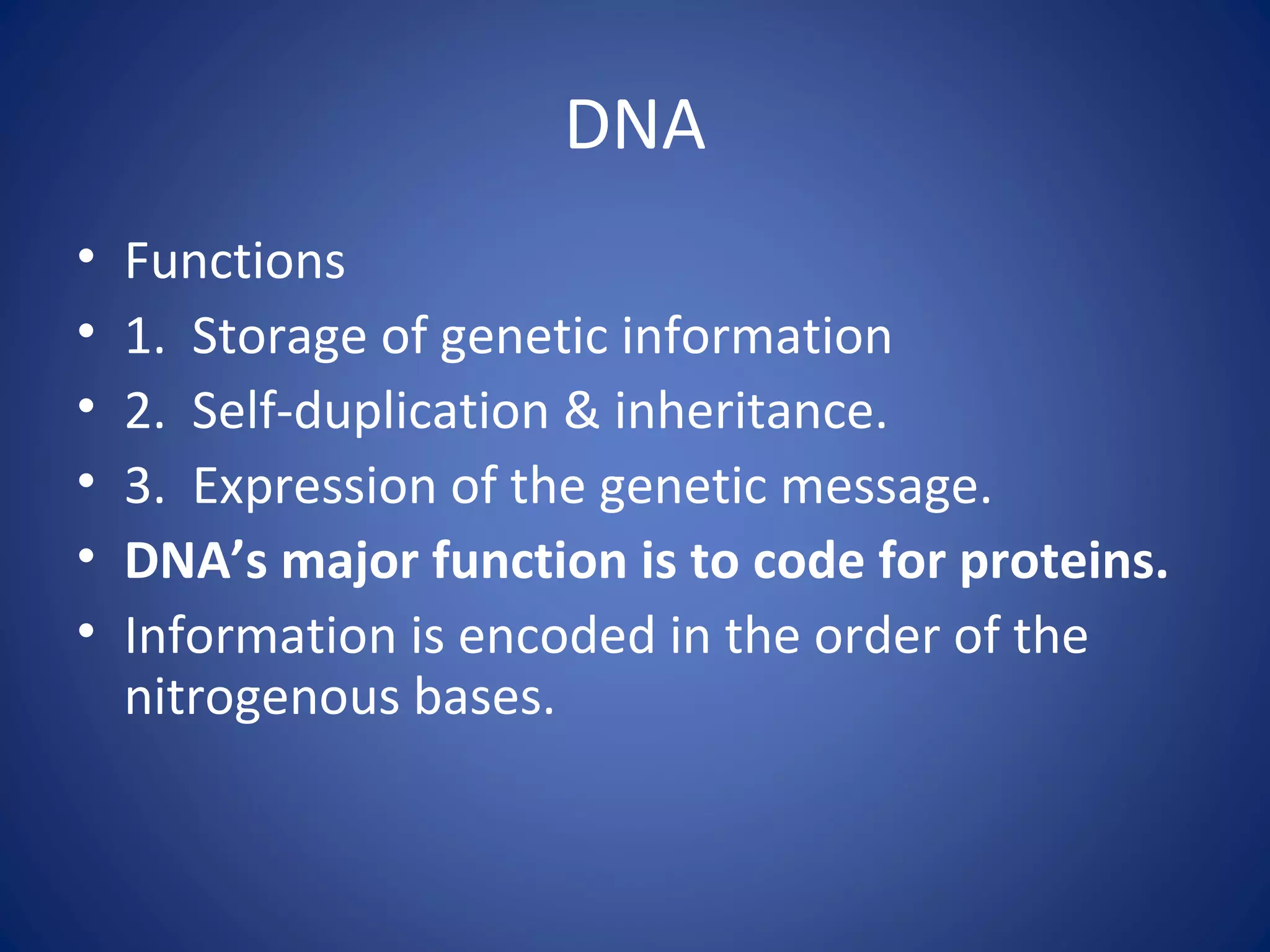
V.S.RAVIKIRAN, MSc.
DEOXYNUCLEIC ACID (DNA)
V.S.RAVIKIRAN, MSc.,
Department of Biochemistry,
ASRAM Medical college,
Eluru-534005.AP, India.
vsravikiran2013@gmail.com
NUCLEIC ACIDS
V.S.RAVIKIRAN
The “central dogma” of Molecular Biology
12
NUCLEIC ACIDS: INFORMATIONAL
POLYMER
• Nucleic acids are polymers of monomer
units called nucleotides
• Nucleic acids store and transmit hereditary
information: gene
• Two major forms of nucleic acid polymers:
DNA and RNA
DNA
(Deoxyribonucleic Acid)
DNA Structure
DNA is a nucleic acid.
The building blocks of DNA are nucleotides,
each composed of:
– a 5-carbon sugar called deoxyribose
– a phosphate group (PO4)
– a nitrogenous base
• adenine, thymine, cytosine, guanine
17
DNA Structure
The nucleotide structure consists of
– the nitrogenous base attached to the 1’ carbon of
deoxyribose
– the phosphate group attached to the 5’ carbon of
deoxyribose
– a free hydroxyl group (-OH) at the 3’ carbon of
deoxyribose
18
DNA Structure
Nucleotides are connected to each other to
form a long chain
phosphodiester bond: bond between adjacent
nucleotides
– formed between the phosphate group of one
nucleotide and the 3’ –OH of the next nucleotide
The chain of nucleotides has a 5’ to 3’
orientation.
20
21
DNA Structure
Determining the 3-dimmensional structure of DNA
involved the work of a few scientists:
– Erwin Chargaff determined that
• amount of adenine = amount of thymine
• amount of cytosine = amount of guanine
This is known as Chargaff’s Rules
Chargaff
DNA Structure
Rosalind Franklin and Maurice Wilkins
– Franklin performed X-ray diffraction studies to
identify the 3-D structure
– discovered that DNA is helical
– discovered that the molecule has a diameter of
2nm and makes a complete turn of the helix every
3.4 nm
Franklin
26
DNA Structure
James Watson and Francis Crick, 1953
– deduced the structure of DNA using evidence
from Chargaff, Franklin, and others
– proposed a double helix structure
28
DNA Structure
The double helix consists of:
– 2 sugar-phosphate backbones
– nitrogenous bases toward the interior of the
molecule
– bases form hydrogen bonds with complementary
bases on the opposite sugar-phosphate backbone
29
30
DNA Structure
The two strands of nucleotides are antiparallel
to each other
– one is oriented 5’ to 3’, the other 3’ to 5’
The two strands wrap around each other to
create the helical shape of the molecule.
Double Helix
• 2 complementary strands of DNA
– sugar-phosphate backbone
– nitrogenous bases stacked in the center
– antiparallel
• 5’ end
• 3’ end
– twists to right
33
Base Pairing in DNA: The Watson-Crick
Model
According to the Watson–Crick model, a DNA molecule
consists of two polynucleotide strands coiled around
each other in a helical, screwlike fashion.
The sugar–phosphate backbone is on the outside of this
right-handed double helix, and the heterocyclic bases
are on the inside, so that a base on one strand points
directly toward a base on the second strand.
The double helix resembles a twisted ladder, with the
sugar–phosphate backbone making up the sides and the
hydrogen-bonded base pairs, the rungs.
AT
GC
TA
CG
CG
GC
AT
Chargaff’s Rules
• base pairing rules
– A=T
– C G
Prentice Hall © 2007 Chapter Twenty Six 40
Hydrogen bonds connect the pairs of bases;
thymine with adenine, cytosine with guanine.
Structure of DNA
DNA double Helix
Chargaff’s rules: the amount of adenine equals the
amount of thymine, and the amount of guanine
equals the amount of cytosine, and the total
amount of purines equals the total amount of
pyrimidine.
Hydrogen bonds between complementary bases
FORMAMIDE, ANNEALING
The three helical forms of DNA (and RNA)
Physiological
DNA
Very unusual
DNA
RNA
High-salt
DNA
Type Shape Helix Base pair
per turn
Pitch
per bp
Width Occurrence
A Broadest Right
handed
11 0.256 nm 2.3 nm High salt
Medium
B Intermediat
e
Right
handed
10 0.338 nm 1.9 nm Normal
Form
Z Elongated Left
handed
12 0.571 nm 1.8 nm Some of
DNA
B-form DNA consists of a right-handed double helix with antiparallel strands
34 Å (10 bp)
per turn
major groove
minor groove
major
groove
minor
3.4 Å
per bp
These dimensions are for DNA fibers. In solution, there are ~10.5 base-pairs per turn.
5’ 3’
5’3’
Pharm201 Lecture 2 2007 50
Canonical A DNA
Pharm201 Lecture 2 2007 51
Z-DNA
Summary of the main structural features of B-form DNA
•Right-handed helix
•Two antiparallel strands held together by
Watson-Crick hydrogen bonds
•Pitch (repeat length) = 34 Å (3.4 nm)
•36o
rotation between residues
•Helix diameter of 20 Å (2.0 nm)
•Wide major groove, narrow minor groove
•Chargaff’s Rules: A = T; G = C
•Charged phosphates
•Bases in anti configuration
•The strands separate at high temperatures
•The solution structure is dynamic
DNA
• Functions
• 1. Storage of genetic information
• 2. Self-duplication & inheritance.
• 3. Expression of the genetic message.
• DNA’s major function is to code for proteins.
• Information is encoded in the order of the
nitrogenous bases.
Figure 11.6
DNA
double
helix
(2-nm
diameter)
Metaphase chromosome
700
nm
Tight helical fiber
(30-nm diameter)
Nucleosome
(10-nm diameter)
Histones
“Beads on
a string”
Supercoil
(200-nm diameter)
HISTONES
H1, H2A, H2B, H3, H4
Structure of DNA for medical school

Structure of DNA for medical school

  • 1.
  • 2.
    V.S.RAVIKIRAN, MSc., Department ofBiochemistry, ASRAM Medical college, Eluru-534005.AP, India. vsravikiran2013@gmail.com
  • 3.
  • 11.
    The “central dogma”of Molecular Biology
  • 12.
  • 13.
    NUCLEIC ACIDS: INFORMATIONAL POLYMER •Nucleic acids are polymers of monomer units called nucleotides • Nucleic acids store and transmit hereditary information: gene • Two major forms of nucleic acid polymers: DNA and RNA
  • 14.
  • 15.
    DNA Structure DNA isa nucleic acid. The building blocks of DNA are nucleotides, each composed of: – a 5-carbon sugar called deoxyribose – a phosphate group (PO4) – a nitrogenous base • adenine, thymine, cytosine, guanine
  • 17.
    17 DNA Structure The nucleotidestructure consists of – the nitrogenous base attached to the 1’ carbon of deoxyribose – the phosphate group attached to the 5’ carbon of deoxyribose – a free hydroxyl group (-OH) at the 3’ carbon of deoxyribose
  • 18.
  • 19.
    DNA Structure Nucleotides areconnected to each other to form a long chain phosphodiester bond: bond between adjacent nucleotides – formed between the phosphate group of one nucleotide and the 3’ –OH of the next nucleotide The chain of nucleotides has a 5’ to 3’ orientation.
  • 20.
  • 21.
    21 DNA Structure Determining the3-dimmensional structure of DNA involved the work of a few scientists: – Erwin Chargaff determined that • amount of adenine = amount of thymine • amount of cytosine = amount of guanine This is known as Chargaff’s Rules
  • 22.
  • 23.
    DNA Structure Rosalind Franklinand Maurice Wilkins – Franklin performed X-ray diffraction studies to identify the 3-D structure – discovered that DNA is helical – discovered that the molecule has a diameter of 2nm and makes a complete turn of the helix every 3.4 nm
  • 24.
  • 26.
    26 DNA Structure James Watsonand Francis Crick, 1953 – deduced the structure of DNA using evidence from Chargaff, Franklin, and others – proposed a double helix structure
  • 28.
    28 DNA Structure The doublehelix consists of: – 2 sugar-phosphate backbones – nitrogenous bases toward the interior of the molecule – bases form hydrogen bonds with complementary bases on the opposite sugar-phosphate backbone
  • 29.
  • 30.
    30 DNA Structure The twostrands of nucleotides are antiparallel to each other – one is oriented 5’ to 3’, the other 3’ to 5’ The two strands wrap around each other to create the helical shape of the molecule.
  • 32.
    Double Helix • 2complementary strands of DNA – sugar-phosphate backbone – nitrogenous bases stacked in the center – antiparallel • 5’ end • 3’ end – twists to right
  • 33.
  • 35.
    Base Pairing inDNA: The Watson-Crick Model According to the Watson–Crick model, a DNA molecule consists of two polynucleotide strands coiled around each other in a helical, screwlike fashion. The sugar–phosphate backbone is on the outside of this right-handed double helix, and the heterocyclic bases are on the inside, so that a base on one strand points directly toward a base on the second strand. The double helix resembles a twisted ladder, with the sugar–phosphate backbone making up the sides and the hydrogen-bonded base pairs, the rungs.
  • 36.
  • 39.
    Chargaff’s Rules • basepairing rules – A=T – C G
  • 40.
    Prentice Hall ©2007 Chapter Twenty Six 40 Hydrogen bonds connect the pairs of bases; thymine with adenine, cytosine with guanine.
  • 41.
  • 42.
    DNA double Helix Chargaff’srules: the amount of adenine equals the amount of thymine, and the amount of guanine equals the amount of cytosine, and the total amount of purines equals the total amount of pyrimidine.
  • 44.
    Hydrogen bonds betweencomplementary bases FORMAMIDE, ANNEALING
  • 46.
    The three helicalforms of DNA (and RNA) Physiological DNA Very unusual DNA RNA High-salt DNA
  • 48.
    Type Shape HelixBase pair per turn Pitch per bp Width Occurrence A Broadest Right handed 11 0.256 nm 2.3 nm High salt Medium B Intermediat e Right handed 10 0.338 nm 1.9 nm Normal Form Z Elongated Left handed 12 0.571 nm 1.8 nm Some of DNA
  • 49.
    B-form DNA consistsof a right-handed double helix with antiparallel strands 34 Å (10 bp) per turn major groove minor groove major groove minor 3.4 Å per bp These dimensions are for DNA fibers. In solution, there are ~10.5 base-pairs per turn. 5’ 3’ 5’3’
  • 50.
    Pharm201 Lecture 22007 50 Canonical A DNA
  • 51.
    Pharm201 Lecture 22007 51 Z-DNA
  • 52.
    Summary of themain structural features of B-form DNA •Right-handed helix •Two antiparallel strands held together by Watson-Crick hydrogen bonds •Pitch (repeat length) = 34 Å (3.4 nm) •36o rotation between residues •Helix diameter of 20 Å (2.0 nm) •Wide major groove, narrow minor groove •Chargaff’s Rules: A = T; G = C •Charged phosphates •Bases in anti configuration •The strands separate at high temperatures •The solution structure is dynamic
  • 53.
    DNA • Functions • 1.Storage of genetic information • 2. Self-duplication & inheritance. • 3. Expression of the genetic message. • DNA’s major function is to code for proteins. • Information is encoded in the order of the nitrogenous bases.
  • 54.
    Figure 11.6 DNA double helix (2-nm diameter) Metaphase chromosome 700 nm Tighthelical fiber (30-nm diameter) Nucleosome (10-nm diameter) Histones “Beads on a string” Supercoil (200-nm diameter) HISTONES H1, H2A, H2B, H3, H4