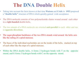
Download to read offline


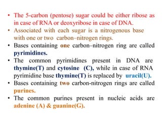
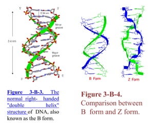
![It contains two polynucleotide strands wound
around each other.
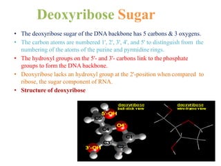
The backbone of each consists of alternating
deoxyribose and phosphate groups.
The phosphate group bonded to the 5' carbon atom of
one deoxyribose is covalently bonded to the 3' carbon of
the next.
The two strands are "antiparallel“ ; that is, one strand
runs 5′ to 3′ while the other runs 3′ to 5′.
The DNA strands are assembled in the 5′ to 3′ direction
[More] and, by convention, we "read" them the same way.
The purine or pyrimidine attached to each
deoxyribose projects in toward the axis of the helix.
Each base forms hydrogen bonds with the one directly
opposite it, forming base pairs (also called nucleotide
pairs).
3.4 Å separate the planes in which adjacent base
pairs are located.
The double helix makes a complete turn in just over 10
nucleotide pairs, so each turn takes a little more (35.7 Å
to be exact) than the 34 Å.](https://image.slidesharecdn.com/unit-vdnastructureandfunction-201029141015/85/Unit-v-dnastructureand-function-12-320.jpg)


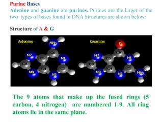
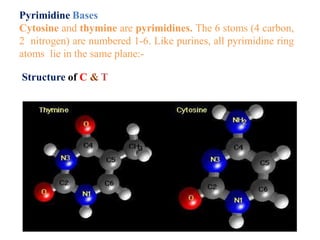
DNA is made up of nucleotides, each containing a nitrogenous base, a sugar (deoxyribose), and a phosphate group. The bases are either purines (adenine and guanine) or pyrimidines (cytosine, thymine, and in RNA, uracil). Watson and Crick discovered that DNA exists as a double helix with the bases pairing together (A with T and C with G) between antiparallel strands. The structure allows DNA to store and replicate genetic information accurately and transmit it to daughter cells.


