Downloaded 742 times
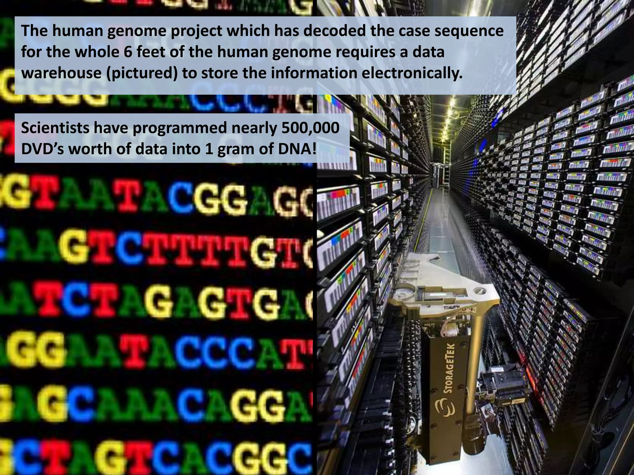
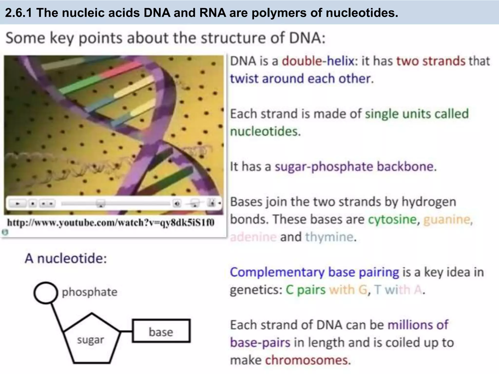
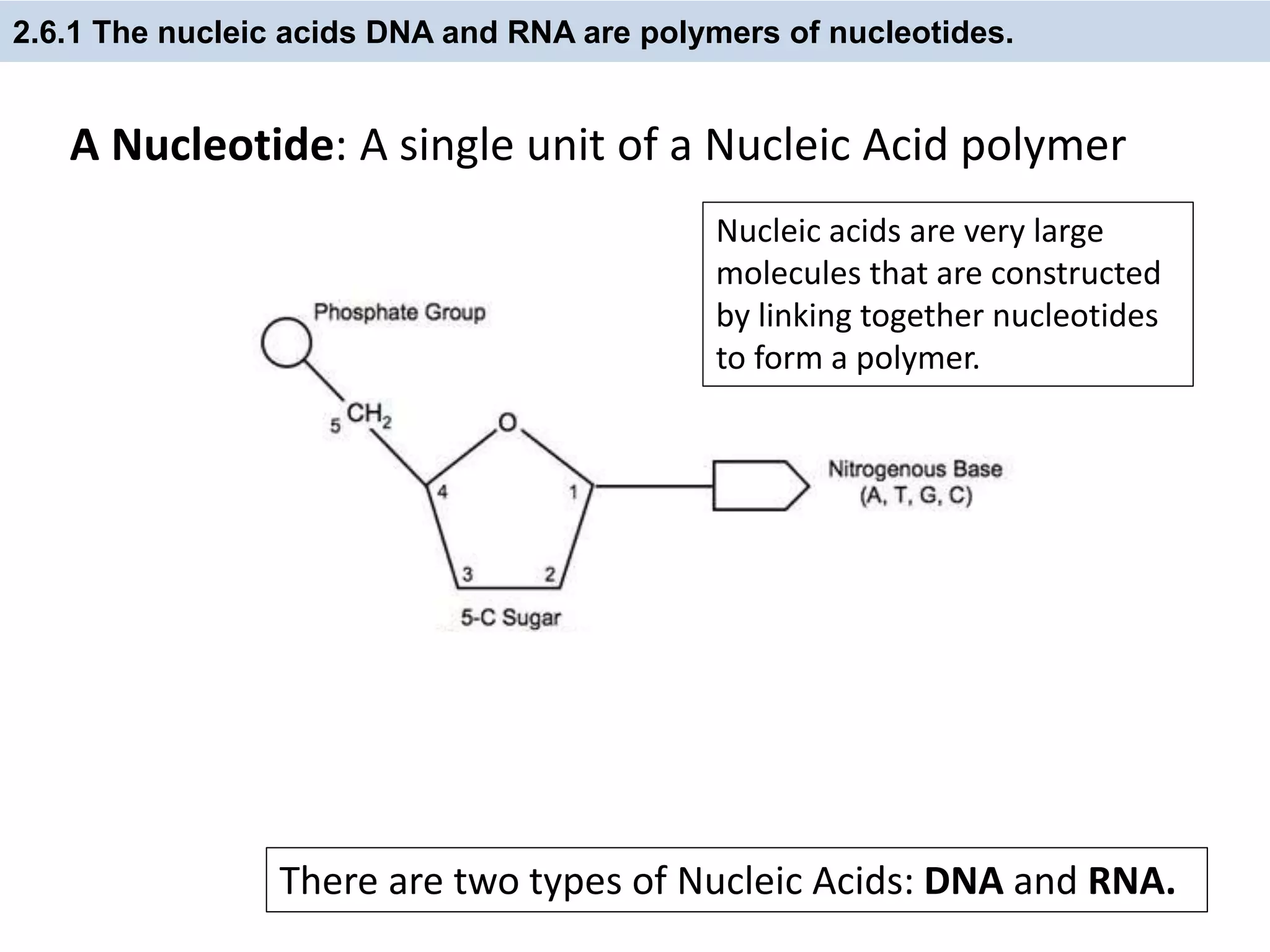
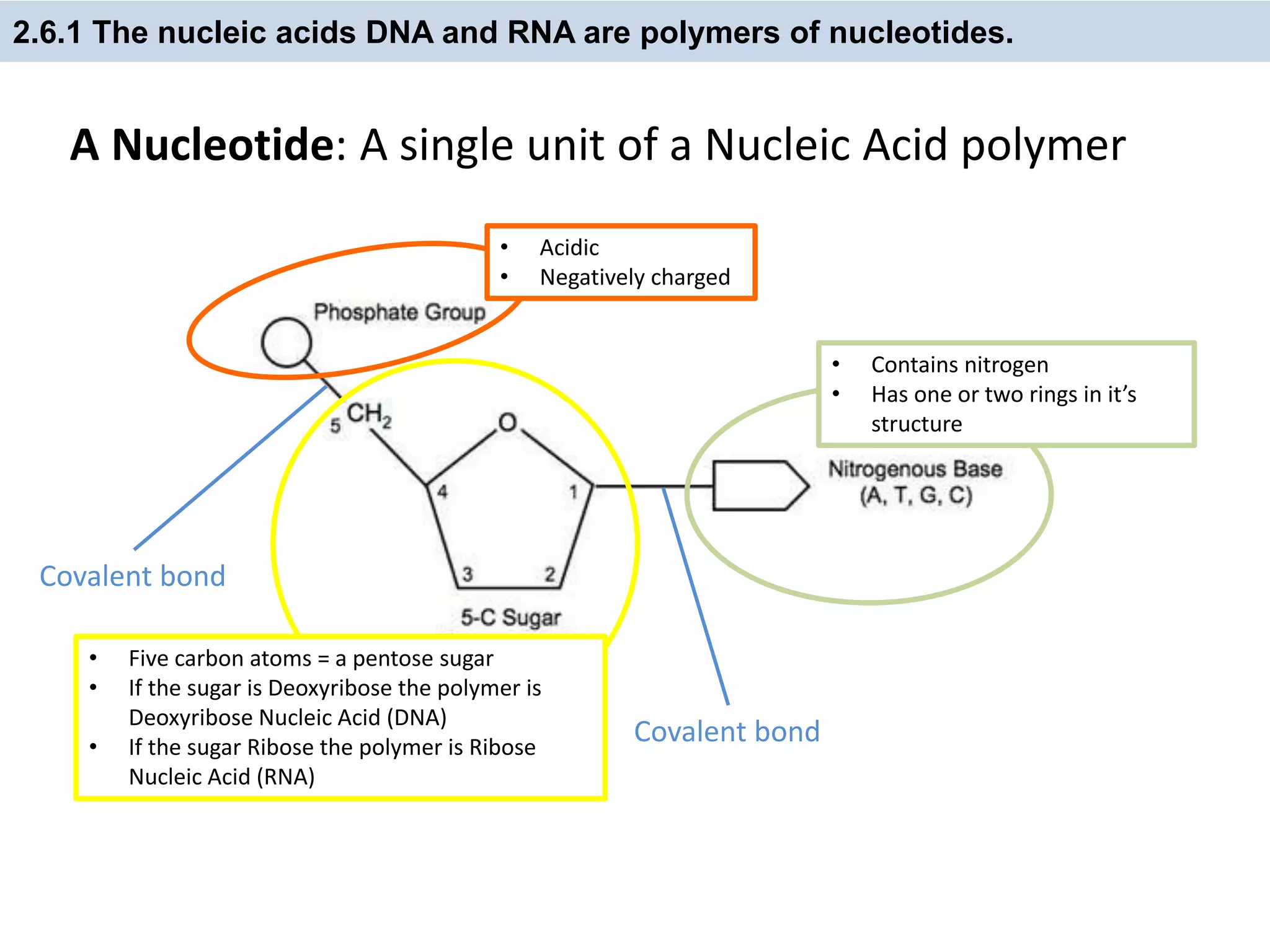
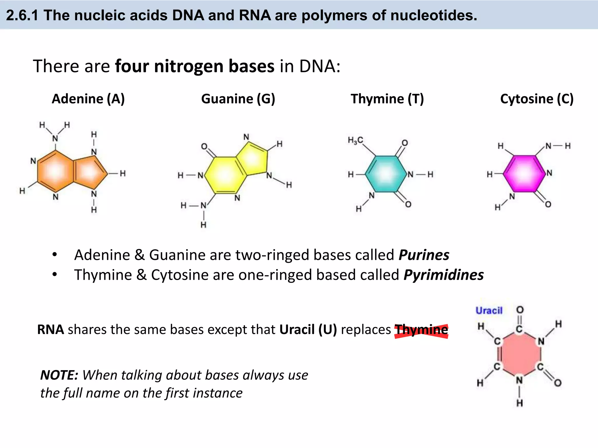
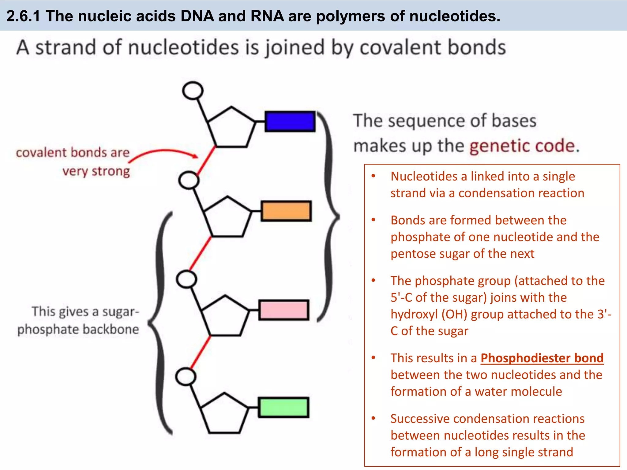
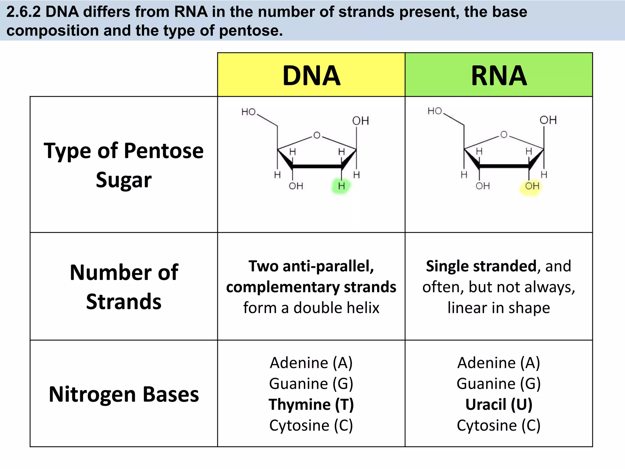
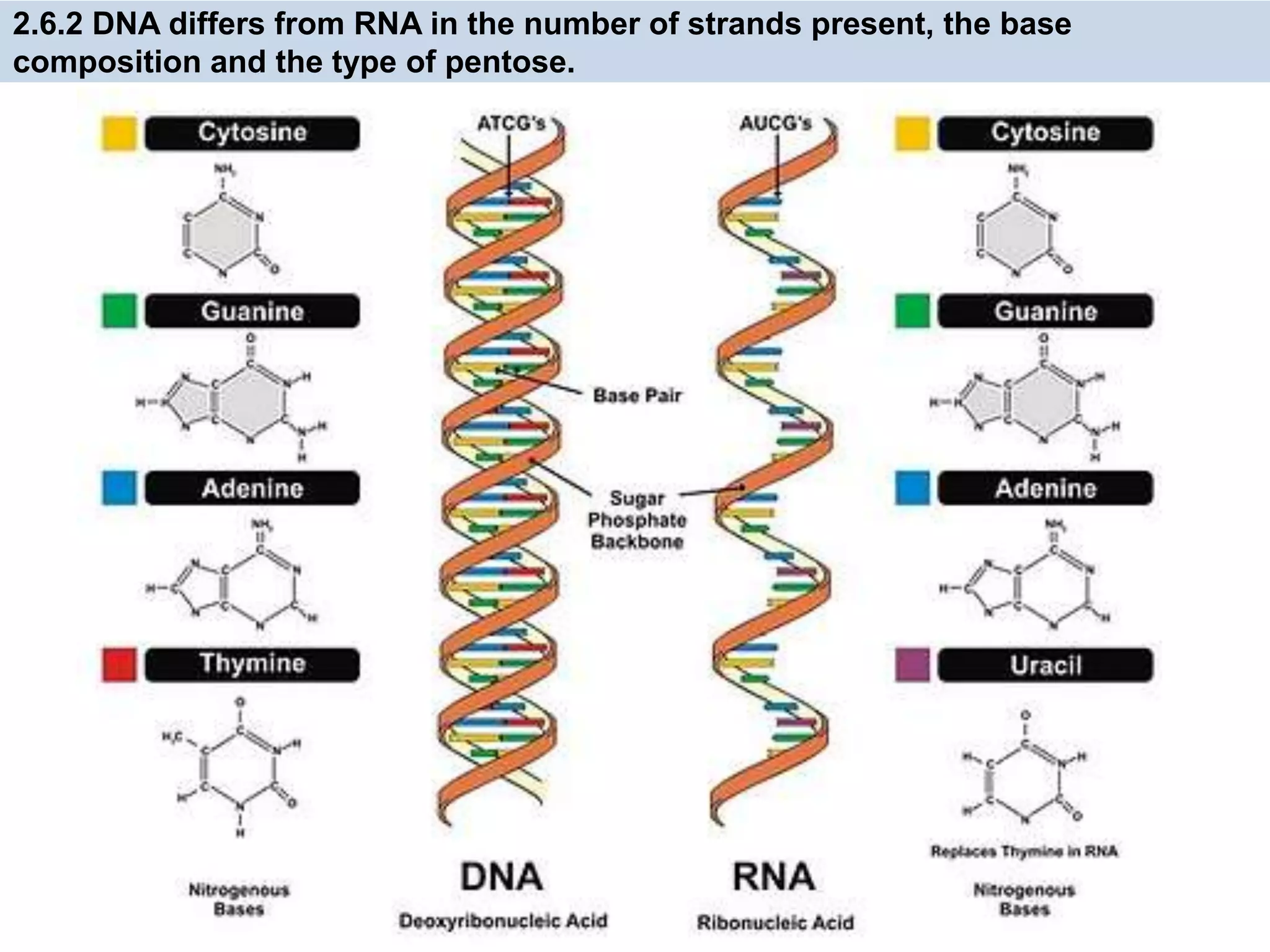
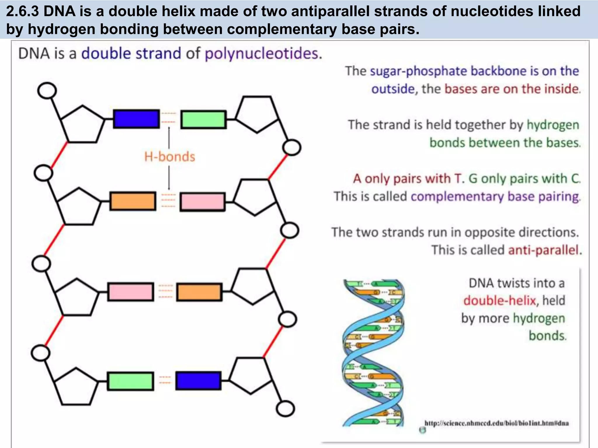
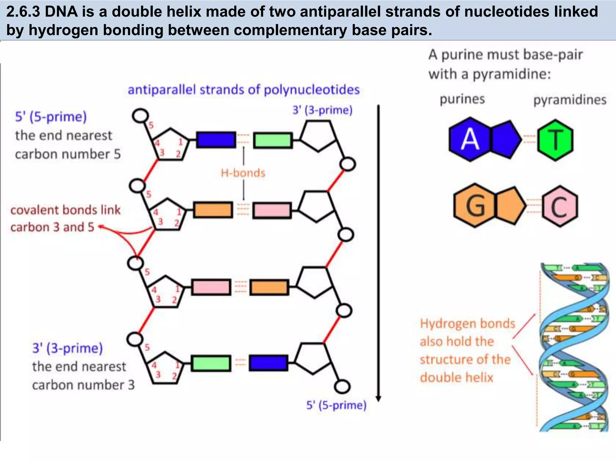
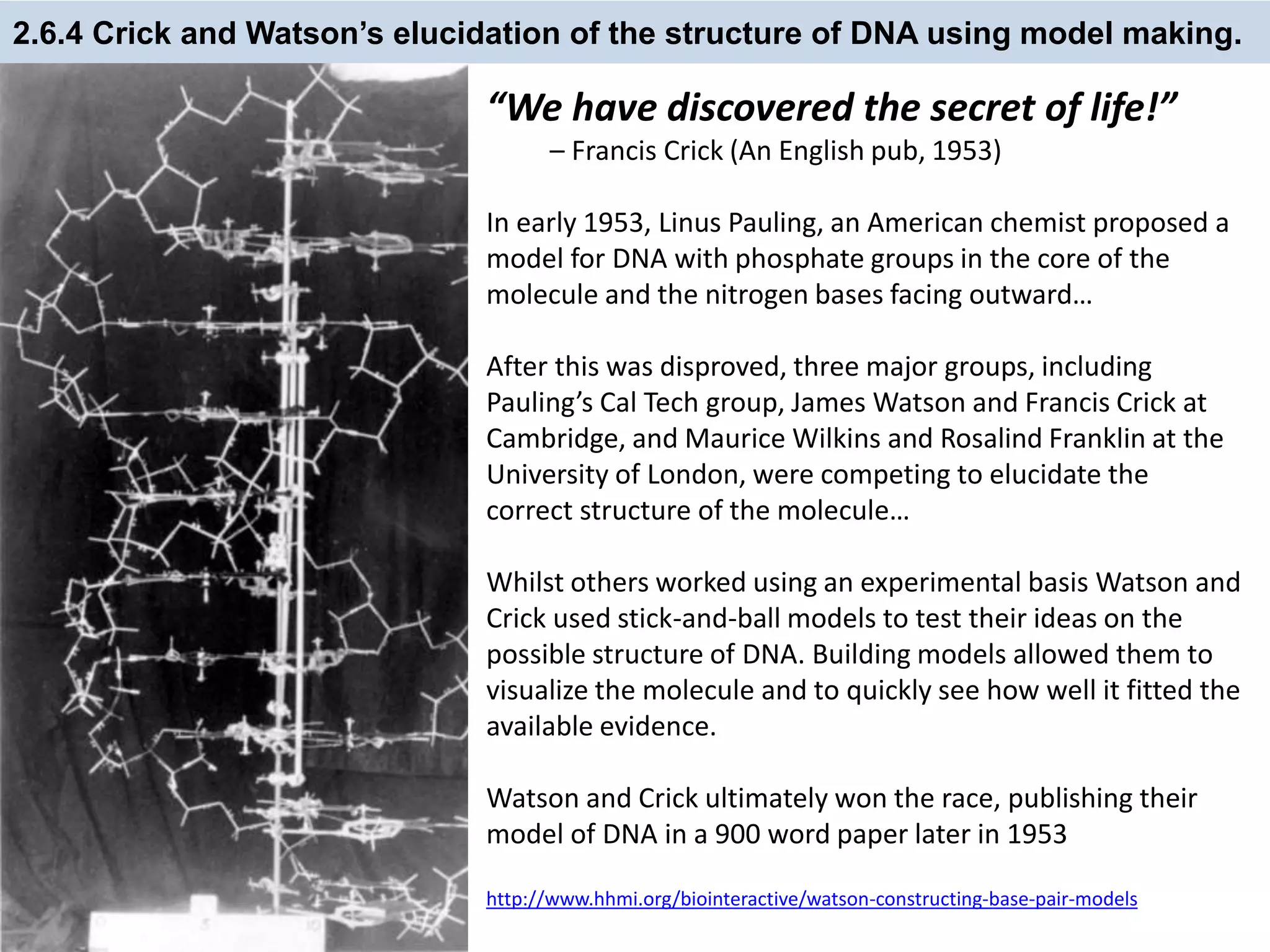
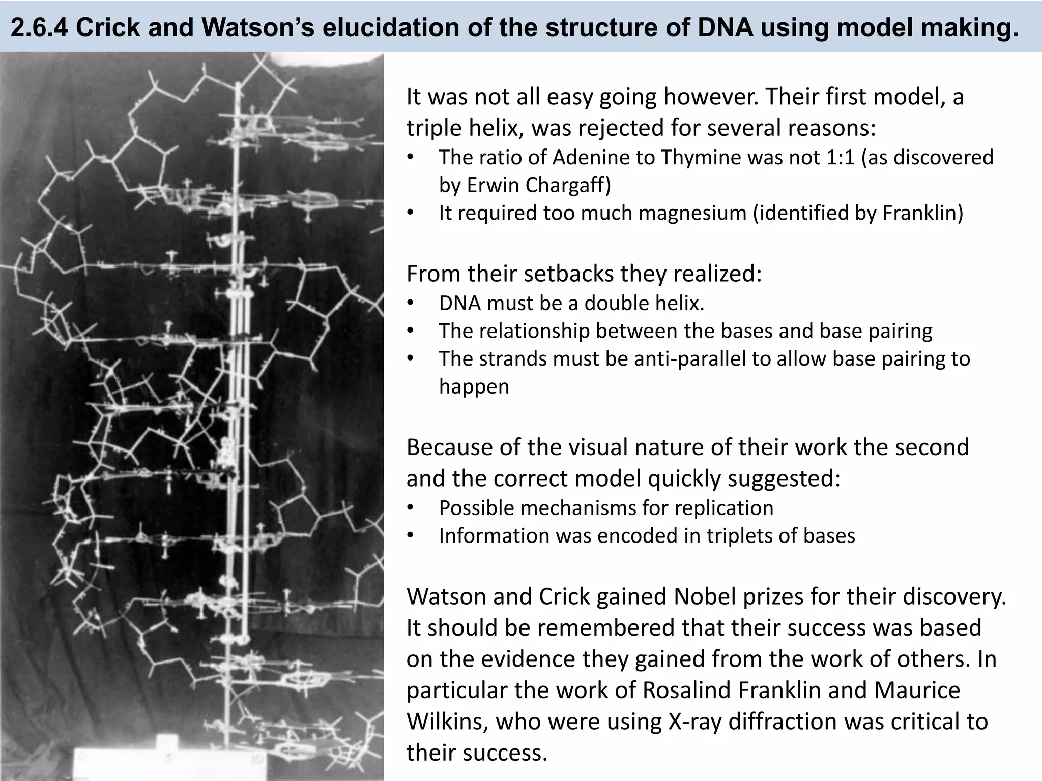
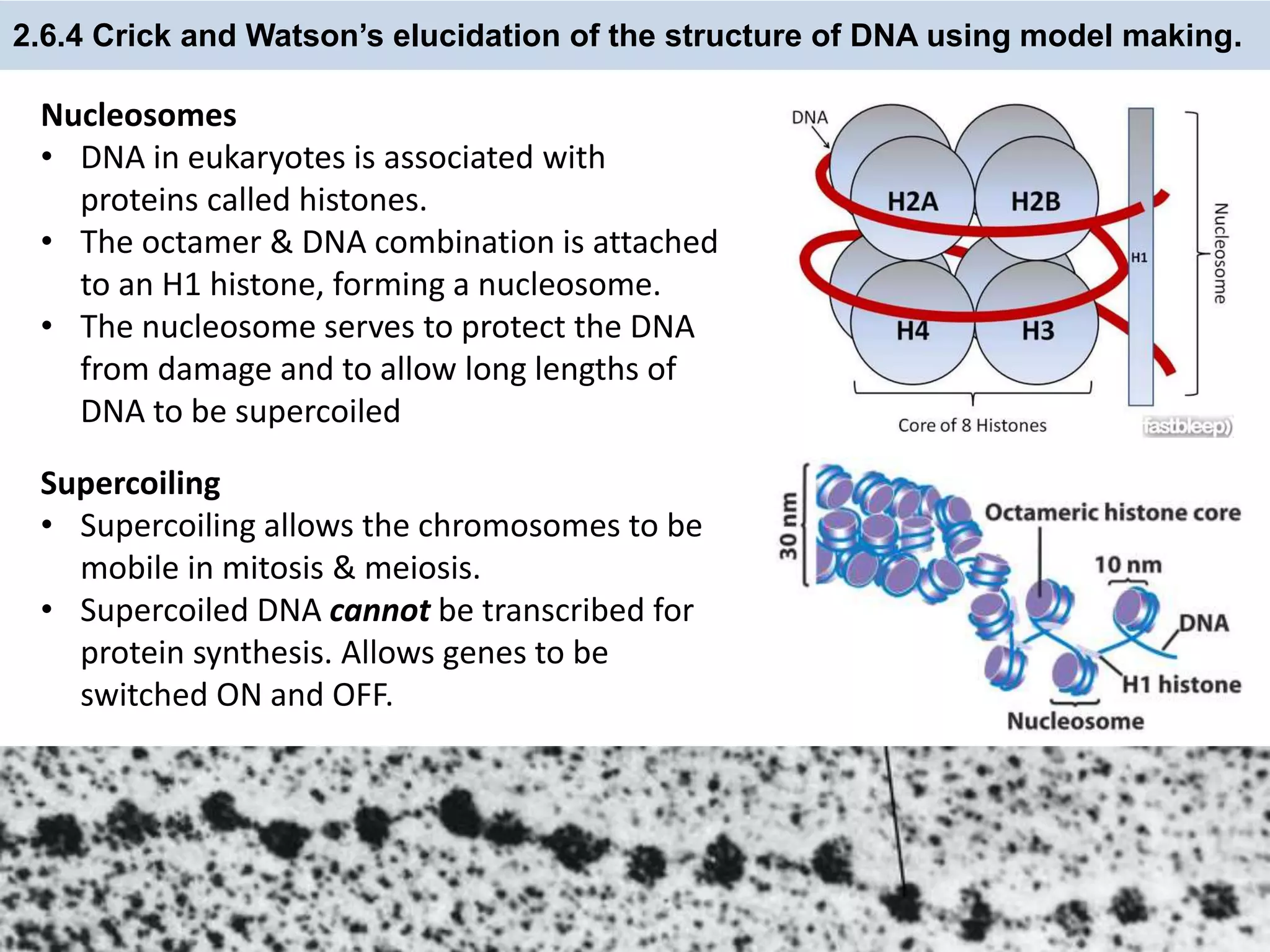
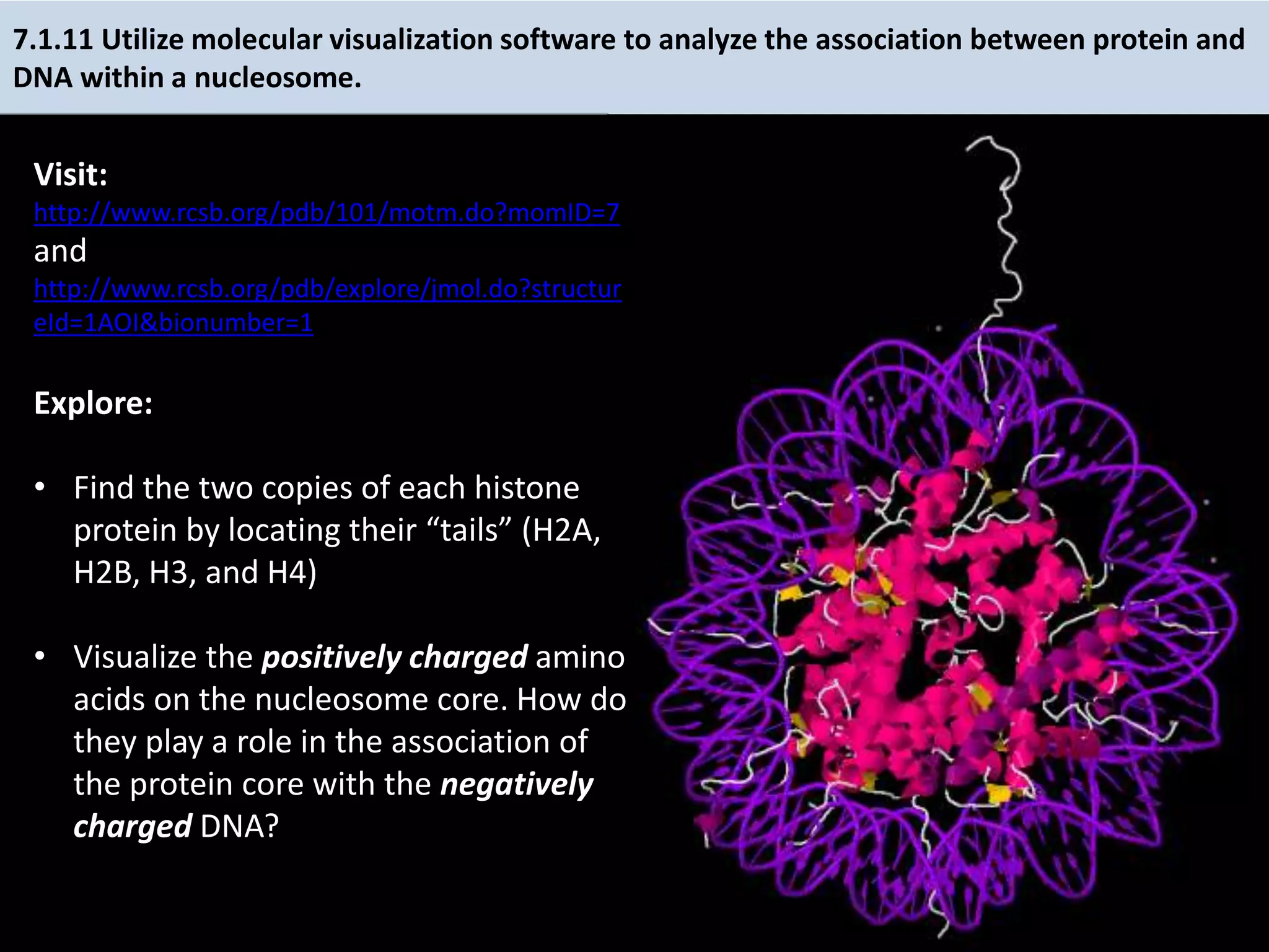
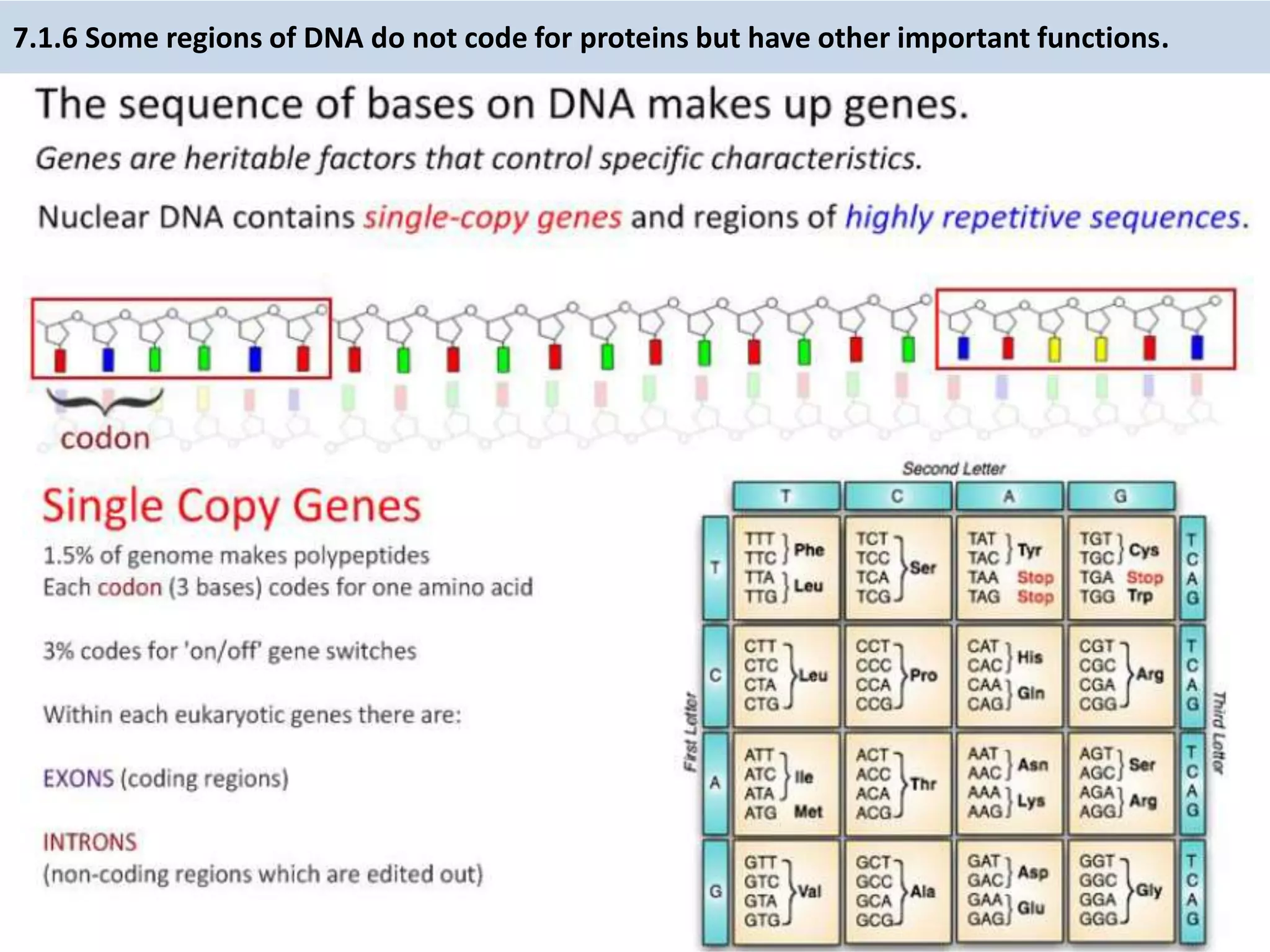
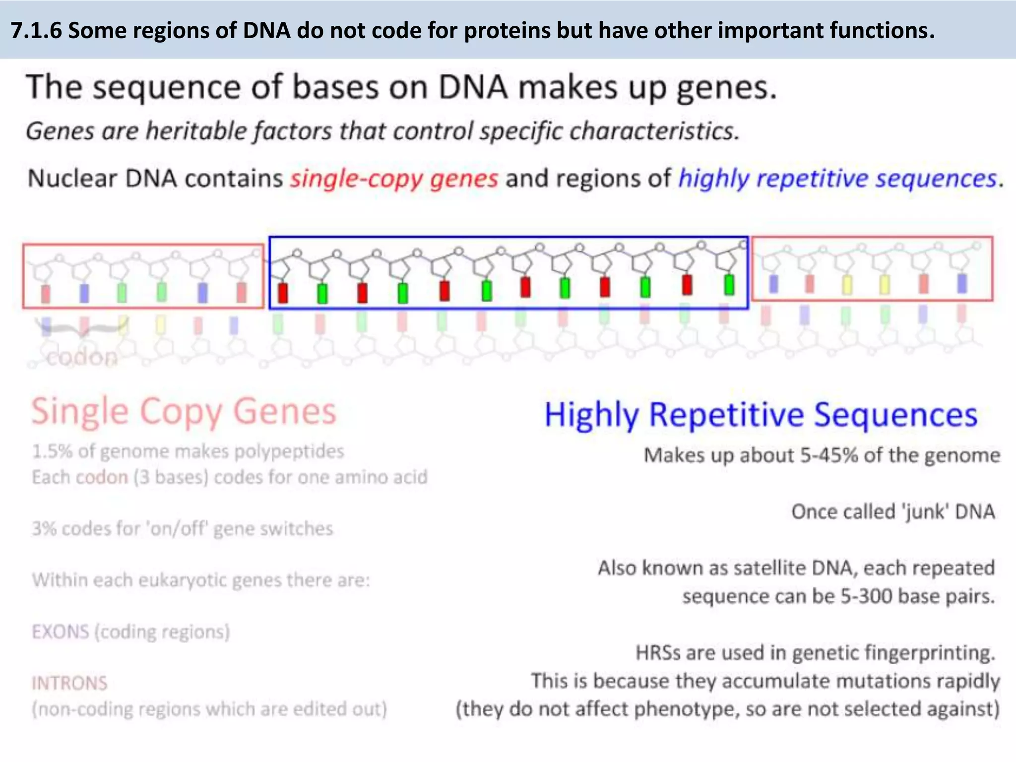
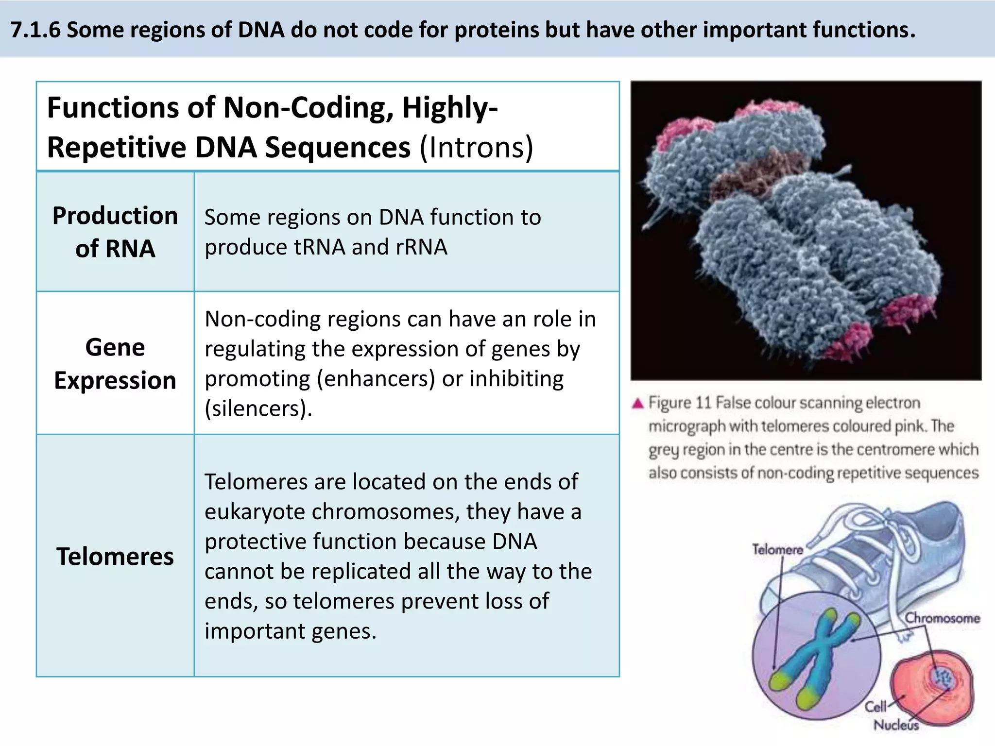
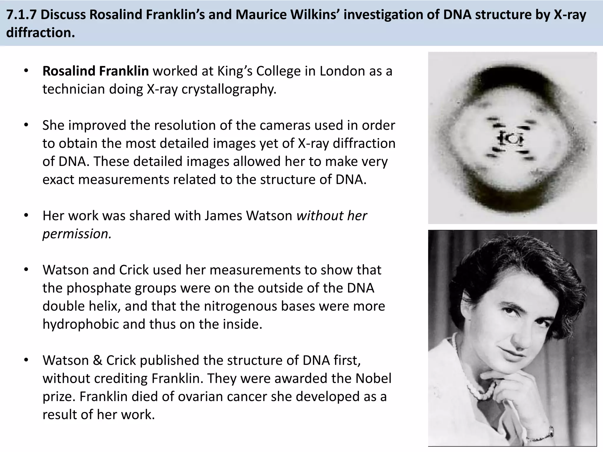
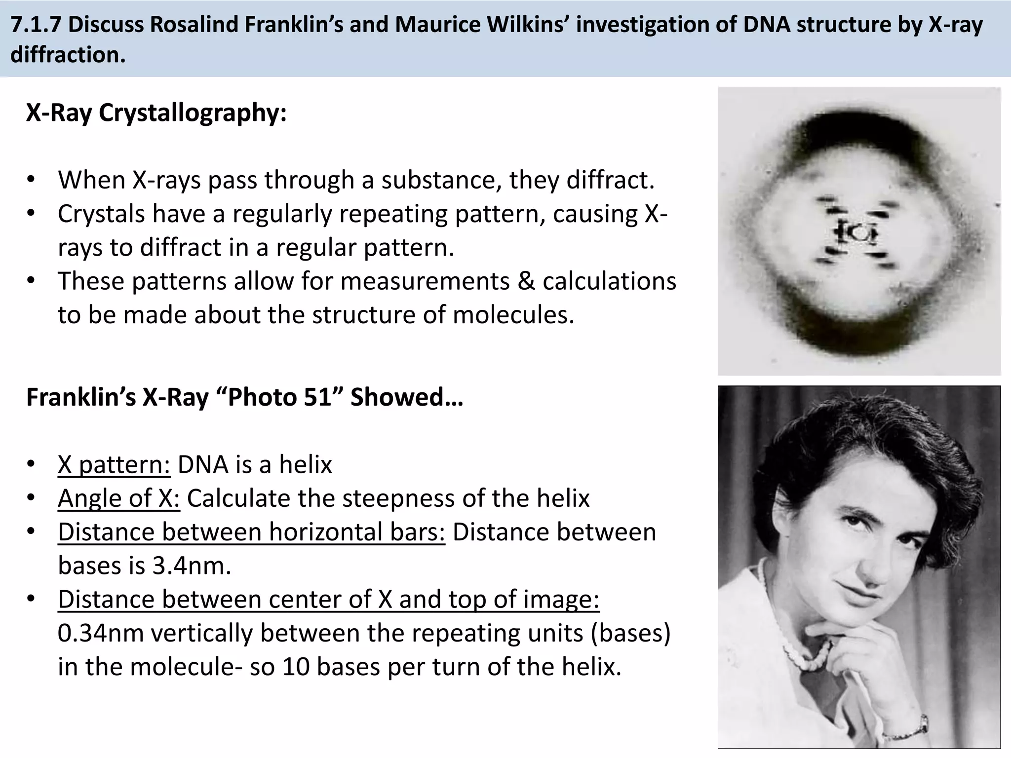
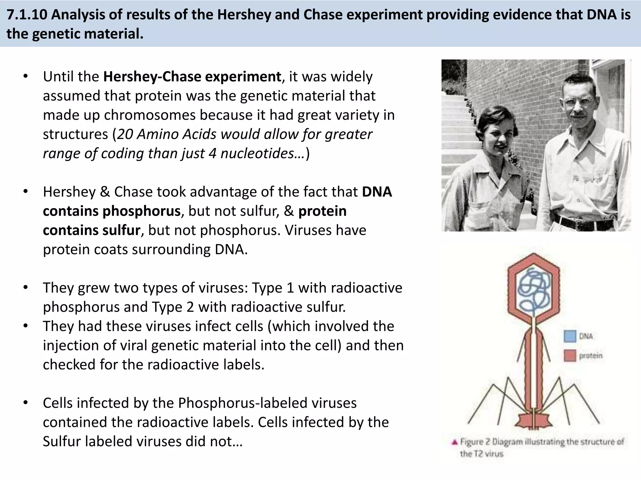
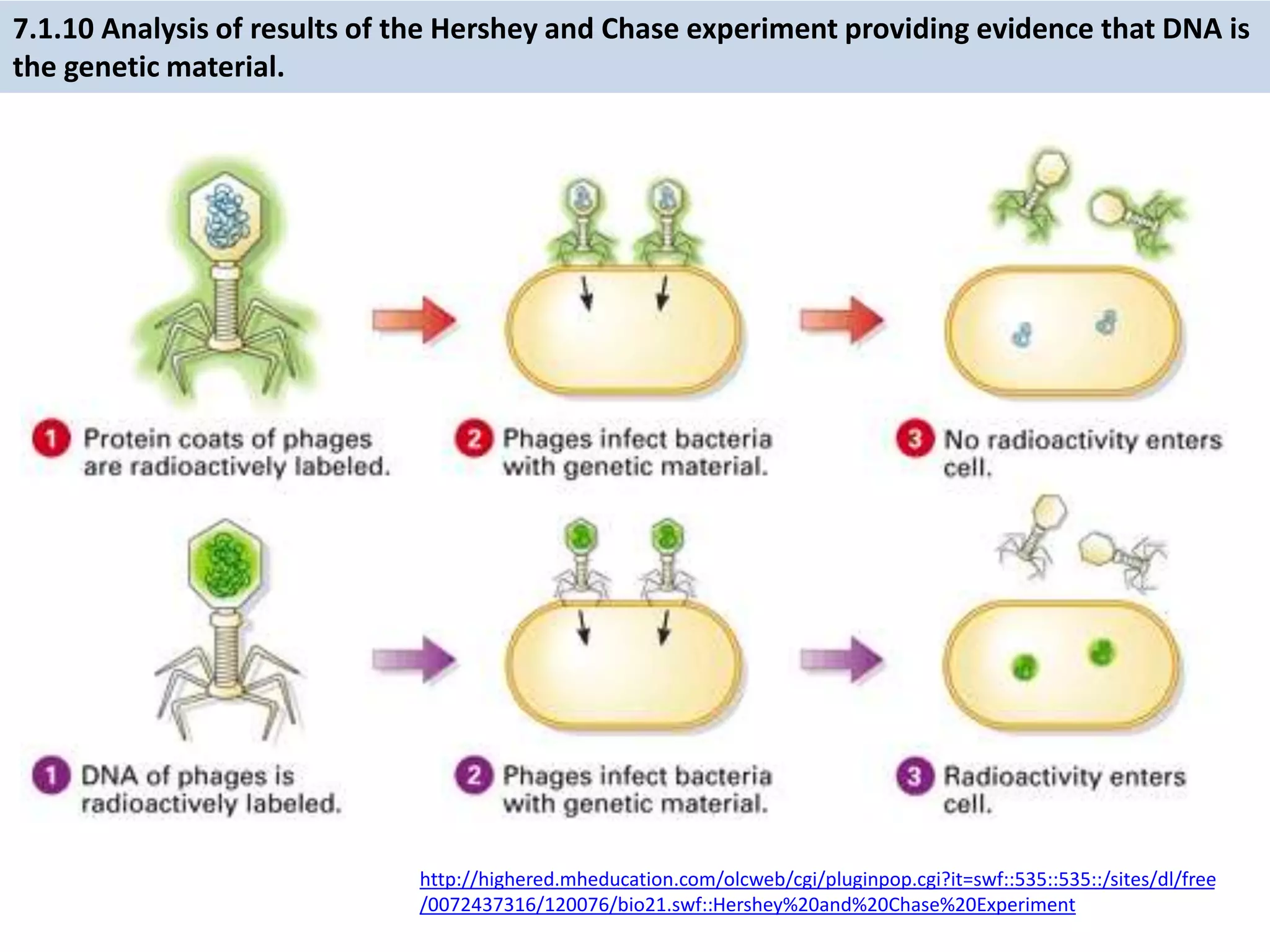


The document outlines the structure and function of DNA and RNA, emphasizing that DNA's design enables efficient genetic information storage. Key differences between DNA and RNA, such as the number of strands and nitrogenous bases, are highlighted, alongside historical insights into the DNA structure elucidation by Watson and Crick, and contributions from Franklin and Wilkins. Additionally, it discusses the cellular organization of DNA, including nucleosomes, telomeres, and the Hershey-Chase experiment that confirmed DNA as genetic material.