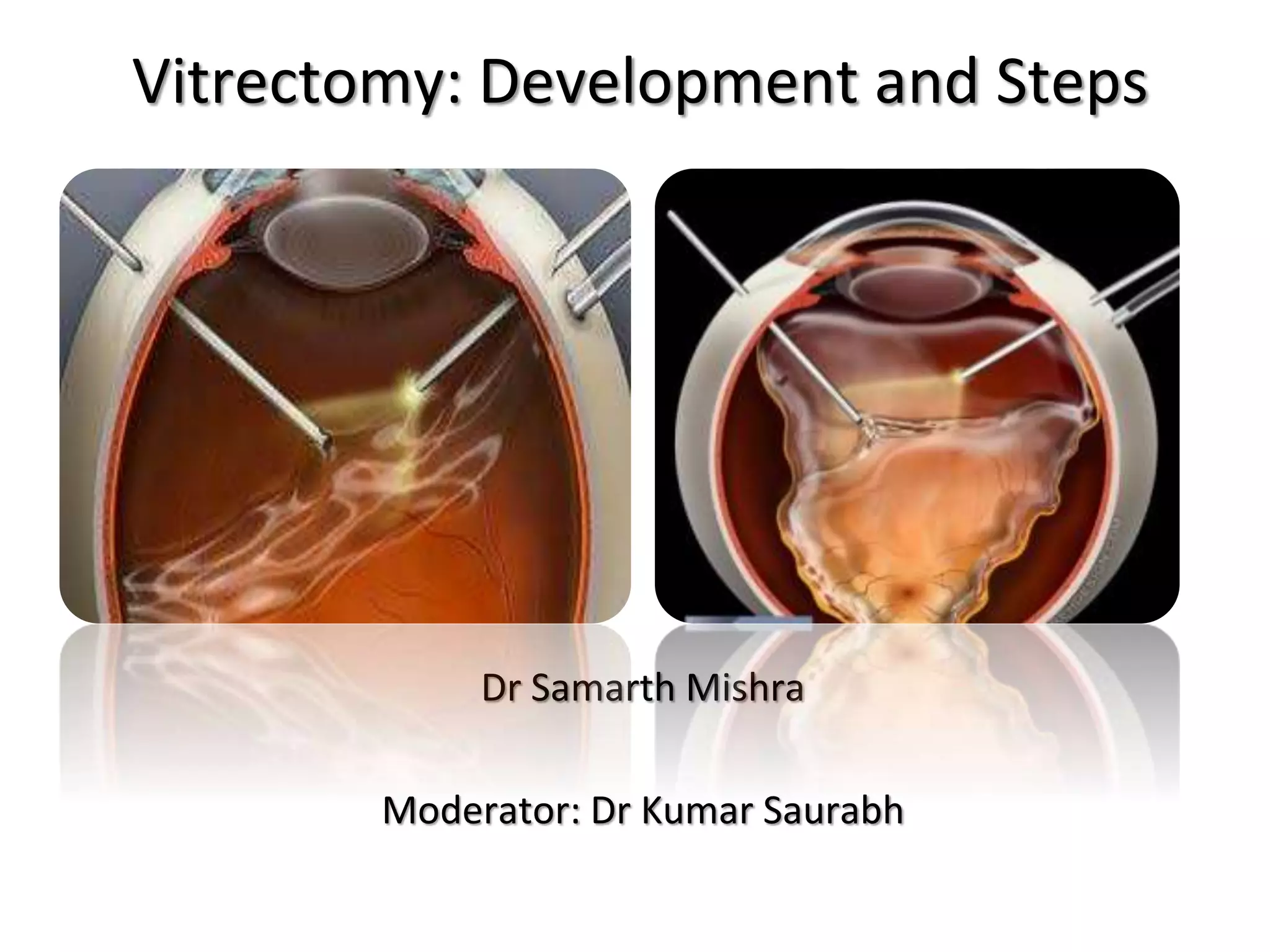
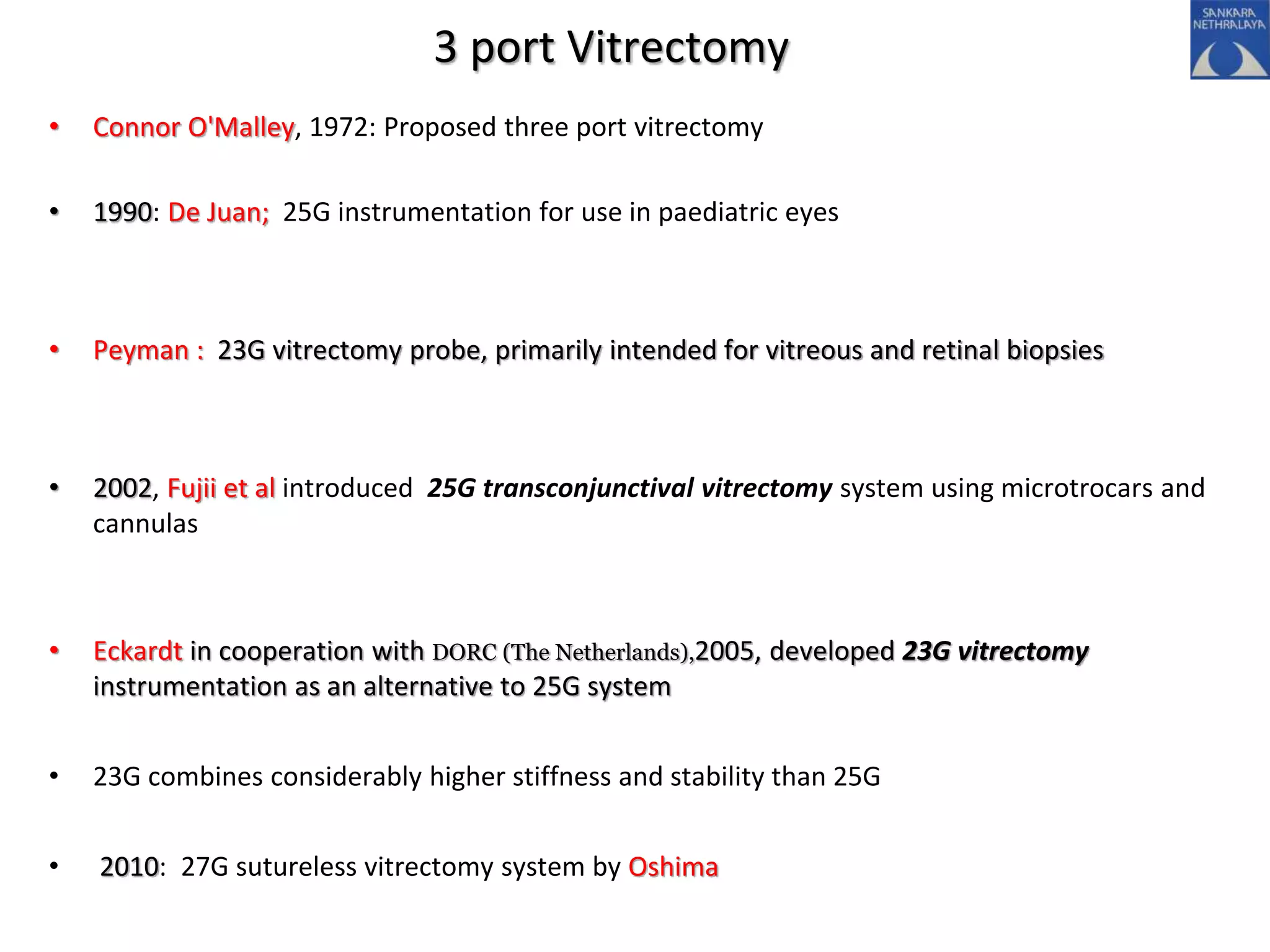
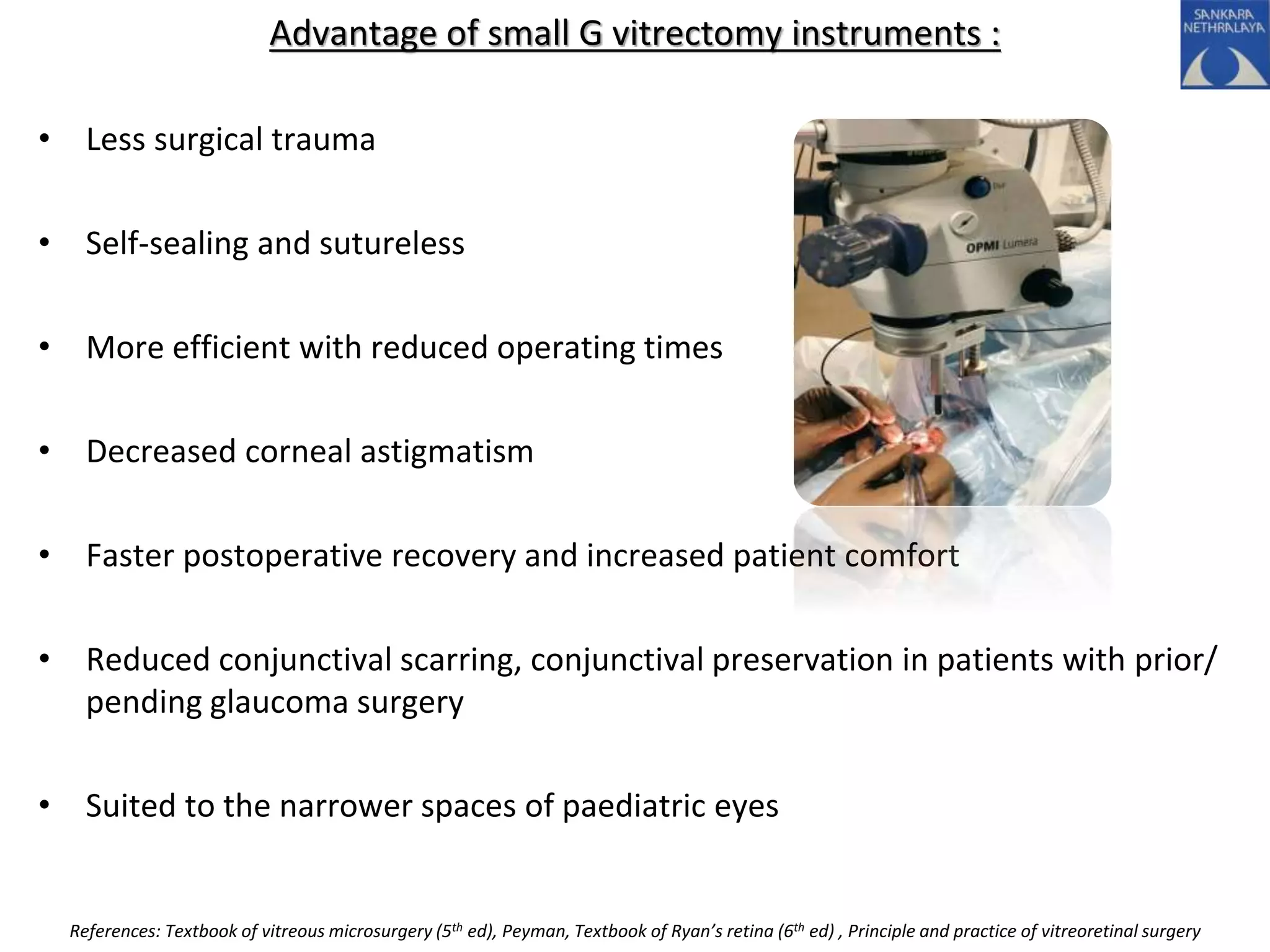
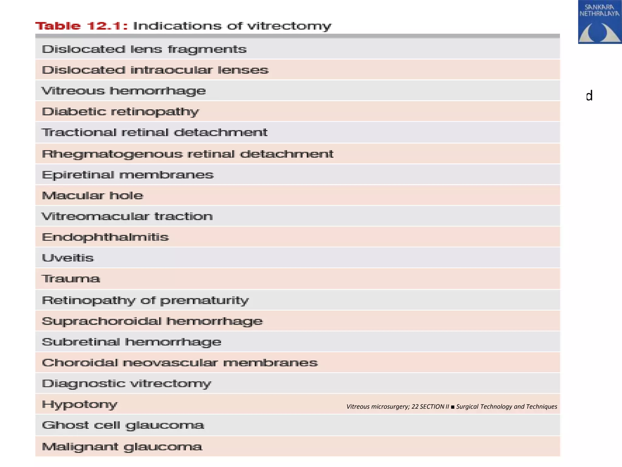
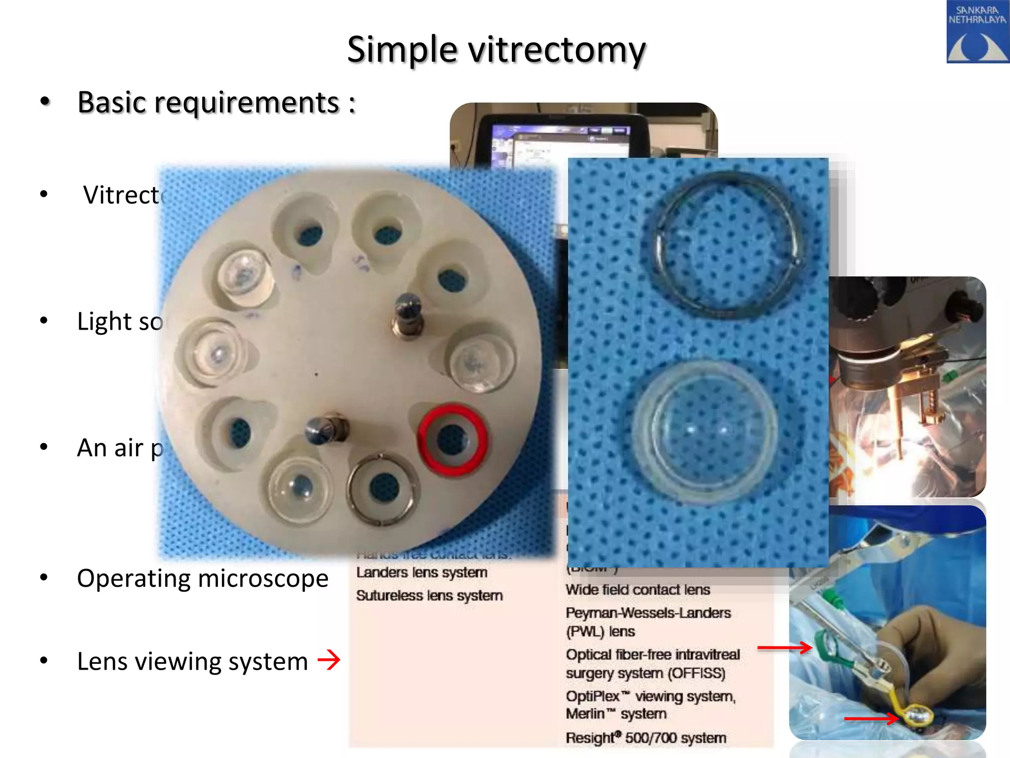
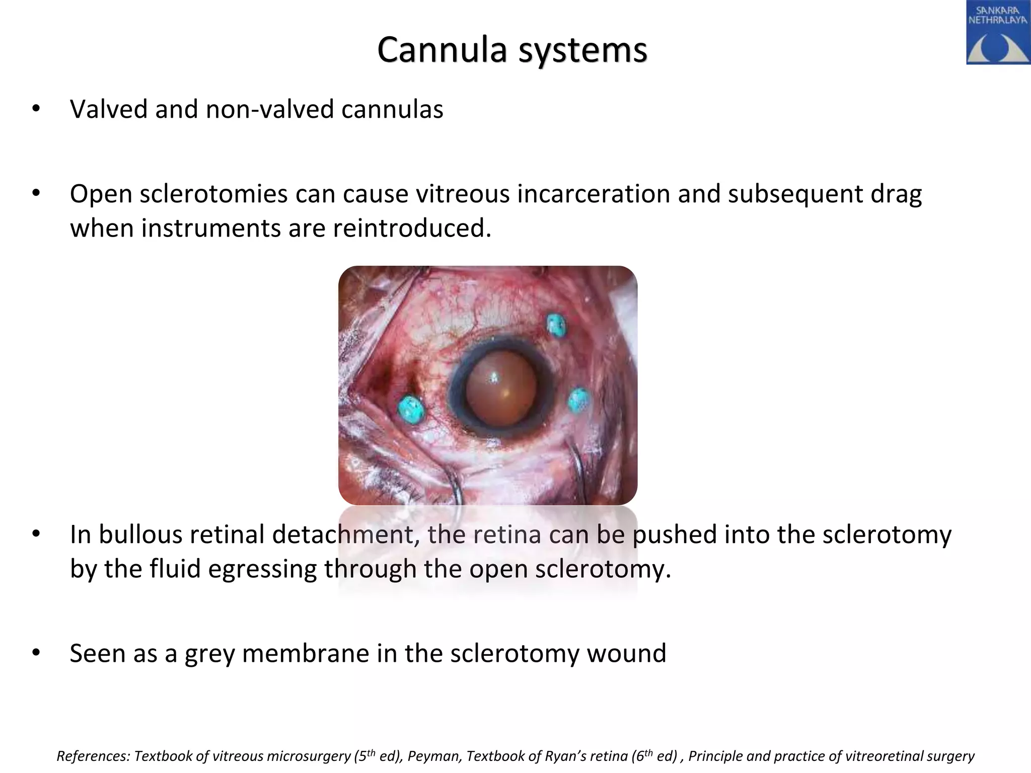
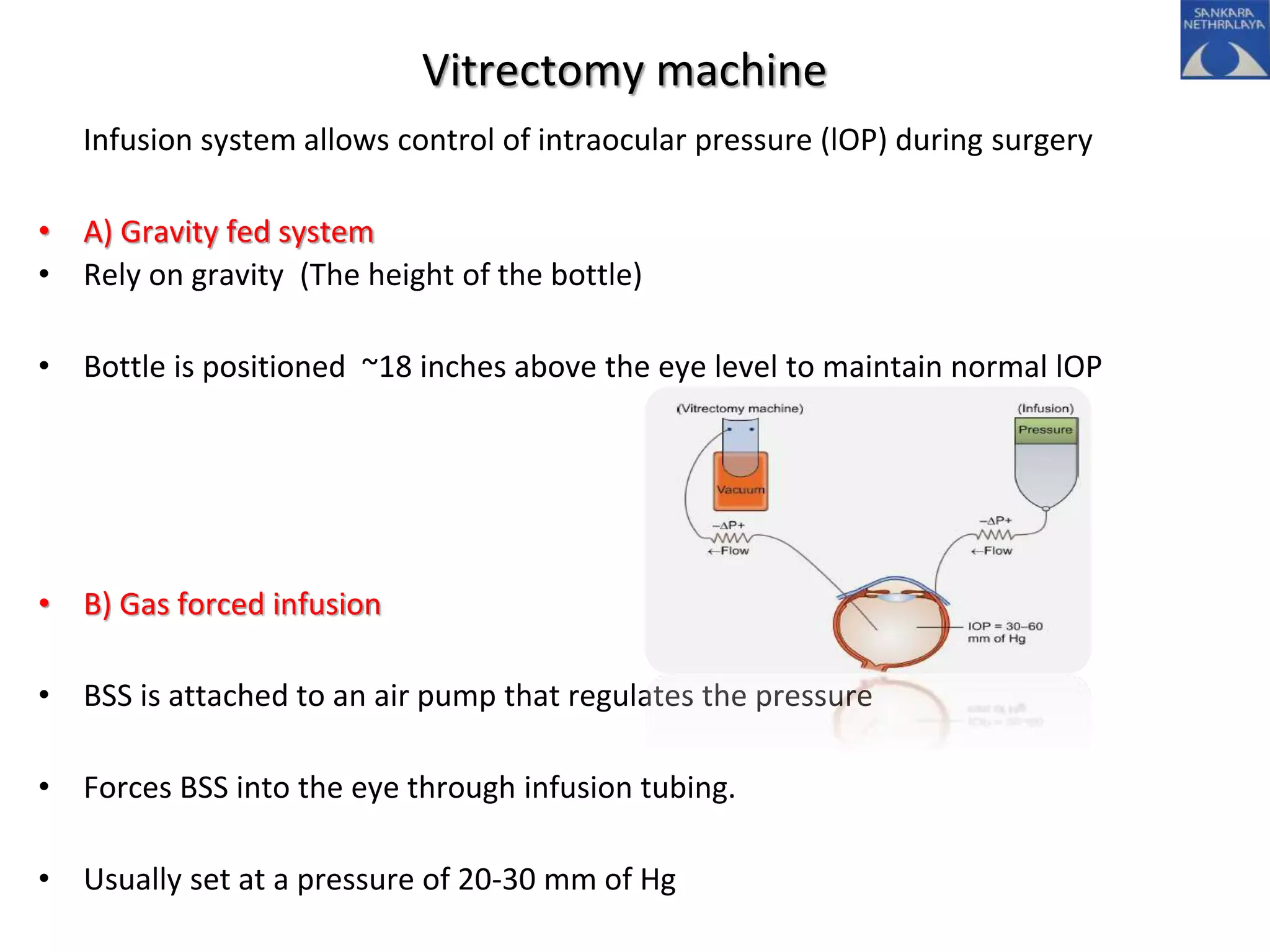
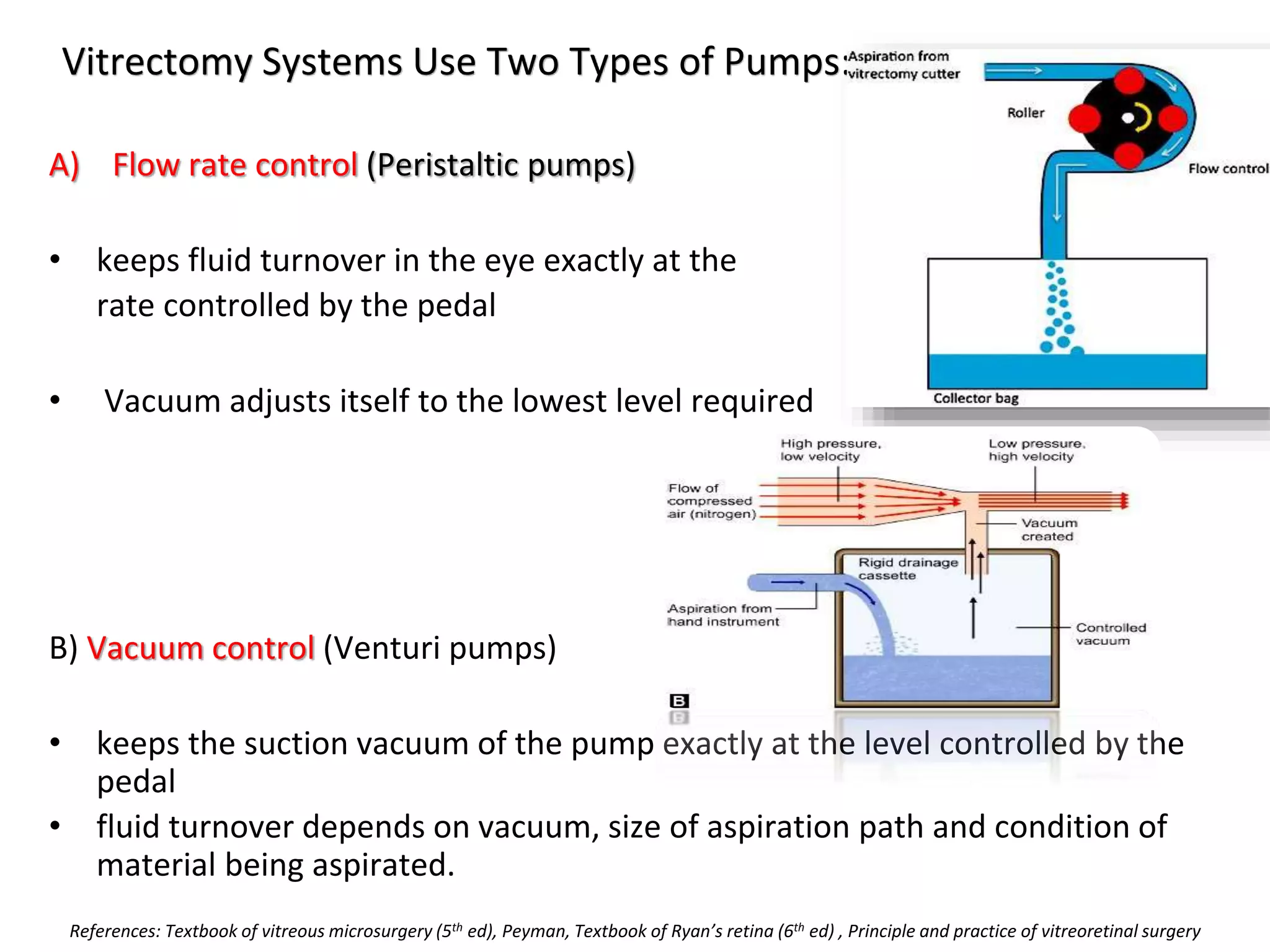
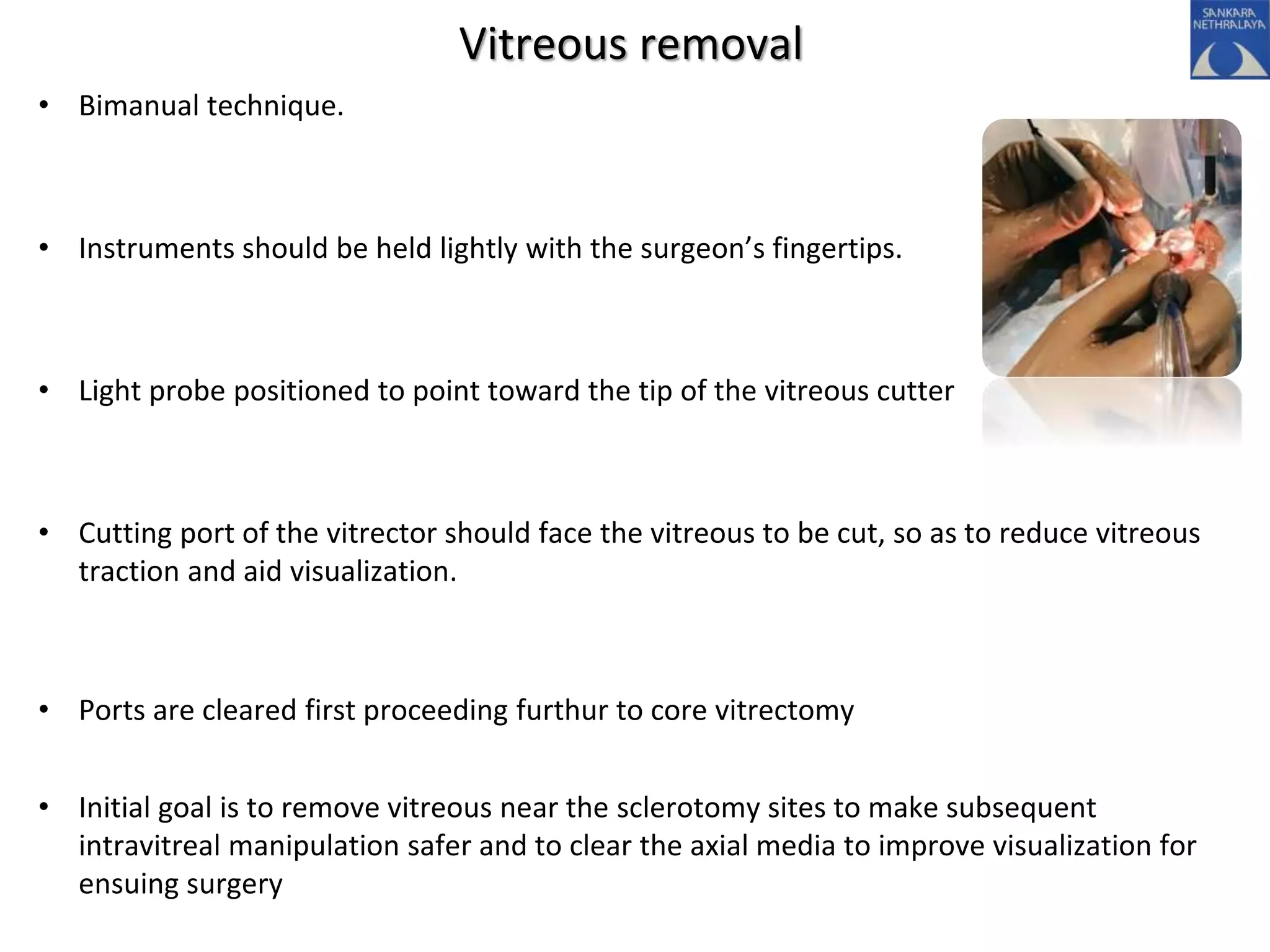
This document provides an overview of vitrectomy, including a brief history, the key steps in a simple vitrectomy procedure, and descriptions of the equipment used. It discusses trocars and cannulas, sclerotomies, illumination sources, viewing systems, vitreous cutters, and vitreous removal. Advantages of smaller gauge vitrectomy instruments are also summarized.
Basic overview of phaco dynamics along with all the Newer phacoemulsification techniques available in current practice - a video-assisted the presentation
Update knowledge about Muntifocal IOL made by Asaduzzaman
Working as Associate Optometrist in Ispahani Islamia Eye Institute &Hospita, Dhaka 1215
Email:asad.optom92@yaho. com
Retinal vasculitis refers to the inflammation of the retinal vessel resulting in evident clinical manifestations i.e. vascular sheathing, leakage and occlusion. This presentation covers the etiology, pathogenesis, clinical features, diagnosis and management of this spectrum of retinal disease.
Principles and technique of pneumatic retinopexy (Dr. Avuru C.J).pptxAVURUCHUKWUNALUJAMES1
Retinal detachment surgeries, principles and technique of pneumatic retinopexy, current trend in retinal detachment surgeries, development of skills in binocular indirect ophthalmoscopic examination, residency traning presentation, University college hospital Ibadan, Oyo state Nigeria, Vitreo-retinal subspecialty training, West african college of surgeons, federal teaching hospital, Lokoja, Kogi state, Nigeria.
LASIK or Lasik (laser-assisted in situ keratomileusis), commonly referred to as laser eye surgery or laser vision correction, is a type of refractive surgery for the correction of myopia, hyperopia, and an actual cure for astigmatism, since it is in the cornea. LASIK surgery is performed by an ophthalmologist who uses a laser or microkeratome to reshape the eye's cornea in order to improve visual acuity. For most people, LASIK provides a long-lasting alternative to eyeglasses or contact lenses.
The planning and analysis of corneal reshaping techniques such as LASIK have been standardized by the American National Standards Institute, an approach based on the Alpins method of astigmatism analysis. The FDA website on LASIK states,
"Before undergoing a refractive procedure, you should carefully weigh the risks and benefits based on your own personal value system, and try to avoid being influenced by friends that have had the procedure or doctors encouraging you to do so."
The procedure involves creating a thin flap on the eye, folding it to enable remodeling of the tissue beneath with a laser and repositioning the flap.
Report Back from SGO 2024: What’s the Latest in Cervical Cancer?bkling
Are you curious about what’s new in cervical cancer research or unsure what the findings mean? Join Dr. Emily Ko, a gynecologic oncologist at Penn Medicine, to learn about the latest updates from the Society of Gynecologic Oncology (SGO) 2024 Annual Meeting on Women’s Cancer. Dr. Ko will discuss what the research presented at the conference means for you and answer your questions about the new developments.
micro teaching on communication m.sc nursing.pdfAnurag Sharma
Microteaching is a unique model of practice teaching. It is a viable instrument for the. desired change in the teaching behavior or the behavior potential which, in specified types of real. classroom situations, tends to facilitate the achievement of specified types of objectives.
Lung Cancer: Artificial Intelligence, Synergetics, Complex System Analysis, S...Oleg Kshivets
RESULTS: Overall life span (LS) was 2252.1±1742.5 days and cumulative 5-year survival (5YS) reached 73.2%, 10 years – 64.8%, 20 years – 42.5%. 513 LCP lived more than 5 years (LS=3124.6±1525.6 days), 148 LCP – more than 10 years (LS=5054.4±1504.1 days).199 LCP died because of LC (LS=562.7±374.5 days). 5YS of LCP after bi/lobectomies was significantly superior in comparison with LCP after pneumonectomies (78.1% vs.63.7%, P=0.00001 by log-rank test). AT significantly improved 5YS (66.3% vs. 34.8%) (P=0.00000 by log-rank test) only for LCP with N1-2. Cox modeling displayed that 5YS of LCP significantly depended on: phase transition (PT) early-invasive LC in terms of synergetics, PT N0—N12, cell ratio factors (ratio between cancer cells- CC and blood cells subpopulations), G1-3, histology, glucose, AT, blood cell circuit, prothrombin index, heparin tolerance, recalcification time (P=0.000-0.038). Neural networks, genetic algorithm selection and bootstrap simulation revealed relationships between 5YS and PT early-invasive LC (rank=1), PT N0—N12 (rank=2), thrombocytes/CC (3), erythrocytes/CC (4), eosinophils/CC (5), healthy cells/CC (6), lymphocytes/CC (7), segmented neutrophils/CC (8), stick neutrophils/CC (9), monocytes/CC (10); leucocytes/CC (11). Correct prediction of 5YS was 100% by neural networks computing (area under ROC curve=1.0; error=0.0).
CONCLUSIONS: 5YS of LCP after radical procedures significantly depended on: 1) PT early-invasive cancer; 2) PT N0--N12; 3) cell ratio factors; 4) blood cell circuit; 5) biochemical factors; 6) hemostasis system; 7) AT; 8) LC characteristics; 9) LC cell dynamics; 10) surgery type: lobectomy/pneumonectomy; 11) anthropometric data. Optimal diagnosis and treatment strategies for LC are: 1) screening and early detection of LC; 2) availability of experienced thoracic surgeons because of complexity of radical procedures; 3) aggressive en block surgery and adequate lymph node dissection for completeness; 4) precise prediction; 5) adjuvant chemoimmunoradiotherapy for LCP with unfavorable prognosis.
ARTIFICIAL INTELLIGENCE IN HEALTHCARE.pdfAnujkumaranit
Artificial intelligence (AI) refers to the simulation of human intelligence processes by machines, especially computer systems. It encompasses tasks such as learning, reasoning, problem-solving, perception, and language understanding. AI technologies are revolutionizing various fields, from healthcare to finance, by enabling machines to perform tasks that typically require human intelligence.
New Drug Discovery and Development .....NEHA GUPTA
The "New Drug Discovery and Development" process involves the identification, design, testing, and manufacturing of novel pharmaceutical compounds with the aim of introducing new and improved treatments for various medical conditions. This comprehensive endeavor encompasses various stages, including target identification, preclinical studies, clinical trials, regulatory approval, and post-market surveillance. It involves multidisciplinary collaboration among scientists, researchers, clinicians, regulatory experts, and pharmaceutical companies to bring innovative therapies to market and address unmet medical needs.
New Directions in Targeted Therapeutic Approaches for Older Adults With Mantl...i3 Health
i3 Health is pleased to make the speaker slides from this activity available for use as a non-accredited self-study or teaching resource.
This slide deck presented by Dr. Kami Maddocks, Professor-Clinical in the Division of Hematology and
Associate Division Director for Ambulatory Operations
The Ohio State University Comprehensive Cancer Center, will provide insight into new directions in targeted therapeutic approaches for older adults with mantle cell lymphoma.
STATEMENT OF NEED
Mantle cell lymphoma (MCL) is a rare, aggressive B-cell non-Hodgkin lymphoma (NHL) accounting for 5% to 7% of all lymphomas. Its prognosis ranges from indolent disease that does not require treatment for years to very aggressive disease, which is associated with poor survival (Silkenstedt et al, 2021). Typically, MCL is diagnosed at advanced stage and in older patients who cannot tolerate intensive therapy (NCCN, 2022). Although recent advances have slightly increased remission rates, recurrence and relapse remain very common, leading to a median overall survival between 3 and 6 years (LLS, 2021). Though there are several effective options, progress is still needed towards establishing an accepted frontline approach for MCL (Castellino et al, 2022). Treatment selection and management of MCL are complicated by the heterogeneity of prognosis, advanced age and comorbidities of patients, and lack of an established standard approach for treatment, making it vital that clinicians be familiar with the latest research and advances in this area. In this activity chaired by Michael Wang, MD, Professor in the Department of Lymphoma & Myeloma at MD Anderson Cancer Center, expert faculty will discuss prognostic factors informing treatment, the promising results of recent trials in new therapeutic approaches, and the implications of treatment resistance in therapeutic selection for MCL.
Target Audience
Hematology/oncology fellows, attending faculty, and other health care professionals involved in the treatment of patients with mantle cell lymphoma (MCL).
Learning Objectives
1.) Identify clinical and biological prognostic factors that can guide treatment decision making for older adults with MCL
2.) Evaluate emerging data on targeted therapeutic approaches for treatment-naive and relapsed/refractory MCL and their applicability to older adults
3.) Assess mechanisms of resistance to targeted therapies for MCL and their implications for treatment selection
Title: Sense of Smell
Presenter: Dr. Faiza, Assistant Professor of Physiology
Qualifications:
MBBS (Best Graduate, AIMC Lahore)
FCPS Physiology
ICMT, CHPE, DHPE (STMU)
MPH (GC University, Faisalabad)
MBA (Virtual University of Pakistan)
Learning Objectives:
Describe the primary categories of smells and the concept of odor blindness.
Explain the structure and location of the olfactory membrane and mucosa, including the types and roles of cells involved in olfaction.
Describe the pathway and mechanisms of olfactory signal transmission from the olfactory receptors to the brain.
Illustrate the biochemical cascade triggered by odorant binding to olfactory receptors, including the role of G-proteins and second messengers in generating an action potential.
Identify different types of olfactory disorders such as anosmia, hyposmia, hyperosmia, and dysosmia, including their potential causes.
Key Topics:
Olfactory Genes:
3% of the human genome accounts for olfactory genes.
400 genes for odorant receptors.
Olfactory Membrane:
Located in the superior part of the nasal cavity.
Medially: Folds downward along the superior septum.
Laterally: Folds over the superior turbinate and upper surface of the middle turbinate.
Total surface area: 5-10 square centimeters.
Olfactory Mucosa:
Olfactory Cells: Bipolar nerve cells derived from the CNS (100 million), with 4-25 olfactory cilia per cell.
Sustentacular Cells: Produce mucus and maintain ionic and molecular environment.
Basal Cells: Replace worn-out olfactory cells with an average lifespan of 1-2 months.
Bowman’s Gland: Secretes mucus.
Stimulation of Olfactory Cells:
Odorant dissolves in mucus and attaches to receptors on olfactory cilia.
Involves a cascade effect through G-proteins and second messengers, leading to depolarization and action potential generation in the olfactory nerve.
Quality of a Good Odorant:
Small (3-20 Carbon atoms), volatile, water-soluble, and lipid-soluble.
Facilitated by odorant-binding proteins in mucus.
Membrane Potential and Action Potential:
Resting membrane potential: -55mV.
Action potential frequency in the olfactory nerve increases with odorant strength.
Adaptation Towards the Sense of Smell:
Rapid adaptation within the first second, with further slow adaptation.
Psychological adaptation greater than receptor adaptation, involving feedback inhibition from the central nervous system.
Primary Sensations of Smell:
Camphoraceous, Musky, Floral, Pepperminty, Ethereal, Pungent, Putrid.
Odor Detection Threshold:
Examples: Hydrogen sulfide (0.0005 ppm), Methyl-mercaptan (0.002 ppm).
Some toxic substances are odorless at lethal concentrations.
Characteristics of Smell:
Odor blindness for single substances due to lack of appropriate receptor protein.
Behavioral and emotional influences of smell.
Transmission of Olfactory Signals:
From olfactory cells to glomeruli in the olfactory bulb, involving lateral inhibition.
Primitive, less old, and new olfactory systems with different path
Flu Vaccine Alert in Bangalore Karnatakaaddon Scans
As flu season approaches, health officials in Bangalore, Karnataka, are urging residents to get their flu vaccinations. The seasonal flu, while common, can lead to severe health complications, particularly for vulnerable populations such as young children, the elderly, and those with underlying health conditions.
Dr. Vidisha Kumari, a leading epidemiologist in Bangalore, emphasizes the importance of getting vaccinated. "The flu vaccine is our best defense against the influenza virus. It not only protects individuals but also helps prevent the spread of the virus in our communities," he says.
This year, the flu season is expected to coincide with a potential increase in other respiratory illnesses. The Karnataka Health Department has launched an awareness campaign highlighting the significance of flu vaccinations. They have set up multiple vaccination centers across Bangalore, making it convenient for residents to receive their shots.
To encourage widespread vaccination, the government is also collaborating with local schools, workplaces, and community centers to facilitate vaccination drives. Special attention is being given to ensuring that the vaccine is accessible to all, including marginalized communities who may have limited access to healthcare.
Residents are reminded that the flu vaccine is safe and effective. Common side effects are mild and may include soreness at the injection site, mild fever, or muscle aches. These side effects are generally short-lived and far less severe than the flu itself.
Healthcare providers are also stressing the importance of continuing COVID-19 precautions. Wearing masks, practicing good hand hygiene, and maintaining social distancing are still crucial, especially in crowded places.
Protect yourself and your loved ones by getting vaccinated. Together, we can help keep Bangalore healthy and safe this flu season. For more information on vaccination centers and schedules, residents can visit the Karnataka Health Department’s official website or follow their social media pages.
Stay informed, stay safe, and get your flu shot today!
Title: Sense of Taste
Presenter: Dr. Faiza, Assistant Professor of Physiology
Qualifications:
MBBS (Best Graduate, AIMC Lahore)
FCPS Physiology
ICMT, CHPE, DHPE (STMU)
MPH (GC University, Faisalabad)
MBA (Virtual University of Pakistan)
Learning Objectives:
Describe the structure and function of taste buds.
Describe the relationship between the taste threshold and taste index of common substances.
Explain the chemical basis and signal transduction of taste perception for each type of primary taste sensation.
Recognize different abnormalities of taste perception and their causes.
Key Topics:
Significance of Taste Sensation:
Differentiation between pleasant and harmful food
Influence on behavior
Selection of food based on metabolic needs
Receptors of Taste:
Taste buds on the tongue
Influence of sense of smell, texture of food, and pain stimulation (e.g., by pepper)
Primary and Secondary Taste Sensations:
Primary taste sensations: Sweet, Sour, Salty, Bitter, Umami
Chemical basis and signal transduction mechanisms for each taste
Taste Threshold and Index:
Taste threshold values for Sweet (sucrose), Salty (NaCl), Sour (HCl), and Bitter (Quinine)
Taste index relationship: Inversely proportional to taste threshold
Taste Blindness:
Inability to taste certain substances, particularly thiourea compounds
Example: Phenylthiocarbamide
Structure and Function of Taste Buds:
Composition: Epithelial cells, Sustentacular/Supporting cells, Taste cells, Basal cells
Features: Taste pores, Taste hairs/microvilli, and Taste nerve fibers
Location of Taste Buds:
Found in papillae of the tongue (Fungiform, Circumvallate, Foliate)
Also present on the palate, tonsillar pillars, epiglottis, and proximal esophagus
Mechanism of Taste Stimulation:
Interaction of taste substances with receptors on microvilli
Signal transduction pathways for Umami, Sweet, Bitter, Sour, and Salty tastes
Taste Sensitivity and Adaptation:
Decrease in sensitivity with age
Rapid adaptation of taste sensation
Role of Saliva in Taste:
Dissolution of tastants to reach receptors
Washing away the stimulus
Taste Preferences and Aversions:
Mechanisms behind taste preference and aversion
Influence of receptors and neural pathways
Impact of Sensory Nerve Damage:
Degeneration of taste buds if the sensory nerve fiber is cut
Abnormalities of Taste Detection:
Conditions: Ageusia, Hypogeusia, Dysgeusia (parageusia)
Causes: Nerve damage, neurological disorders, infections, poor oral hygiene, adverse drug effects, deficiencies, aging, tobacco use, altered neurotransmitter levels
Neurotransmitters and Taste Threshold:
Effects of serotonin (5-HT) and norepinephrine (NE) on taste sensitivity
Supertasters:
25% of the population with heightened sensitivity to taste, especially bitterness
Increased number of fungiform papillae
The prostate is an exocrine gland of the male mammalian reproductive system
It is a walnut-sized gland that forms part of the male reproductive system and is located in front of the rectum and just below the urinary bladder
Function is to store and secrete a clear, slightly alkaline fluid that constitutes 10-30% of the volume of the seminal fluid that along with the spermatozoa, constitutes semen
A healthy human prostate measures (4cm-vertical, by 3cm-horizontal, 2cm ant-post ).
It surrounds the urethra just below the urinary bladder. It has anterior, median, posterior and two lateral lobes
It’s work is regulated by androgens which are responsible for male sex characteristics
Generalised disease of the prostate due to hormonal derangement which leads to non malignant enlargement of the gland (increase in the number of epithelial cells and stromal tissue)to cause compression of the urethra leading to symptoms (LUTS
Tom Selleck Health: A Comprehensive Look at the Iconic Actor’s Wellness Journeygreendigital
Tom Selleck, an enduring figure in Hollywood. has captivated audiences for decades with his rugged charm, iconic moustache. and memorable roles in television and film. From his breakout role as Thomas Magnum in Magnum P.I. to his current portrayal of Frank Reagan in Blue Bloods. Selleck's career has spanned over 50 years. But beyond his professional achievements. fans have often been curious about Tom Selleck Health. especially as he has aged in the public eye.
Follow us on: Pinterest
Introduction
Many have been interested in Tom Selleck health. not only because of his enduring presence on screen but also because of the challenges. and lifestyle choices he has faced and made over the years. This article delves into the various aspects of Tom Selleck health. exploring his fitness regimen, diet, mental health. and the challenges he has encountered as he ages. We'll look at how he maintains his well-being. the health issues he has faced, and his approach to ageing .
Early Life and Career
Childhood and Athletic Beginnings
Tom Selleck was born on January 29, 1945, in Detroit, Michigan, and grew up in Sherman Oaks, California. From an early age, he was involved in sports, particularly basketball. which played a significant role in his physical development. His athletic pursuits continued into college. where he attended the University of Southern California (USC) on a basketball scholarship. This early involvement in sports laid a strong foundation for his physical health and disciplined lifestyle.
Transition to Acting
Selleck's transition from an athlete to an actor came with its physical demands. His first significant role in "Magnum P.I." required him to perform various stunts and maintain a fit appearance. This role, which he played from 1980 to 1988. necessitated a rigorous fitness routine to meet the show's demands. setting the stage for his long-term commitment to health and wellness.
Fitness Regimen
Workout Routine
Tom Selleck health and fitness regimen has evolved. adapting to his changing roles and age. During his "Magnum, P.I." days. Selleck's workouts were intense and focused on building and maintaining muscle mass. His routine included weightlifting, cardiovascular exercises. and specific training for the stunts he performed on the show.
Selleck adjusted his fitness routine as he aged to suit his body's needs. Today, his workouts focus on maintaining flexibility, strength, and cardiovascular health. He incorporates low-impact exercises such as swimming, walking, and light weightlifting. This balanced approach helps him stay fit without putting undue strain on his joints and muscles.
Importance of Flexibility and Mobility
In recent years, Selleck has emphasized the importance of flexibility and mobility in his fitness regimen. Understanding the natural decline in muscle mass and joint flexibility with age. he includes stretching and yoga in his routine. These practices help prevent injuries, improve posture, and maintain mobilit
These simplified slides by Dr. Sidra Arshad present an overview of the non-respiratory functions of the respiratory tract.
Learning objectives:
1. Enlist the non-respiratory functions of the respiratory tract
2. Briefly explain how these functions are carried out
3. Discuss the significance of dead space
4. Differentiate between minute ventilation and alveolar ventilation
5. Describe the cough and sneeze reflexes
Study Resources:
1. Chapter 39, Guyton and Hall Textbook of Medical Physiology, 14th edition
2. Chapter 34, Ganong’s Review of Medical Physiology, 26th edition
3. Chapter 17, Human Physiology by Lauralee Sherwood, 9th edition
4. Non-respiratory functions of the lungs https://academic.oup.com/bjaed/article/13/3/98/278874
Recomendações da OMS sobre cuidados maternos e neonatais para uma experiência pós-natal positiva.
Em consonância com os ODS – Objetivos do Desenvolvimento Sustentável e a Estratégia Global para a Saúde das Mulheres, Crianças e Adolescentes, e aplicando uma abordagem baseada nos direitos humanos, os esforços de cuidados pós-natais devem expandir-se para além da cobertura e da simples sobrevivência, de modo a incluir cuidados de qualidade.
Estas diretrizes visam melhorar a qualidade dos cuidados pós-natais essenciais e de rotina prestados às mulheres e aos recém-nascidos, com o objetivo final de melhorar a saúde e o bem-estar materno e neonatal.
Uma “experiência pós-natal positiva” é um resultado importante para todas as mulheres que dão à luz e para os seus recém-nascidos, estabelecendo as bases para a melhoria da saúde e do bem-estar a curto e longo prazo. Uma experiência pós-natal positiva é definida como aquela em que as mulheres, pessoas que gestam, os recém-nascidos, os casais, os pais, os cuidadores e as famílias recebem informação consistente, garantia e apoio de profissionais de saúde motivados; e onde um sistema de saúde flexível e com recursos reconheça as necessidades das mulheres e dos bebês e respeite o seu contexto cultural.
Estas diretrizes consolidadas apresentam algumas recomendações novas e já bem fundamentadas sobre cuidados pós-natais de rotina para mulheres e neonatos que recebem cuidados no pós-parto em unidades de saúde ou na comunidade, independentemente dos recursos disponíveis.
É fornecido um conjunto abrangente de recomendações para cuidados durante o período puerperal, com ênfase nos cuidados essenciais que todas as mulheres e recém-nascidos devem receber, e com a devida atenção à qualidade dos cuidados; isto é, a entrega e a experiência do cuidado recebido. Estas diretrizes atualizam e ampliam as recomendações da OMS de 2014 sobre cuidados pós-natais da mãe e do recém-nascido e complementam as atuais diretrizes da OMS sobre a gestão de complicações pós-natais.
O estabelecimento da amamentação e o manejo das principais intercorrências é contemplada.
Recomendamos muito.
Vamos discutir essas recomendações no nosso curso de pós-graduação em Aleitamento no Instituto Ciclos.
Esta publicação só está disponível em inglês até o momento.
Prof. Marcus Renato de Carvalho
www.agostodourado.com
Ozempic: Preoperative Management of Patients on GLP-1 Receptor Agonists Saeid Safari
Preoperative Management of Patients on GLP-1 Receptor Agonists like Ozempic and Semiglutide
ASA GUIDELINE
NYSORA Guideline
2 Case Reports of Gastric Ultrasound
2. Brief history of vitrectomy
Steps for simple vitrectomy
Trocars and cannula
Sclerotomy
Illumination source
Viewing systems
Cutters
Vitreous removal
Closure of ports
3. "can we touch the vitreous and go unpunished" ??
• Such was the vitreous sanctity in 1960s and early 1970s
• David Kasner, through cornea removed opaque vitreous with primary amyloidosis (
2 patients )
• He developed open sky vitreous surgery technique
• Machemer introduced pars plana vitrectomy (PPV) in 1971.
• Vitreous infusion suction cutter(VISC), 17-gauge (1.42mm diameter)
4. Experiment to remove the egg white by a rotating drill inserted into a tube.
Machemer: working in garage could remove egg albumin through a small opening in the egg shell
5. 3 port Vitrectomy
• Connor O'Malley, 1972: Proposed three port vitrectomy
• 1990: De Juan; 25G instrumentation for use in paediatric eyes
• Peyman : 23G vitrectomy probe, primarily intended for vitreous and retinal biopsies
• 2002, Fujii et al introduced 25G transconjunctival vitrectomy system using microtrocars and
cannulas
• Eckardt in cooperation with DORC (The Netherlands),2005, developed 23G vitrectomy
instrumentation as an alternative to 25G system
• 23G combines considerably higher stiffness and stability than 25G
• 2010: 27G sutureless vitrectomy system by Oshima
6. Advantage of small G vitrectomy instruments :
• Less surgical trauma
• Self-sealing and sutureless
• More efficient with reduced operating times
• Decreased corneal astigmatism
• Faster postoperative recovery and increased patient comfort
• Reduced conjunctival scarring, conjunctival preservation in patients with prior/
pending glaucoma surgery
• Suited to the narrower spaces of paediatric eyes
References: Textbook of vitreous microsurgery (5th ed), Peyman, Textbook of Ryan’s retina (6th ed) , Principle and practice of vitreoretinal surgery
7. Disadvantage:
• Reducing the instrument diameter reduced the infusion and aspiration rates obtained
• Hagen-Poiseuille’s Law : The volume flow rate along a pipe is directly proportional to
the fourth power of the pipe’s radius.
• Affects the functionality and efficiency
Vitreous microsurgery; 22 SECTION II ■ Surgical Technology and Techniques
8. Simple vitrectomy
• Basic requirements :
• Vitrectomy machine
• Light source
• An air pump
• Operating microscope
• Lens viewing system
9. SET UP
Positioning of the patient
• The first critical step
• Eye should be in primary position.
• Small shoulder roll under the patient facilitates slight extension of the neck
doughnut-shaped rubber ring (‘tennikoit’ ring)
• Surgeon rests the wrists on the support frame around the patients head
throughout the operation
10. • Level of support is just above the patient’s ears
• Eye should be cleaned with povidone-iodine (5%) and draped as for any
surgery.
• The sterile adhesive drape used to cover the patient is pushed down into the
space between the patient’s head and the hand support.
• This forms a trough to collect fluid that could otherwise spill onto the floor.
11. Surgical preparation of the eye
• 20G vitrectomy system requires a conjunctival incision performed at the limbus,
both nasal and temporal
• Nasal incision extends from the horizontal approximately 1.5-2 clock-hours
superiorly
• Temporal incision usually extends for 4 clock-hours
• If a sew-in contact lens system is used, a suture is placed at the 3 and 9 o'clock
positions at the limbus.
• 4-0 silk/ Mersilene suture is used.
References: Textbook of vitreous microsurgery (5th ed), Peyman, Textbook of Ryan’s retina (6th ed) , Principle and practice of vitreoretinal surgery
12. Trocar/Cannula System
• The outer diameter of vitrectomy instruments is given in “Gauge”.
• Higher the gauge number, the smaller the outer diameter of an instrument.
• The trocar/cannula system theoretically creates less traction on the vitreous base
during instrument entry and exit.
• Cannulas maintain the alignment between the conjunctiva and sclera.
• Less traumatic to wound borders (than the repeated insertion and withdrawal of
instruments)
• 20G: 0.89mm
• 23G: 0.64mm
• 25G :0.51mm
• 27G: 0.4mm
Source: Alcon laboratories
13. Cannula systems
• Valved and non-valved cannulas
• Open sclerotomies can cause vitreous incarceration and subsequent drag
when instruments are reintroduced.
• In bullous retinal detachment, the retina can be pushed into the sclerotomy
by the fluid egressing through the open sclerotomy.
• Seen as a grey membrane in the sclerotomy wound
References: Textbook of vitreous microsurgery (5th ed), Peyman, Textbook of Ryan’s retina (6th ed) , Principle and practice of vitreoretinal surgery
14. Sclerotomy
• 20-gauge vitrectomy requires limited peritomy
• Incisions are made with a 20G MVR blade perpendicular to sclera and aimed towards
center of vitreous cavity to avoid damaging lens and retina
• In higher gauge, the conjunctiva above the sclerotomy is typically displaced and in
• Aphakic 3.0 mm from limbus
• Pseudophakic and phakic: 3.5 - 4.0 mm
• It is anterior to the ora serrata to prevent a retinal tear and posterior to the
vascularized pars plicata to avoid hemorrhage
References: Textbook of vitreous microsurgery (5th ed), Peyman, Textbook of Ryan’s retina (6th ed) , Principle and practice of vitreoretinal surgery
15. • ZORRO’S INCISION : Blade is inserted at an angle of 10 to 15 ° and enters vitreous without
straightening
• POLLACK: Suggested a biplanar incision, where the trocar is inserted at an angle of 5° to the
sclera until 50% depth, and then raised to an angle of 30° to sclera
• All blades used for incisions must be sharp, so that when introduced into the eye, they do not
push or drag pars plana epithelium or vitreous base ahead of them, causing a retinal tear or
dialysis,
• Sclerotomies should ideally be ~160 degrees apart to facilitate manipulation in the vitreous
cavity
References: Textbook of vitreous microsurgery (5th ed), Peyman, Textbook of Ryan’s retina (6th ed) , Principle and practice of vitreoretinal surgery
16. Video
• The first incision is for the infusion cannula(ITQ), just inferior to the lateral rectus
• If the cannula is placed too far inferiorly, it presses against the lid speculum
• In 20G , 7-0 vicryl mattress suture, which secures the base flanges of the infusion
cannula, is placed before entering the eye
References: Textbook of vitreous microsurgery (5th ed), Peyman, Textbook of Ryan’s retina (6th ed) , Principle and practice of vitreoretinal surgery
17. Illumination source
• To illuminate the material being removed
• Employs yellow light but may use white light
• A fibreoptic cable encased in a plastic handpiece connected to the vitrectomy light
source
• Intensity of light pipe should be set at the minimum that allows visualization
• Minimises the risk of phototoxicity
• Should be placed as far to the retina as possible to reduce the intensity of the light at
the retinal surface
References: Textbook of vitreous microsurgery (5th ed), Peyman, Textbook of Ryan’s retina (6th ed) , Principle and practice of vitreoretinal surgery
18. • Light probe is held just inside the sclerotomy for a
wider area
• The farther into the eye the endoilluminator is held ,
the smaller is its cone of illumination
• Light pipe positioned to point towards tip of vitreous
cutter such that the light illuminates the tip and the
material ahead of it.
19. Fixed (chandelier/ torpedo) illumination
• Inserted at the pars plana and fixed to the sclera
• Torpedo minilight (Insight Instruments, Stuart, Florida, USA), the Awh 25-gauge
chandelier (Synergetics, Inc, St Charles, Missouri, USA) and the Twinlight (DORC Zuidland,
The Netherlands).
29/30G Dual chandeliers
Sakaguchi, Hirokazu, et al. "A 29/30-gauge dual-chandelier
illumination system for panoramic viewing during microincision
vitrectomy surgery." Retina 31.6 (2011): 1231-1233.
20. Infusion cannula
• ITQ
• Used to deliver the BSS into the vitreous cavity.
• The cannula is usually 2, 4, or 6 mm in length
• Standard pars plana vitrectomy will employ a 4 mm infusion cannula.
References: Textbook of vitreous microsurgery (5th ed), Peyman, Textbook of Ryan’s retina (6th ed) , Principle and practice of vitreoretinal surgery
21. • Longer infusion cannula preferred in:
• Aphakic / pseudophakic patients/
• Patients with high myopia/
• Peripheral retinal elevations (such as choroidal detachments)
• Reduces the risk of the cannula entering the subretinal space
• A 2-mm cannula is employed to reduce the risk of lens damage.
Taken from : https://www.retinalphysician.com/issues/2011/september-2011/surgical-precision
References: Textbook of vitreous microsurgery (5th ed), Peyman, Textbook of Ryan’s retina (6th ed) , Principle and practice of vitreoretinal surgery
22. • Infusion cannula is placed through a sclerotomy ITQ
• MVR blade is kept perpendicular to the sclera and aimed towards the
midvitreous cavity
• Reduces the risk of hitting the lens or damaging the retina .
• MVR blade is visualized directly in the vitreous as it passes through the pars
plana.
• The 6-0 Vicryl suture can be left in place following removal the infusion
cannula at the conclusion of surgery
• Some prefer to use a 6-0 nylon or Mersilene suture, which is cut at the
conclusion of the surgical case and then replaced with a 6-0 Vicryl suture
following removal of the infusion cannula.
23. • Slip knot can be used so that the knot can be loosened and the infusion cannula removed at
the conclusion of the procedure without replacing the suture
• Visualize the position of cannula in the vitreous cavity before turning on the infusion.
• Grasp the infusion cannula with smooth forceps and gently push the cannula toward the
central vitreous caviry while tilting the eye toward the cannula itself
• Light pipe is used to see of the infusion tip
References: Textbook of vitreous microsurgery (5th ed), Peyman, Textbook of Ryan’s retina (6th ed) , Principle and practice of vitreoretinal surgery
24. • If tip not visualized, the indirect ophthalmoscope can be tried
In cases of opaque media or where the infusion cannula tip is not visualized:
• Initial vitrectomy is commenced with infusion line connected to a hand-held infusion
needle (20-gauge bent cannula/ bent butterfly needle) whose tip can be seen, until the media
is clear enough to see the cannula
• Infusion needle is held in the non-dominant hand through a superior sclerotomy
• In a choroidal detachment, initial infusion can be provided through a 22-gauge
needle and the detachment drained prior to fixing the infusion cannula.
References: Textbook of vitreous microsurgery (5th ed), Peyman, Textbook of Ryan’s retina (6th ed) , Principle and practice of vitreoretinal surgery
25. If choroidal tissue is noted completely/ partially covering the infusion
cannula tip after fixing:
• The infusion is withheld and this tissue is cleared with an MVR knife prior to starting the
fluid.
• After the infusion is turned on, incisions are made in SNQ and STQ for the vitrectomy
instrument and fiberoptic endoilluminator probe
26. Viewing system
• Employs a neutralising lens
• Most commonly used is handheld contact lens attached to an irrigating
handpiece ( held on the cornea by the assistant )
• Sew-on contact lens : Held in place by sutures sewn at the limbus
❖ Disadvantage: blood may migrate under it and reduce visualization
• BSS / viscoelastic solution couples the lenses to cornea
• Lenses of various strengths and configurations allow viewing of the macula or
with the aid of prism lenses the retinal periphery
• High minus lenses (Biconcave lenses ) are used for Air – Fluid exchage
Macular Lens
References: Textbook of vitreous microsurgery (5th ed), Peyman, Textbook of Ryan’s retina (6th ed) , Principle and practice of vitreoretinal surgery
27. Vitreous cutter
• Allows high-speed cutting of formed vitreous and controlled removal of
the vitreous gel with low suction
• Electrical: Use electric energy to drive the motor, heavier
• Pneumatic:Utilize pressurized air pulses to drive the diaphragm and inner tube
forward, light weight
A) Guillotine-type mechanism, which in most cases is driven pneumatically
by the vitrectomy machine
B) Rotatory cutters the inner tube rotates within the outer tube to cut the
vitreous.
C) Oscillatory cutters are similar to rotatory type, but the rotation is not
360°. They rotate 180° to one side to cut the vitreous and again 180° to
other side again
28. `
• Most currently available vitreous cutters feature a side-cutting port with the guillotine
action in the vertical plane
• Some newer vitreous cutters have side-cutting port closer to the tip of the steel and use
horizontal cutting mechanism.
• Cut rates on older systems go upto 1,500cpm.
• Current vitreous cutters are capable of delivering cut rates of up to 16,000 cpm.
29. • The vitreous cutter is connected to the vitrectomy machine through two lines.
• First goes from the cutter to the cutting drive on the machine
• This line plugs into the vitrectomy machine at the port labeled 'cutter'.
• The second line is the aspiration line from the vitreous cutter to the vitrectomy
machine
• Priming mode allows to prime the cutter before its use
• Higher the cut rate, the smaller the amount of vitreous (“bite size”) aspirated into
the cutter, reducing both vitreous and retinal traction
30. Vitrectomy machine
Infusion system allows control of intraocular pressure (lOP) during surgery
• A) Gravity fed system
• Rely on gravity (The height of the bottle)
• Bottle is positioned ~18 inches above the eye level to maintain normal lOP
• B) Gas forced infusion
• BSS is attached to an air pump that regulates the pressure
• Forces BSS into the eye through infusion tubing.
• Usually set at a pressure of 20-30 mm of Hg
31. Vitrectomy Systems Use Two Types of Pumps:
A) Flow rate control (Peristaltic pumps)
• keeps fluid turnover in the eye exactly at the
rate controlled by the pedal
• Vacuum adjusts itself to the lowest level required
B) Vacuum control (Venturi pumps)
• keeps the suction vacuum of the pump exactly at the level controlled by the
pedal
• fluid turnover depends on vacuum, size of aspiration path and condition of
material being aspirated.
References: Textbook of vitreous microsurgery (5th ed), Peyman, Textbook of Ryan’s retina (6th ed) , Principle and practice of vitreoretinal surgery
32. Vitreous removal
• Bimanual technique.
• Instruments should be held lightly with the surgeon’s fingertips.
• Light probe positioned to point toward the tip of the vitreous cutter
• Cutting port of the vitrector should face the vitreous to be cut, so as to reduce vitreous
traction and aid visualization.
• Ports are cleared first proceeding furthur to core vitrectomy
• Initial goal is to remove vitreous near the sclerotomy sites to make subsequent
intravitreal manipulation safer and to clear the axial media to improve visualization for
ensuing surgery
33. • Instruments is held steady in the vitreous cavity
• Moved only systematically and purposefully to the minimal extent required
• Vitreous is allowed to come to the vitreous cutter.
• When the vitreous appears to cease migrating toward the vitreous cutter, the cutter
is advanced further posteriorly in the central vitreous cavity to engage any
remaining posterior vitreous.
• Excessive wandering movements can lead to vitreous traction and peripheral retinal
breaks
34. • Vitrectomy proceeds in an anteroposterior fashion.
• If there are media opacities, they should be removed from the central
vitreous.
• Any reflection of the light off the cutter can be minimized by changing the
angle of illumination from the fiberoptic source.
References: Textbook of vitreous microsurgery (5th ed), Peyman, Textbook of Ryan’s retina (6th ed) , Principle and practice of vitreoretinal surgery
35. • After anterior and central core of the vitreous are sufficiently removed, the
posterior hyaloid status is noted.
• If there is a CPVD: Opening is created in the posterior hyaloid at one side and this is
followed out to the periphery and circumferentially
• In partial PVD; a complete PVD should be induced from the disc outward, over
attached retina and away from the macula.
References: Textbook of vitreous microsurgery (5th ed), Peyman, Textbook of Ryan’s retina (6th ed) , Principle and practice of vitreoretinal surgery
36. Truncation of cone
• After removal of the anterior vitreous cortex, the second objective is removal of the
posterior vitreous cortex
• Eyes requiring vitrectomy usually have total vitreoretinal contact/ partial PVD with
conical vitreous configuration, or total PVD with frontal plane configuration.
Entry is made nasally
• The portions of posterior cortex extending between areas of vitreoretinal adherence
are known as bridging
• Here tangential traction occurs
• Both the conical surface and the bridging portions, must be removed to relieve
traction
37. Removal of the adherent posterior hyaloid
• The posterior hyaloid is attached to the optic nerve, the retinal vessels, over the
macula, and to areas of old retinal scarring
• Extreme caution is necessary when removing the posterior hyaloid.
• Any undue traction on the hyaloid will be transmitted to the vitreous base and can
result in retinal tears or detachment.
• Anteroposterior pulling has to be avoided as it can cause retinal tears.
• The posterior hyaloid is typically engaged in the peripapillary region, where the
potential for damage to the retina is least.
References: Textbook of vitreous microsurgery (5th ed), Peyman, Textbook of Ryan’s retina (6th ed) , Principle and practice of vitreoretinal surgery
39. • A soft-tipped extrusion cannula with active suction used for this
• A silicone-tipped cannula cut to approximately 3 mm is attached to suction
through the aspiration line of the vitrectomy machine and is brought to the
peripapillary region
• May use vitreous cutter on low aspiration to engage the posterior hyaloid.
• Barbed MVR blade or other pick may also be used.
References: Textbook of vitreous microsurgery (5th ed), Peyman, Textbook of Ryan’s retina (6th ed) , Principle and practice of vitreoretinal surgery
40. How to confirm the engagement of the posterior hyaloid—
• 1.) The infusion line will stop dripping. No fluid will be seen to enter the eye and the BSS
bottle will not drip
• 2. ) ‘Fish-strike' sign : Bending of the soft tipped cannula as it is moved to and fro over
the retinal surface.
• When the posterior hyaloid is completely detached: Weiss ring will be present in the
central vitreous cavity.
• Undue traction to the elevated hyaloid is avoided, because this may increase the risk of
peripheral retinal tears
References: Textbook of vitreous microsurgery (5th ed), Peyman, Textbook of Ryan’s retina (6th ed) , Principle and practice of vitreoretinal surgery
41. • The posterior hyaloid should be removed from posterior to anterior using the vitreous
cutter.
• The peripheral vitrectomy should proceed toward the vitreous base, with or without
scleral depression, depending on the underlying pathologic condition.
• Triamcinolone acetonide staining helps to visualize the posterior cortex
Taken from: Retinal Reattachment: General Surgical
Principles and Techniques
42. Shaving and trimming of the vitreous base
• Scleral depression with a cotton-tipped applicator or scleral indentors
• Done by the assistant
• Easiest and safest at the crest of the indentation mound where it is visible
• Attention should be paid to the infusion tip intermittently.
References: Textbook of vitreous microsurgery (5th ed), Peyman, Textbook of Ryan’s retina (6th ed) , Principle and practice of vitreoretinal surgery
43. • Inadvertent displacement of the infusion from depression can cause
complications
• It is important for assistant to reduce the amount of depression as the
suction increases,
• This is because of the propensity of the peripheral retina to collapse toward
the vitreous cutter as the eye becomes soft.
• Vitrector suction should be reduced in the periphery to avoid retinal pull and
inadvertent breaks.
• High cut rates of 2,500–5,000 cpm are safer for trimming the peripheral
vitreous
44. Closure of sclerotomy ports
• Superonasal and superotemporal sclerotomies are closed first
• For 20G or leaking ports, 7-0 Vicryl suture used.
• Permanent closure with nylon/ Mersilene suture can be done.
• Infusion to be clamped temporarily while the sclerotomies are being sutured.
• The infusion cannula is then removed from its sclerotomy and the preplaced 6-0 Vicryl
suture is pulled up and tied down tight
• If a temporary suture was not used, the infusion cannula should be removed and a
suture placed in the sclerotomy to ensure permanent closure.
• 25-gauge vitrectomy, a transconjunctival self-retaining cannula system, creates self-
sealing incisions for vitreoretinal surgery.
References: Textbook of vitreous microsurgery (5th ed), Peyman, Textbook of Ryan’s retina (6th ed) , Principle and practice of vitreoretinal surgery
45. Hypersonic Vitrectomy (VITESSE ) (B+L)
• Ultrasound-based
• 27 kHz to 31 kHz
• 100% open 100% of the time
• Liquefies tissue in a highly-localized zone at the
edge of the port
• Rather than being cut, the vitreous is reduced to the viscosity of water and
aspirated, using much lower energy with greater efficiency
• Causes less traction and turbulence
Source: Bausch + Lomb Surgical , St Louis, MO