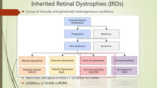
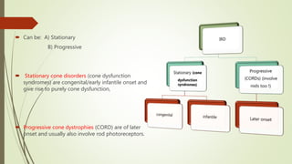
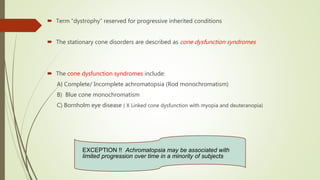
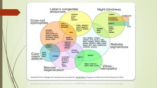
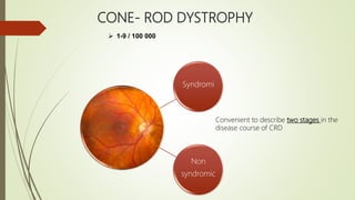
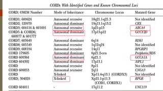
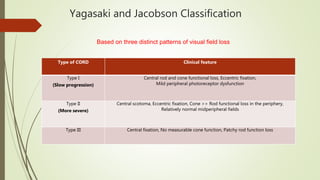
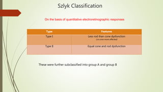
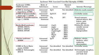
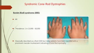
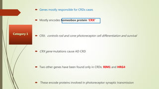
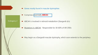
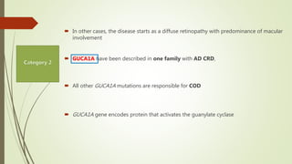
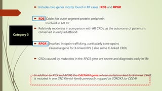
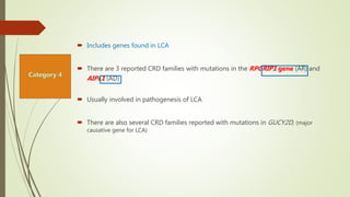
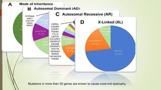
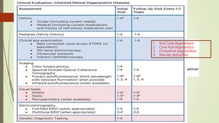
Cone and Rod Dystrophy
Presenter : Dr Samarth Mishra
Moderator: Dr Parveen Sen
Inherited Retinal Dystrophies (IRDs)
 Group of clinically and genetically heterogeneous conditions
 More than 250 genes in total ( > 32 Genes for CORD)
 Incidence: 1/ 20,000–1,00,000
 Can be: A) Stationary
B) Progressive
 Stationary cone disorders (cone dysfunction
syndromes) are congenital/early infantile onset and
give rise to purely cone dysfunction,
 Progressive cone dystrophies (CORD) are of later
onset and usually also involve rod photoreceptors.
 Term “dystrophy” reserved for progressive inherited conditions
 The stationary cone disorders are described as cone dysfunction syndromes
 The cone dysfunction syndromes include:
A) Complete/ Incomplete achromatopsia (Rod monochromatism)
B) Blue cone monochromatism
C) Bornholm eye disease ( X Linked cone dysfunction with myopia and deuteranopia)
EXCEPTION !! Achromatopsia may be associated with
limited progression over time in a minority of subjects
Achromatopsia
 “Rod monochromatism” / “Oligocone trichromacy“
 Presentation by 6 months, Incidence: 1 in 30,000
 Photophobia and nystagmus
 V/A < 20/200 for those with complete achromatopsia
 May be as good as 20/80 for those with incomplete achromatopsia
 V/A is usually stable overtime ( non progressive )
 This is in contrast to other cone dystrophies in which visual acuity progressively worsens
 In cone dystrophies, the cone cells are present and functional at birth. Unlike achromatopsia
 Central scotoma / Eccentric fixation
 Fundus : Narrowing of blood vessels, retinal pigment epithelium (RPE) mottling, or alteration of the
foveal reflex
 Scotopic FF-ERG: Normal (~75%) ( Limited number of photoreceptors within the fovea compared
with the entire retina )
 (mfERG) allows for more precise diagnostics ( Eliminates contribution of the extramacular retina )
Incomplete achromatopsia may have a phenotypic appearance similar to that of young patients with
early cone dystrophy
OCT:
Achromatopsia patients generally exhibit three features:
 Loss of the photoreceptor layer in the foveal region and disruption of the IS/OS junction
 Foveal hypoplasia
 Macular thinning
Genes responsible : CNGA3, CNGB3, GNAT2, and PDE6C
CONE- ROD DYSTROPHY
Syndromi
Non
syndromic
 1-9 / 100 000
Convenient to describe two stages in the
disease course of CRD
First Stage :
Symptoms
 Decreased V/A
 Discovered at school (In the first decade of life)
 Does not significantly improve with spectacles
 Deviated gaze (To project images on parafoveal regions that are less damaged)
 Intense photophobia ( Can be even before visual loss )
 Dyschromatopsia / Impaired colour vision
 Night blindness is not mentioned by patients
 Central scotomas, while periphery is spared (patients have no difficulties to move)
 Fundus shows pigment deposits and retinal atrophy in the macular region
 Retinal vessels normal/ moderately attenuated
 Temporal pallor ( accounts for the macular fibre bundle )
 Cone responses are more severely affected than rod responses
 ERG: Shift in implicit time of cone responses
 FFA/ FAF: May show involvement of peripheral retina
 At this stage, the question is to differentiate CRDs from macular dystrophies such as
Stargardt disease, cone dystrophies and other rare macular conditions !!
Second stage !!
 Nyctalopia becomes apparent
 Peripheral visual field loss progresses
 Difficulties to move autonomously
 V/A decrease to a level where reading is no longer possible
 Nystagmus (often present)
 At this stage, patients are legally blind (V/A <6/60), even though large parts of the peripheral
visual field remain preserved
Clinicalfeaturesingeneral
Visual field
 Central scotoma appears first,
preventing fluent reading
 Loss of peripheral vision follow
 Severe vision loss earlier than in
retinitis pigmentosa
 Visual field defects begin in the
pericentral region between 5-30
degrees from fixation
Fundus
 Bull’s-eye maculopathy
 Normal looking macula/ fine macular lesions
 There may be minor macular RPE atrophy
 Pigmentary deposits (resembling bone spicules), frequently in macular area
 Attenuation of the retinal vessels
• Temporal pallor/ Waxy pallor of the optic disc
• Various degrees of retinal atrophy
17
1
4
3
2
5
SCOTOPIC RESPONSES
ROD SYSTEM
PHOTOPIC RESPONSES
CONE SYSTEM
ERG REPORT
Rods
Photoreceptors+
Bipolar cells+ Muller
cells
Amacrine cells
Cones +
Bipolar cells
Cones
30 Hz Flicker
Rod response
Maximal response
Oscillatory potential
 Predominant involvement of photopic (cones) over scotopic (rods)
responses
 Earliest ERG finding: Delayed 30 Hz flicker implicit time
 Followed by deterioration of the 30 Hz flicker ERG amplitude
 Reduced a-wave and b-wave amplitudes of the single flash photopic
ERG
 Scotopic function is preserved in early disease, but is usually affected
in late disease.
19
CONE DYSTROPHY
NORMAL
CONE ROD DYSTROPHY
A B
=Achromatopsia
It is important to ascertain the diagnosis by repeating the examination one or two
years after it has been first established
 Full field ERG is the key test (Remember achromatopisa ERG normal ~ 75%)
 mfERG useful to follow the functionality of the central retina
Yagasaki and Jacobson Classification
Type of CORD Clinical feature
Type I
(Slow progression)
Central rod and cone functional loss, Eccentric fixation,
Mild peripheral photoreceptor dysfunction
Type II
(More severe)
Central scotoma, Eccentric fixation, Cone >> Rod functional loss in the periphery,
Relatively normal midperipheral fields
Type III Central fixation, No measurable cone function, Patchy rod function loss
Based on three distinct patterns of visual field loss
Szlyk Classification
Type Features
Type I Less rod than cone dysfunction
(i.e cone more affected)
Type II Equal cone and rod dysfunction
On the basis of quantitative electroretinographic responses
These were further subclassified into group A and group B
v
Syndromic Cone-Rod Dystrophies
Bardet Biedl syndrome (BBS)
 AR
 Prevalence: 1 in 13,000 - 60,000
 Classically described as a Rod-COD but many variants have been reported with a
prominent macular involvement indicating a Cone-Rod dystrophy
Bardet Biedl
syndrome
Retinal
dystrophy
Post-axial
Polydactyly
Obesity
Hypogonadism
Mental
retardation
Renal
abnormality
 Macular involvement with decreased VA and photophobia
 Foveo-macular hyperfluorescence on FFA
 Diagnosis of retinal dystrophy is often established in the first decade of life
 Legal blindness is reached before 20 years of age
 12 BBS genes encoding proteins involved in the cilium structure have been reported
Spinocerebellar Ataxia (Type 7)
 AD
 Spinocerebellar degeneration due to expansions of polyglutamine in the ataxin protein
 Initially, the disease often presents as an isolated retinal dystrophy
 Begins with granular macula
 Progressively spreads out to the whole retina, while the macula becomes atrophic
Ectodermal diseases
 Amelogenesis imperfecta
 Tooth enamel is abnormal.
 One form of amelogenesis imperfecta with AR inheritance is
associated with CRD and abnormally shaped teeth
 First reported in a large consanguineous Arabic family from the Gaza Strip
 AR
 The causative gene is CNNM4 (2q11) , which encodes a metal transporter
 The protein is localized in retina and teeth (specifically mainly in the enamel and
ameloblasts)
Hypotrichosis with Juvenile Macular Dystrophy:
 Rare form of AR alopecia a/w macular dystrophy
 In a few instances it has been reported to be a CRD
Dysmorphic syndromes
 CRDs reported in spondylometaphyseal dysplasia and cleft lip
Metabolic Dysfunctions
 CRD occurs in several metabolic disease (thiamine-responsive megaloblastic anemia )
 A case with a mitochondrial mutation (T8993G) also reported
Alport syndrome:
Fundus shows whitish flecks looking like crystals
around the macula rather than an authentic
pigmentary retinopathy
Mechanismofthedisease
Classification according to the genes involved
 Non syndromic CRDs are genetically heterogeneous
 3 Mendelian types of inheritance have been reported
 13 genes responsible for non syndromic CRDs
(10 cloned, 3 mapped)
 These genes can be classified into 4 categories
Etiology of Non
Syndromic
CRDs
Category
3
Category
2
Category
4
Category
1
CRX ABCA4
GUCA1A
RDS
RPGR
RPGRIP
AIPL1
 Genes mostly responsible for CRDs cases
 Mostly encodes homeobox protein ‘CRX’
 CRX: controls rod and cone photoreceptor cell differentiation and survival
 CRX gene mutations cause AD CRD
 Two other genes have been found only in CRDs: RIM1 and HRG4
 These encode proteins involved in photoreceptor synaptic transmission
 Genes mostly found in macular dystrophies
 Comprises essentially ABCA4
 ABCA4 is involved in retinoid metabolism (Stargardt d/s)
 Mutations in ABCA4: Responsible for 30-60% of AR CRDs
 May begin as a Stargardt macular dystrophy, which soon extends to the periphery
 In other cases, the disease starts as a diffuse retinopathy with predominance of macular
involvement
 GUCA1A have been described in one family with AD CRD,
 All other GUCA1A mutations are responsible for COD
 GUCA1A gene encodes protein that activates the guanylate cyclase
 Includes two genes mostly found in RP cases : RDS and RPGR
 RDS: Codes for outer segment protein peripherin
Involved in AD RP
 Relatively moderate in comparison with AR CRDs, as the autonomy of patients is
conserved in early adulthood
 RPGR : Involved in opsin trafficking, particularly cone opsins
Causative gene for X-linked RPs ( also some X-linked CRD)
 CRDs caused by mutations in the RPGR gene are severe and diagnosed early in life
In addition to RDS and RPGR, the CACNA1F gene, whose mutations lead to X-linked CSNB,
is mutated in one CRD Finnish family previously mapped as CORDX3 (or COD4)
 Includes genes found in LCA
 There are 3 reported CRD families with mutations in the RPGRIP1 gene (AR) and
AIPL1 (AD)
 Usually involved in pathogenesis of LCA
 There are also several CRD families reported with mutations in GUCY2D, (major
causative gene for LCA)
It seems that most genes responsible for CRDs are involved in other types of retinal
dystrophies, including RPs, macular dystrophies and cone dystrophies, therefore we can
place CRDs in the center of the vast panel of retinal dystrophies
So, any gene causing retinal dystrophy may potentially be involved in CRD pathogenesis
!!!
 Challenge is to understand the underlying mechanisms
Mutations in more than 30 genes are known to cause cone-rod dystrophy.
Specific examples of COD / CORD
 Molecular genetics
GUCA1A: 4 Exon gene
Encodes GCAP1(Guanylate cyclase activator protein)
GCAP1 activates retGC1 (retinal guanylate cyclase)
Required for cGMP regeneration
 Requires regulation in a Ca2+-dependent manner
 GCAP1 contains three Ca2+-binding EF-hand motifs, structural alterations to which occur in
most disease-causing GUCA1A sequence variants
 These include the gain-in-function variants p.(Tyr99Cys) ,16 p.(Glu155Gly) and p.(Asp100Gly)
GUCA1A associated Cod/Cord (AD)
 Large phenotypic variability seen in patients harbouring identical GUCA1A mutations
 Michaelides et al demonstrated the p.(Tyr99Cys) missense variant resulting in three
different dominantly inherited phenotypes in a non-consanguineous British family: COD,
CORD and isolated macular
How do GUCA1A associated COD/CORD present clinically ??
 2nd – 3rd decade
 Central vision reduction
 photophobia
 Color vision impairment / Dyschromatopsia
 ERG: Reduced cone single-flash and flicker amplitudes with a normal implicit time (an
unusual finding in generalised retinal disease)
 Rod function typically remains normal : Attributed to greater GCAP1 expression in cones
 Funduscopy: Mild RPE disturbance to extensive macular atrophy
 FAF: An increased signal at the fovea may be seen in early disease
 AOSLO: Has identified cellular variability between two related patients harbouring a
single 428delTinsAC AC insertion/ deletion variant
PRPH2-associated CORD ( AD)
 PRPH2: Five-exon gene
Encodes peripherin-2
A cell surface glycoprotein in OS with an essential role in disc
morphogenesis
 Function in disc stabilisation and maintenance of rim curvature
 CORD associated variants in PRPH2 can be attributed to the region
encoding the second intradiscal loop between its four transmembrane
components
 This contains cysteine residues essential for intraprotein folding and
interprotein interactions
 Identified missense variants in this region include p.(Asn244His), p.(Val200Glu)
and p.(Arg172Trp)
 p.(Asn244His) or p.(Val200Glu) variant: Present with early central RPE atrophy that advances
peripherally on disease progression, with little intrafamilial variability
 p.(Arg172Trp) variant: A) Vary substantially, ranging from non-penetrance to severe CORD
B) A/W other phenotypes, including RP, macular dystrophy
and central areolar choroidal dystrophy
How do PRPH2 associated COD/CORD present ??
 2nd -3rddecade with
 Reduced central vision,
 Photophobia and
 Nyctalopia.
 Certain genotype–phenotype correlations have been observed, including
p.(Arg172Trp)-PRPH2 retinopathy being associated with faster loss of visual acuity
than the p.(Arg172Gln) variant
 Fundus: Ranges from a bull’s eye maculopathy to macular atrophy
 FAF: Characteristic speckled macular appearance in most patients
 AOSLO: Reveals increased cone spacing throughout the macula with corresponding loss
of outer retinal structures
ABCA4-associated COD/CORD (AR)
 This protein has an essential role in the removal of N-retinylidene-
phosphatidyl-ethanolamine (PE) from the luminal to cytoplasmic
aspect of the OS disc membrane
If not exported and dissociated !!
 N-retinylidene-PE can accumulate in the OS to form the toxic
fluorophore, N-retinylidene-N-ret- inylethanolamine (A2E), a
component of lipofuscin
Molecular genetics
50-exon gene
Encodes a retina-specific ATP dependent cassette transporter
Located in the curved rim of the OS disc membrane
 ABCA4 : Most common genetic causes of IRD
Associated with vast phenotypic variability.
 Over 1000 disease- causing variants in ABCA4 have been identified to date, with
resulting phenotype varying between COD, CORD and Stargardt disease (STGD)
How do ABCA4 associated COD/ CORD present ??
 Symptomatic onset usually occurs in childhood
 Central scotoma and rapidly progressing macular atrophy
 Majority of patients have rod involvement at presentation (CORD)
 Associated with a worse prognosis
 Funduscopy: Initially normal fundus or mild retinal abnormalities (such as loss of foveal
reflex)
 peripheral degenerative changes occur later
 FAF: Bull’s eye maculopathy-like appearance with yellow-white retinal flecks
Increasing macular atrophy over time
 OCT : Loss of outer retinal architecture at the central macula
 AOSLO: Longitudinal increase in abnormal AF regions correlates with both visual
functional decline and abnormal cone spacing
RPGR-associated COD/CORD (XL)
Molecular genetics
19-exon gene
 Gives rise to two alternatively spliced retinal isoforms, encoded by exons 1–19 and
1–15
 The latter isoform, also known as exon open reading frame 15 (ORF15), is the
most highly expressed retinal variant and a mutational hot spot that accounts for
most XL COD and CORD cases
 C-terminal ORF15 sequence is implicated in intraflagellar protein transport
 Most disease-causing variants in RPGR result in RP, but those leading to
COD/CORD are preferentially sequestered at the 3’ end of the ORF15 region.
How do RPGR associated COD/CORD present ??
 2nd – 4th decade in affected men
 Central visual loss
 Mild photophobia
 Myopia
Higher levels of legal blindness was seen among RPGR associated COD/CORD than RPGR
associated RP by the age of 40 years
High myopia was predictive of faster visual decline in this study
FAF: parafoveal rings of increased signal
Management
 No proven treatments for COD/CORD that halt progression or restore lost vision
 Current management consists of:
 Symptomatic alleviation
 Refractive correction
 Use of tinted spectacles/contact lenses for photophobia
 LVA ( Hand held magnifiers, telescopes )
 Eccentric viewing
 Fresnel prisms
 Preferred retinal locus (PRL)
 White cane training
 Vocational counselling and training
 Educate patient and family on progression of disease
 Genetic counselling . . .
Molecular genetics:
 Retina has several key characteristics that render it as an ideal tissue for gene therapy
 Highly accessible for imaging and surgery
 It is enclosed, allowing administration of small amounts of therapeutic product
 It is sequestered from the systemic circulation by the blood-retinal-barrier, thus providing
it with an immune-privileged status
To facilitate genetic counselling,
Advice on prognosis
Participation in anticipated clinical trials
 Recent evidence has demonstrated the potential of gene therapy for long-term improvement in
COD/CORD visual outcomes
 Gene therapy encompasses :
gene replacement / gene editing / gene silencing
 Human treatment trials of gene replacement therapy are already under way for retinal disease
associated with mutations in ABCA4 ( ClinicalTrials.gov identifier: NCT01367444) and RPGR (NCT03252847,
NCT03116113 and NCT03316560), with results awaited
Points to remember !!
 In GUCA1A-associated retinopathy, sleeping with the lights on is advocated by
some clinicians for preventing accumulation of cGMP, which otherwise occurs at
night and causes photoreceptor damage
 In contrast, light avoidance using tinted spectacles may confer benefit in ABCA4-
associated retinopathy by inhibiting A2E production,which produces DNA-
damaging epoxides
 Vitamin A should also be avoided in ABCA4-associated retinopathy as it may
enhance A2E production and, therefore, disease progression.
OCT features !!!
 Oishi et al demonstrated an association between abnormal AF and the severity of functional
impairment
 On OCT, an absent interdigitation zone (IZ) is an early occurrence
 IZ is a band representing the interaction between apical processes of the RPE and
photoreceptor OS
 Progressive disruption and loss of the ellipsoid zone (EZ)
(EZ corresponds to the ellipsoid portion of the photoreceptor inner segment)
Irregular foveal loss of the ellipsoid portion of the IS
band
Central retinal thinning and segmental foveal loss of the IS
band
Central foveal thickening and irregular perifoveal loss of the IS band
Advanced disease : Outer retinal atrophy including the RPE is observed
Application of adaptive optics in CORD
 Exploration of phenotypic diversity in CORD has been transformed with the application of adaptive
optics (AO)
 AO Images human retina at a cellular
resolution by real time measurement
 Used to characterise and quantify the central macular photoreceptor mosaic
 Longitudinal AO imaging offers utility in measuring the rate of cone cell loss in progressive disease with
precision
Pre-natal diagnosis
 Include amniocentesis / chorionic biopsy
 Families in which the responsible gene has been identified
 Concerns:
Raises an ethical issue
Are invasive prenatal procedures justified in a non life-threatening disease ? ?
D/D of non syndromic CRDs with other pigmentary retinopathies
 Retinitis pigmentosa
A) Typical RP (rod cone dystrophy, RCD)
 In typical RCD, the diagnosis is easy because the first symptom is night blindness
 Remains isolated for several years with normal V/A before vision loss in day- light
becomes prominent
 Fundus: Pigment deposits located in the periphery
Leber congenital amaurosis (LCA)
 Both macula and peripheral retina are affected and degenerate rapidly from birth
 A/W high degree of visual impairment
 Appears either as a rod or cone predominant disease, or both
 Nystagmus, poor light fixation
 V/A <1/20
 Flat ERG
CRD and LCA share the same clinical signs
Presence of a lapse time of several years before dramatic worsening of the visual
disability will allow to classify the disease as CRD rather than LCA !!
Maculopathies
 Difficult to differentiate from end stage CRD / RP
 FF-ERG is key investigation
 Stargardt disease : Peripheral retina usually remains free of lesions
 However, there are extended lesions in some late stage Stargardt cases,
(Many CRD are caused by the "Stargardt gene", ABCA4)
 Early stage of the CRD may be similar to Stargardt disease, but, in a decade, signs of
peripheral involvement occur.
Pure Cone dystrophies
 Rods remain normal
 clinical signs: loss of V/A, photophobia, dyschromatopsia
 exclusive cone involvement at ERG
 In contrast to CRDs, rods remain at least partly spared at these late stages
(Non recordable in late stage CRD)
 Stationary retinal diseases
 This is essentially achromatopsia, which is diagnosed on the basis of mainly cone
involvement, the lack of disease evolution, and the normal fundus.
Enhanced ‘S’ Cone Syndrome
 Characteristic hypersensitivity of the short (S)‐wavelength‐sensitive cone photoreceptors
 Hemeralopia
 Fundus : Retinoschisis / intraretinal cysts/ yellow dots / RPE deposits
 presentation of ESCS is usually reported to be in the first decade
 similar waveform of the photopic and scotopic responses to the bright flash stimulus because of the
dominance of short‐wavelength sensitive mechanisms
 Delayed 30 Hz flicker ERG amplitude lower than that of the photopic a-wave.
mutations in the NR2E3 gene
Association with Goldmann‐Favre syndrome (GFS)
Mutations in PDE6H and in KCNV2
1. Rod Cone degeneration
2. Cone Rod degeneration
3. Chrioretinal degeneration
4. Macular dystrophies
Nil Financial interest
www.familyconnect.org
Other organisations :
 American Foundation for the Blind
 Retina International
 Foundation Fighting Blindness (Canada)
 Genetic and Rare Diseases (GARD) Information Center
 MD Support – The Eyes of the Macular Degeneration Community
 National Association for Parents of Children with Visual Impairments (NAPVI)
 National Federation of the Blind
 NIH/National Eye Institute

Cone and Rod Dystrophy

  • 1.
    Cone and RodDystrophy Presenter : Dr Samarth Mishra Moderator: Dr Parveen Sen
  • 2.
    Inherited Retinal Dystrophies(IRDs)  Group of clinically and genetically heterogeneous conditions  More than 250 genes in total ( > 32 Genes for CORD)  Incidence: 1/ 20,000–1,00,000
  • 3.
     Can be:A) Stationary B) Progressive  Stationary cone disorders (cone dysfunction syndromes) are congenital/early infantile onset and give rise to purely cone dysfunction,  Progressive cone dystrophies (CORD) are of later onset and usually also involve rod photoreceptors.
  • 4.
     Term “dystrophy”reserved for progressive inherited conditions  The stationary cone disorders are described as cone dysfunction syndromes  The cone dysfunction syndromes include: A) Complete/ Incomplete achromatopsia (Rod monochromatism) B) Blue cone monochromatism C) Bornholm eye disease ( X Linked cone dysfunction with myopia and deuteranopia) EXCEPTION !! Achromatopsia may be associated with limited progression over time in a minority of subjects
  • 6.
    Achromatopsia  “Rod monochromatism”/ “Oligocone trichromacy“  Presentation by 6 months, Incidence: 1 in 30,000  Photophobia and nystagmus  V/A < 20/200 for those with complete achromatopsia  May be as good as 20/80 for those with incomplete achromatopsia  V/A is usually stable overtime ( non progressive )  This is in contrast to other cone dystrophies in which visual acuity progressively worsens
  • 7.
     In conedystrophies, the cone cells are present and functional at birth. Unlike achromatopsia  Central scotoma / Eccentric fixation  Fundus : Narrowing of blood vessels, retinal pigment epithelium (RPE) mottling, or alteration of the foveal reflex  Scotopic FF-ERG: Normal (~75%) ( Limited number of photoreceptors within the fovea compared with the entire retina )  (mfERG) allows for more precise diagnostics ( Eliminates contribution of the extramacular retina ) Incomplete achromatopsia may have a phenotypic appearance similar to that of young patients with early cone dystrophy
  • 8.
    OCT: Achromatopsia patients generallyexhibit three features:  Loss of the photoreceptor layer in the foveal region and disruption of the IS/OS junction  Foveal hypoplasia  Macular thinning Genes responsible : CNGA3, CNGB3, GNAT2, and PDE6C
  • 9.
    CONE- ROD DYSTROPHY Syndromi Non syndromic 1-9 / 100 000 Convenient to describe two stages in the disease course of CRD
  • 10.
    First Stage : Symptoms Decreased V/A  Discovered at school (In the first decade of life)  Does not significantly improve with spectacles  Deviated gaze (To project images on parafoveal regions that are less damaged)  Intense photophobia ( Can be even before visual loss )  Dyschromatopsia / Impaired colour vision  Night blindness is not mentioned by patients  Central scotomas, while periphery is spared (patients have no difficulties to move)  Fundus shows pigment deposits and retinal atrophy in the macular region
  • 11.
     Retinal vesselsnormal/ moderately attenuated  Temporal pallor ( accounts for the macular fibre bundle )  Cone responses are more severely affected than rod responses  ERG: Shift in implicit time of cone responses  FFA/ FAF: May show involvement of peripheral retina  At this stage, the question is to differentiate CRDs from macular dystrophies such as Stargardt disease, cone dystrophies and other rare macular conditions !!
  • 12.
    Second stage !! Nyctalopia becomes apparent  Peripheral visual field loss progresses  Difficulties to move autonomously  V/A decrease to a level where reading is no longer possible  Nystagmus (often present)  At this stage, patients are legally blind (V/A <6/60), even though large parts of the peripheral visual field remain preserved
  • 13.
  • 14.
    Visual field  Centralscotoma appears first, preventing fluent reading  Loss of peripheral vision follow  Severe vision loss earlier than in retinitis pigmentosa  Visual field defects begin in the pericentral region between 5-30 degrees from fixation
  • 15.
    Fundus  Bull’s-eye maculopathy Normal looking macula/ fine macular lesions  There may be minor macular RPE atrophy  Pigmentary deposits (resembling bone spicules), frequently in macular area  Attenuation of the retinal vessels • Temporal pallor/ Waxy pallor of the optic disc • Various degrees of retinal atrophy
  • 17.
    17 1 4 3 2 5 SCOTOPIC RESPONSES ROD SYSTEM PHOTOPICRESPONSES CONE SYSTEM ERG REPORT Rods Photoreceptors+ Bipolar cells+ Muller cells Amacrine cells Cones + Bipolar cells Cones 30 Hz Flicker Rod response Maximal response Oscillatory potential
  • 18.
     Predominant involvementof photopic (cones) over scotopic (rods) responses  Earliest ERG finding: Delayed 30 Hz flicker implicit time  Followed by deterioration of the 30 Hz flicker ERG amplitude  Reduced a-wave and b-wave amplitudes of the single flash photopic ERG  Scotopic function is preserved in early disease, but is usually affected in late disease.
  • 19.
  • 20.
  • 21.
    It is importantto ascertain the diagnosis by repeating the examination one or two years after it has been first established  Full field ERG is the key test (Remember achromatopisa ERG normal ~ 75%)  mfERG useful to follow the functionality of the central retina
  • 22.
    Yagasaki and JacobsonClassification Type of CORD Clinical feature Type I (Slow progression) Central rod and cone functional loss, Eccentric fixation, Mild peripheral photoreceptor dysfunction Type II (More severe) Central scotoma, Eccentric fixation, Cone >> Rod functional loss in the periphery, Relatively normal midperipheral fields Type III Central fixation, No measurable cone function, Patchy rod function loss Based on three distinct patterns of visual field loss
  • 23.
    Szlyk Classification Type Features TypeI Less rod than cone dysfunction (i.e cone more affected) Type II Equal cone and rod dysfunction On the basis of quantitative electroretinographic responses These were further subclassified into group A and group B
  • 24.
  • 25.
    Syndromic Cone-Rod Dystrophies BardetBiedl syndrome (BBS)  AR  Prevalence: 1 in 13,000 - 60,000  Classically described as a Rod-COD but many variants have been reported with a prominent macular involvement indicating a Cone-Rod dystrophy
  • 26.
  • 27.
     Macular involvementwith decreased VA and photophobia  Foveo-macular hyperfluorescence on FFA  Diagnosis of retinal dystrophy is often established in the first decade of life  Legal blindness is reached before 20 years of age  12 BBS genes encoding proteins involved in the cilium structure have been reported
  • 28.
    Spinocerebellar Ataxia (Type7)  AD  Spinocerebellar degeneration due to expansions of polyglutamine in the ataxin protein  Initially, the disease often presents as an isolated retinal dystrophy  Begins with granular macula  Progressively spreads out to the whole retina, while the macula becomes atrophic
  • 30.
    Ectodermal diseases  Amelogenesisimperfecta  Tooth enamel is abnormal.  One form of amelogenesis imperfecta with AR inheritance is associated with CRD and abnormally shaped teeth
  • 31.
     First reportedin a large consanguineous Arabic family from the Gaza Strip  AR  The causative gene is CNNM4 (2q11) , which encodes a metal transporter  The protein is localized in retina and teeth (specifically mainly in the enamel and ameloblasts)
  • 32.
    Hypotrichosis with JuvenileMacular Dystrophy:  Rare form of AR alopecia a/w macular dystrophy  In a few instances it has been reported to be a CRD
  • 33.
    Dysmorphic syndromes  CRDsreported in spondylometaphyseal dysplasia and cleft lip
  • 34.
    Metabolic Dysfunctions  CRDoccurs in several metabolic disease (thiamine-responsive megaloblastic anemia )  A case with a mitochondrial mutation (T8993G) also reported
  • 35.
    Alport syndrome: Fundus showswhitish flecks looking like crystals around the macula rather than an authentic pigmentary retinopathy
  • 36.
  • 39.
    Classification according tothe genes involved  Non syndromic CRDs are genetically heterogeneous  3 Mendelian types of inheritance have been reported  13 genes responsible for non syndromic CRDs (10 cloned, 3 mapped)  These genes can be classified into 4 categories Etiology of Non Syndromic CRDs Category 3 Category 2 Category 4 Category 1 CRX ABCA4 GUCA1A RDS RPGR RPGRIP AIPL1
  • 40.
     Genes mostlyresponsible for CRDs cases  Mostly encodes homeobox protein ‘CRX’  CRX: controls rod and cone photoreceptor cell differentiation and survival  CRX gene mutations cause AD CRD  Two other genes have been found only in CRDs: RIM1 and HRG4  These encode proteins involved in photoreceptor synaptic transmission
  • 41.
     Genes mostlyfound in macular dystrophies  Comprises essentially ABCA4  ABCA4 is involved in retinoid metabolism (Stargardt d/s)  Mutations in ABCA4: Responsible for 30-60% of AR CRDs  May begin as a Stargardt macular dystrophy, which soon extends to the periphery
  • 42.
     In othercases, the disease starts as a diffuse retinopathy with predominance of macular involvement  GUCA1A have been described in one family with AD CRD,  All other GUCA1A mutations are responsible for COD  GUCA1A gene encodes protein that activates the guanylate cyclase
  • 43.
     Includes twogenes mostly found in RP cases : RDS and RPGR  RDS: Codes for outer segment protein peripherin Involved in AD RP  Relatively moderate in comparison with AR CRDs, as the autonomy of patients is conserved in early adulthood  RPGR : Involved in opsin trafficking, particularly cone opsins Causative gene for X-linked RPs ( also some X-linked CRD)  CRDs caused by mutations in the RPGR gene are severe and diagnosed early in life In addition to RDS and RPGR, the CACNA1F gene, whose mutations lead to X-linked CSNB, is mutated in one CRD Finnish family previously mapped as CORDX3 (or COD4)
  • 44.
     Includes genesfound in LCA  There are 3 reported CRD families with mutations in the RPGRIP1 gene (AR) and AIPL1 (AD)  Usually involved in pathogenesis of LCA  There are also several CRD families reported with mutations in GUCY2D, (major causative gene for LCA)
  • 45.
    It seems thatmost genes responsible for CRDs are involved in other types of retinal dystrophies, including RPs, macular dystrophies and cone dystrophies, therefore we can place CRDs in the center of the vast panel of retinal dystrophies So, any gene causing retinal dystrophy may potentially be involved in CRD pathogenesis !!!  Challenge is to understand the underlying mechanisms
  • 46.
    Mutations in morethan 30 genes are known to cause cone-rod dystrophy.
  • 47.
    Specific examples ofCOD / CORD  Molecular genetics GUCA1A: 4 Exon gene Encodes GCAP1(Guanylate cyclase activator protein) GCAP1 activates retGC1 (retinal guanylate cyclase) Required for cGMP regeneration  Requires regulation in a Ca2+-dependent manner  GCAP1 contains three Ca2+-binding EF-hand motifs, structural alterations to which occur in most disease-causing GUCA1A sequence variants  These include the gain-in-function variants p.(Tyr99Cys) ,16 p.(Glu155Gly) and p.(Asp100Gly) GUCA1A associated Cod/Cord (AD)
  • 48.
     Large phenotypicvariability seen in patients harbouring identical GUCA1A mutations  Michaelides et al demonstrated the p.(Tyr99Cys) missense variant resulting in three different dominantly inherited phenotypes in a non-consanguineous British family: COD, CORD and isolated macular
  • 49.
    How do GUCA1Aassociated COD/CORD present clinically ??  2nd – 3rd decade  Central vision reduction  photophobia  Color vision impairment / Dyschromatopsia  ERG: Reduced cone single-flash and flicker amplitudes with a normal implicit time (an unusual finding in generalised retinal disease)  Rod function typically remains normal : Attributed to greater GCAP1 expression in cones
  • 50.
     Funduscopy: MildRPE disturbance to extensive macular atrophy  FAF: An increased signal at the fovea may be seen in early disease  AOSLO: Has identified cellular variability between two related patients harbouring a single 428delTinsAC AC insertion/ deletion variant
  • 51.
    PRPH2-associated CORD (AD)  PRPH2: Five-exon gene Encodes peripherin-2 A cell surface glycoprotein in OS with an essential role in disc morphogenesis  Function in disc stabilisation and maintenance of rim curvature  CORD associated variants in PRPH2 can be attributed to the region encoding the second intradiscal loop between its four transmembrane components  This contains cysteine residues essential for intraprotein folding and interprotein interactions  Identified missense variants in this region include p.(Asn244His), p.(Val200Glu) and p.(Arg172Trp)
  • 52.
     p.(Asn244His) orp.(Val200Glu) variant: Present with early central RPE atrophy that advances peripherally on disease progression, with little intrafamilial variability  p.(Arg172Trp) variant: A) Vary substantially, ranging from non-penetrance to severe CORD B) A/W other phenotypes, including RP, macular dystrophy and central areolar choroidal dystrophy
  • 53.
    How do PRPH2associated COD/CORD present ??  2nd -3rddecade with  Reduced central vision,  Photophobia and  Nyctalopia.  Certain genotype–phenotype correlations have been observed, including p.(Arg172Trp)-PRPH2 retinopathy being associated with faster loss of visual acuity than the p.(Arg172Gln) variant
  • 54.
     Fundus: Rangesfrom a bull’s eye maculopathy to macular atrophy  FAF: Characteristic speckled macular appearance in most patients  AOSLO: Reveals increased cone spacing throughout the macula with corresponding loss of outer retinal structures
  • 55.
    ABCA4-associated COD/CORD (AR) This protein has an essential role in the removal of N-retinylidene- phosphatidyl-ethanolamine (PE) from the luminal to cytoplasmic aspect of the OS disc membrane If not exported and dissociated !!  N-retinylidene-PE can accumulate in the OS to form the toxic fluorophore, N-retinylidene-N-ret- inylethanolamine (A2E), a component of lipofuscin Molecular genetics 50-exon gene Encodes a retina-specific ATP dependent cassette transporter Located in the curved rim of the OS disc membrane
  • 56.
     ABCA4 :Most common genetic causes of IRD Associated with vast phenotypic variability.  Over 1000 disease- causing variants in ABCA4 have been identified to date, with resulting phenotype varying between COD, CORD and Stargardt disease (STGD)
  • 57.
    How do ABCA4associated COD/ CORD present ??  Symptomatic onset usually occurs in childhood  Central scotoma and rapidly progressing macular atrophy  Majority of patients have rod involvement at presentation (CORD)  Associated with a worse prognosis
  • 58.
     Funduscopy: Initiallynormal fundus or mild retinal abnormalities (such as loss of foveal reflex)  peripheral degenerative changes occur later  FAF: Bull’s eye maculopathy-like appearance with yellow-white retinal flecks Increasing macular atrophy over time  OCT : Loss of outer retinal architecture at the central macula  AOSLO: Longitudinal increase in abnormal AF regions correlates with both visual functional decline and abnormal cone spacing
  • 59.
    RPGR-associated COD/CORD (XL) Moleculargenetics 19-exon gene  Gives rise to two alternatively spliced retinal isoforms, encoded by exons 1–19 and 1–15  The latter isoform, also known as exon open reading frame 15 (ORF15), is the most highly expressed retinal variant and a mutational hot spot that accounts for most XL COD and CORD cases  C-terminal ORF15 sequence is implicated in intraflagellar protein transport  Most disease-causing variants in RPGR result in RP, but those leading to COD/CORD are preferentially sequestered at the 3’ end of the ORF15 region.
  • 60.
    How do RPGRassociated COD/CORD present ??  2nd – 4th decade in affected men  Central visual loss  Mild photophobia  Myopia Higher levels of legal blindness was seen among RPGR associated COD/CORD than RPGR associated RP by the age of 40 years High myopia was predictive of faster visual decline in this study FAF: parafoveal rings of increased signal
  • 61.
    Management  No proventreatments for COD/CORD that halt progression or restore lost vision  Current management consists of:  Symptomatic alleviation  Refractive correction  Use of tinted spectacles/contact lenses for photophobia  LVA ( Hand held magnifiers, telescopes )  Eccentric viewing  Fresnel prisms  Preferred retinal locus (PRL)  White cane training  Vocational counselling and training  Educate patient and family on progression of disease  Genetic counselling . . .
  • 62.
    Molecular genetics:  Retinahas several key characteristics that render it as an ideal tissue for gene therapy  Highly accessible for imaging and surgery  It is enclosed, allowing administration of small amounts of therapeutic product  It is sequestered from the systemic circulation by the blood-retinal-barrier, thus providing it with an immune-privileged status To facilitate genetic counselling, Advice on prognosis Participation in anticipated clinical trials
  • 64.
     Recent evidencehas demonstrated the potential of gene therapy for long-term improvement in COD/CORD visual outcomes  Gene therapy encompasses : gene replacement / gene editing / gene silencing  Human treatment trials of gene replacement therapy are already under way for retinal disease associated with mutations in ABCA4 ( ClinicalTrials.gov identifier: NCT01367444) and RPGR (NCT03252847, NCT03116113 and NCT03316560), with results awaited
  • 65.
    Points to remember!!  In GUCA1A-associated retinopathy, sleeping with the lights on is advocated by some clinicians for preventing accumulation of cGMP, which otherwise occurs at night and causes photoreceptor damage  In contrast, light avoidance using tinted spectacles may confer benefit in ABCA4- associated retinopathy by inhibiting A2E production,which produces DNA- damaging epoxides  Vitamin A should also be avoided in ABCA4-associated retinopathy as it may enhance A2E production and, therefore, disease progression.
  • 66.
    OCT features !!! Oishi et al demonstrated an association between abnormal AF and the severity of functional impairment  On OCT, an absent interdigitation zone (IZ) is an early occurrence  IZ is a band representing the interaction between apical processes of the RPE and photoreceptor OS  Progressive disruption and loss of the ellipsoid zone (EZ) (EZ corresponds to the ellipsoid portion of the photoreceptor inner segment)
  • 68.
    Irregular foveal lossof the ellipsoid portion of the IS band Central retinal thinning and segmental foveal loss of the IS band Central foveal thickening and irregular perifoveal loss of the IS band Advanced disease : Outer retinal atrophy including the RPE is observed
  • 69.
    Application of adaptiveoptics in CORD  Exploration of phenotypic diversity in CORD has been transformed with the application of adaptive optics (AO)  AO Images human retina at a cellular resolution by real time measurement  Used to characterise and quantify the central macular photoreceptor mosaic  Longitudinal AO imaging offers utility in measuring the rate of cone cell loss in progressive disease with precision
  • 70.
    Pre-natal diagnosis  Includeamniocentesis / chorionic biopsy  Families in which the responsible gene has been identified  Concerns: Raises an ethical issue Are invasive prenatal procedures justified in a non life-threatening disease ? ?
  • 71.
    D/D of nonsyndromic CRDs with other pigmentary retinopathies  Retinitis pigmentosa A) Typical RP (rod cone dystrophy, RCD)  In typical RCD, the diagnosis is easy because the first symptom is night blindness  Remains isolated for several years with normal V/A before vision loss in day- light becomes prominent  Fundus: Pigment deposits located in the periphery
  • 72.
    Leber congenital amaurosis(LCA)  Both macula and peripheral retina are affected and degenerate rapidly from birth  A/W high degree of visual impairment  Appears either as a rod or cone predominant disease, or both  Nystagmus, poor light fixation  V/A <1/20  Flat ERG CRD and LCA share the same clinical signs Presence of a lapse time of several years before dramatic worsening of the visual disability will allow to classify the disease as CRD rather than LCA !!
  • 73.
    Maculopathies  Difficult todifferentiate from end stage CRD / RP  FF-ERG is key investigation
  • 74.
     Stargardt disease: Peripheral retina usually remains free of lesions  However, there are extended lesions in some late stage Stargardt cases, (Many CRD are caused by the "Stargardt gene", ABCA4)  Early stage of the CRD may be similar to Stargardt disease, but, in a decade, signs of peripheral involvement occur.
  • 75.
    Pure Cone dystrophies Rods remain normal  clinical signs: loss of V/A, photophobia, dyschromatopsia  exclusive cone involvement at ERG  In contrast to CRDs, rods remain at least partly spared at these late stages (Non recordable in late stage CRD)  Stationary retinal diseases  This is essentially achromatopsia, which is diagnosed on the basis of mainly cone involvement, the lack of disease evolution, and the normal fundus.
  • 76.
    Enhanced ‘S’ ConeSyndrome  Characteristic hypersensitivity of the short (S)‐wavelength‐sensitive cone photoreceptors  Hemeralopia  Fundus : Retinoschisis / intraretinal cysts/ yellow dots / RPE deposits  presentation of ESCS is usually reported to be in the first decade  similar waveform of the photopic and scotopic responses to the bright flash stimulus because of the dominance of short‐wavelength sensitive mechanisms  Delayed 30 Hz flicker ERG amplitude lower than that of the photopic a-wave. mutations in the NR2E3 gene Association with Goldmann‐Favre syndrome (GFS)
  • 77.
    Mutations in PDE6Hand in KCNV2
  • 78.
    1. Rod Conedegeneration 2. Cone Rod degeneration 3. Chrioretinal degeneration 4. Macular dystrophies
  • 80.
  • 81.
    Other organisations : American Foundation for the Blind  Retina International  Foundation Fighting Blindness (Canada)  Genetic and Rare Diseases (GARD) Information Center  MD Support – The Eyes of the Macular Degeneration Community  National Association for Parents of Children with Visual Impairments (NAPVI)  National Federation of the Blind  NIH/National Eye Institute