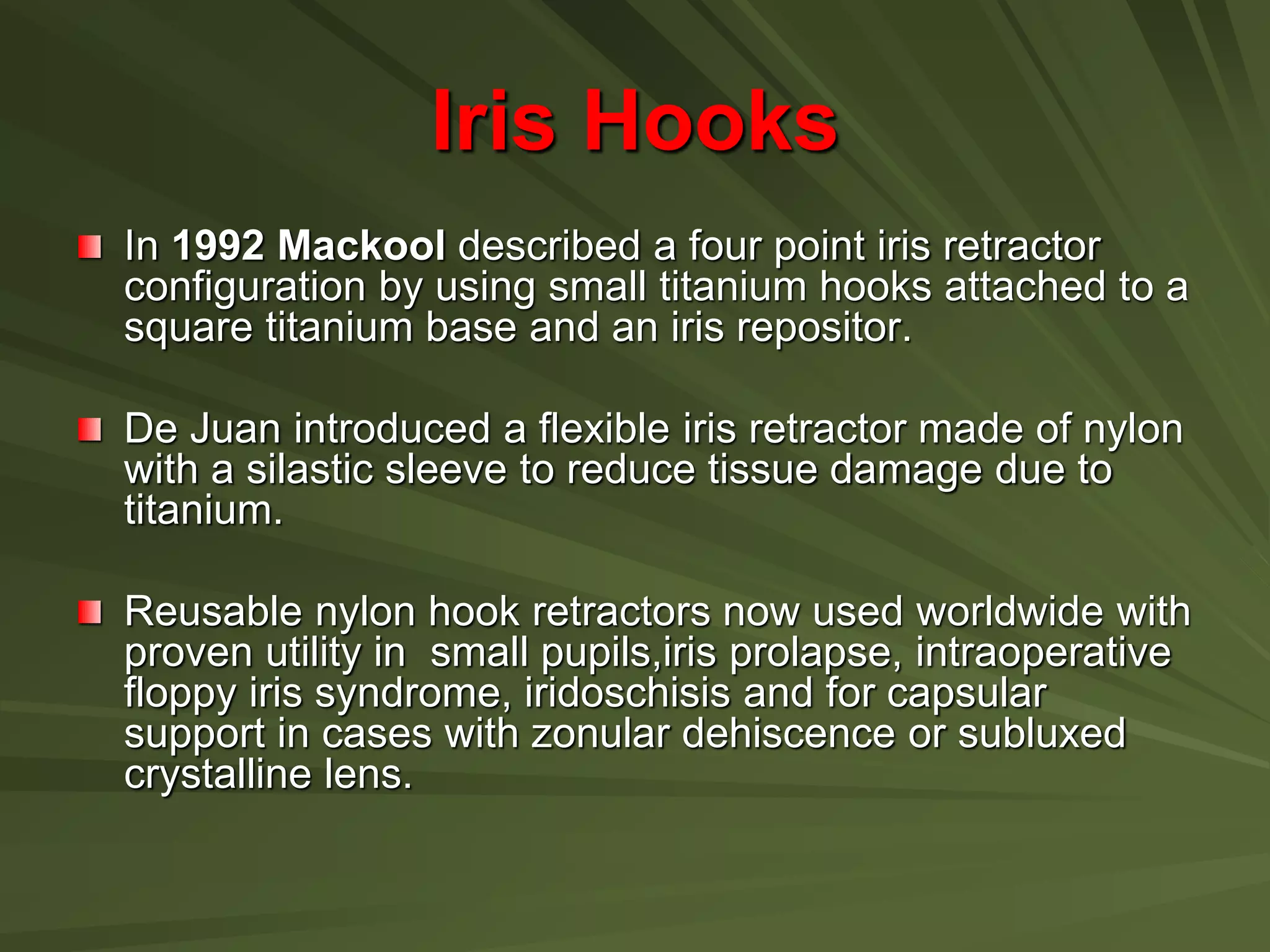
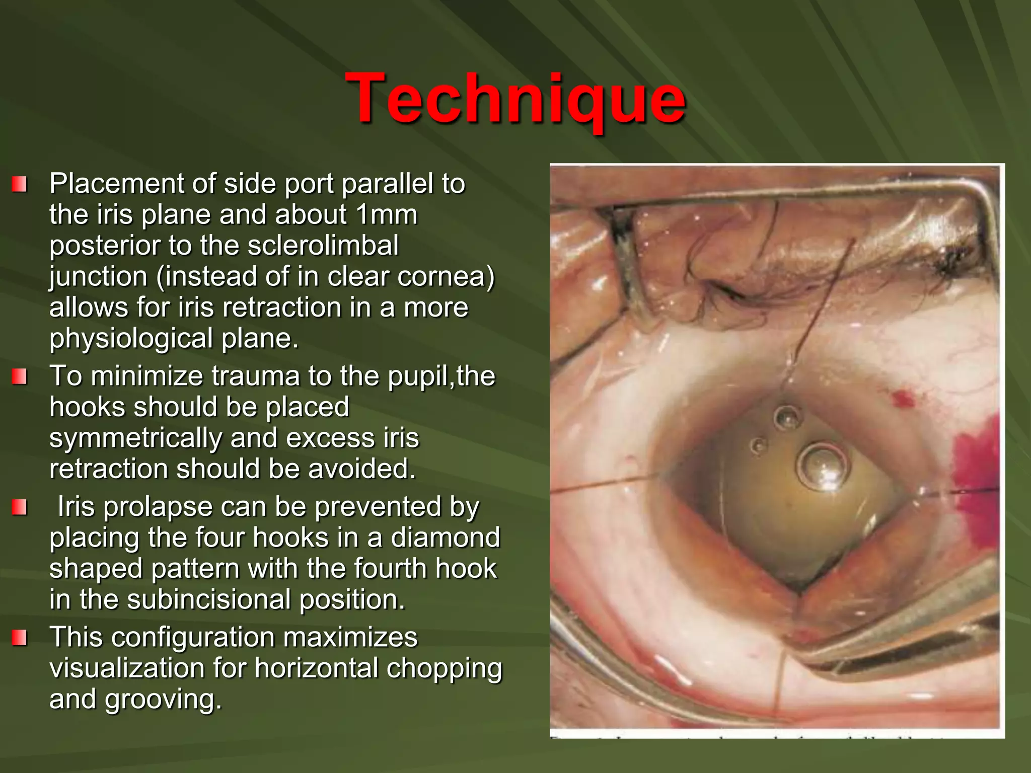
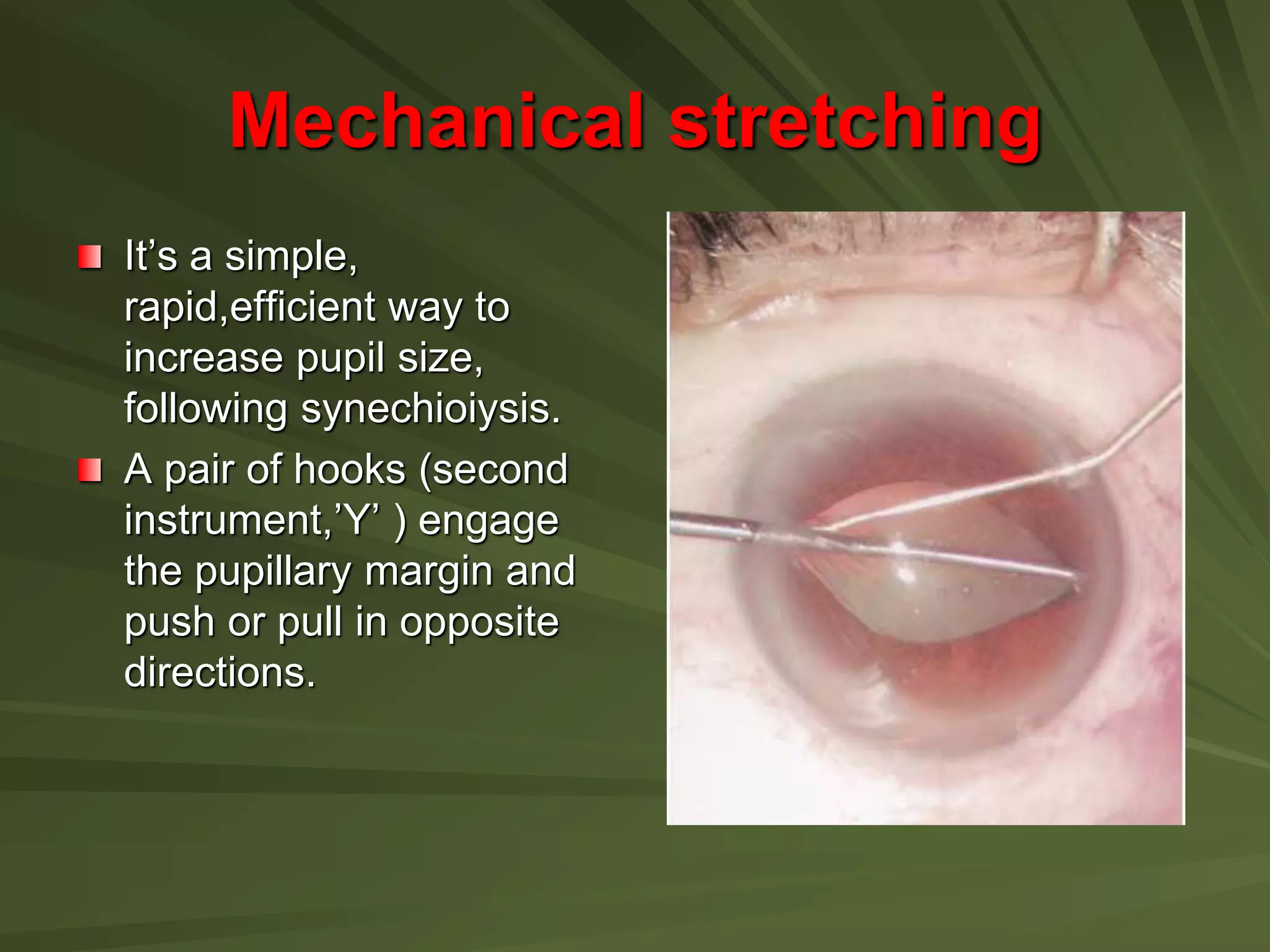
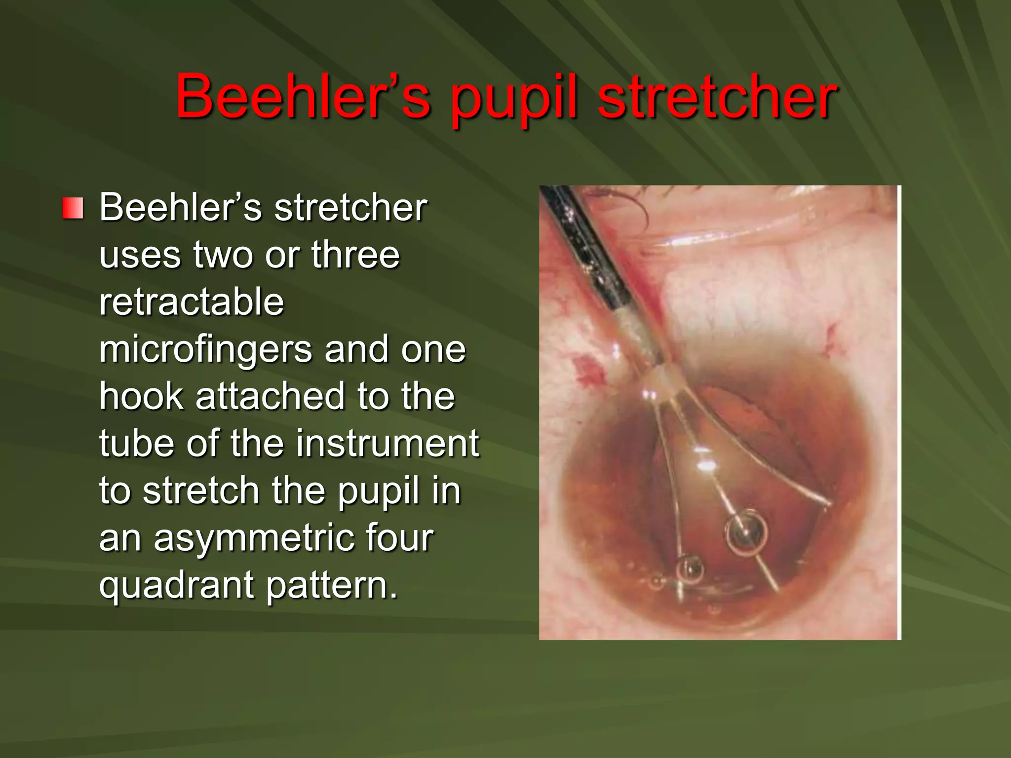
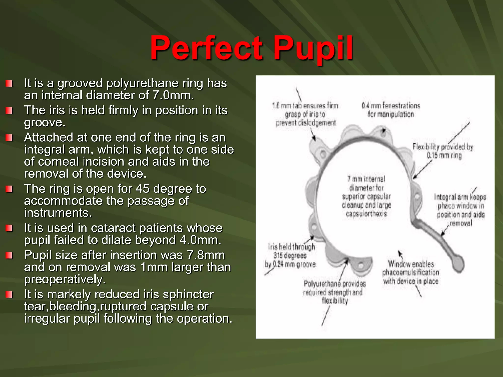
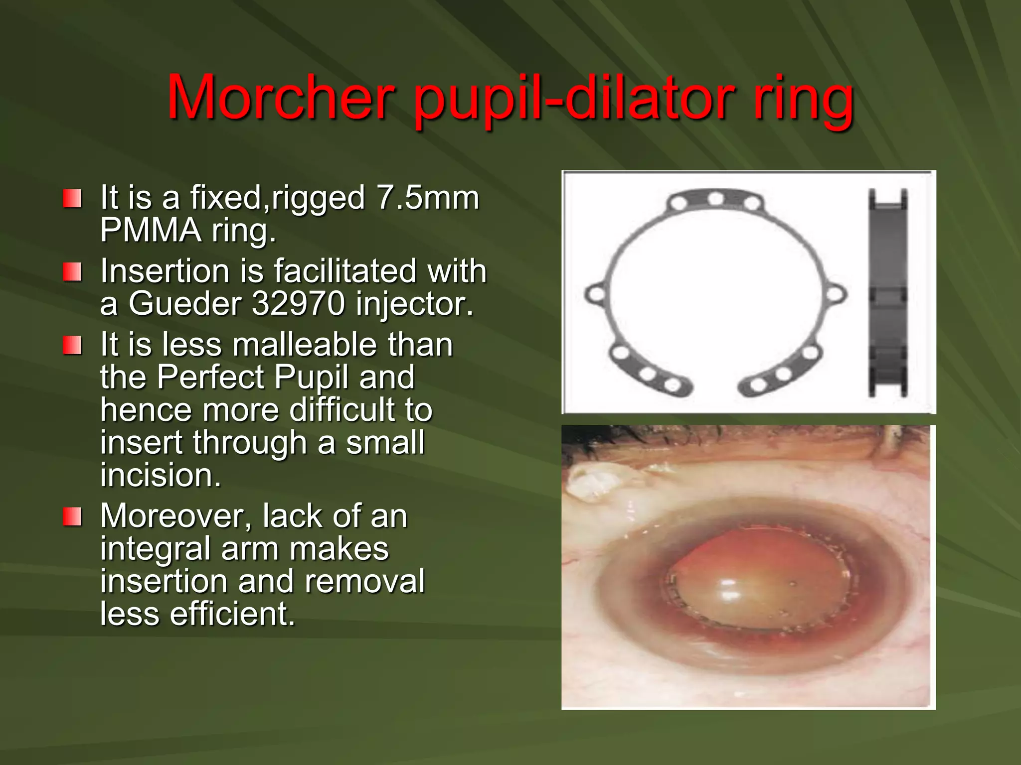
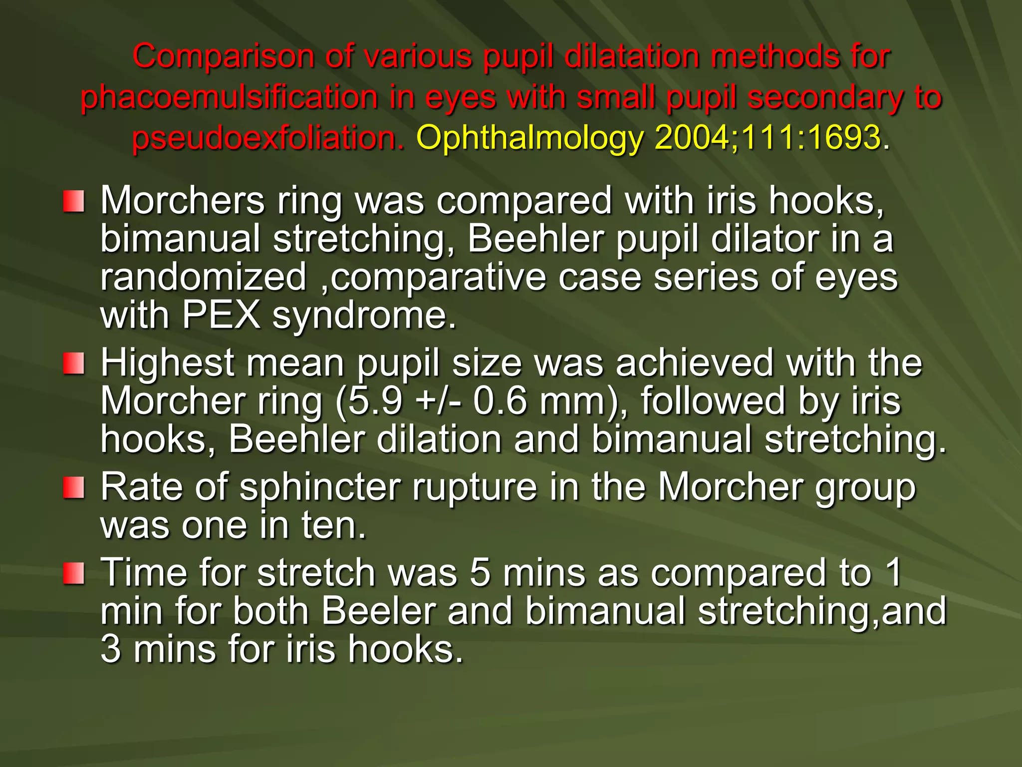
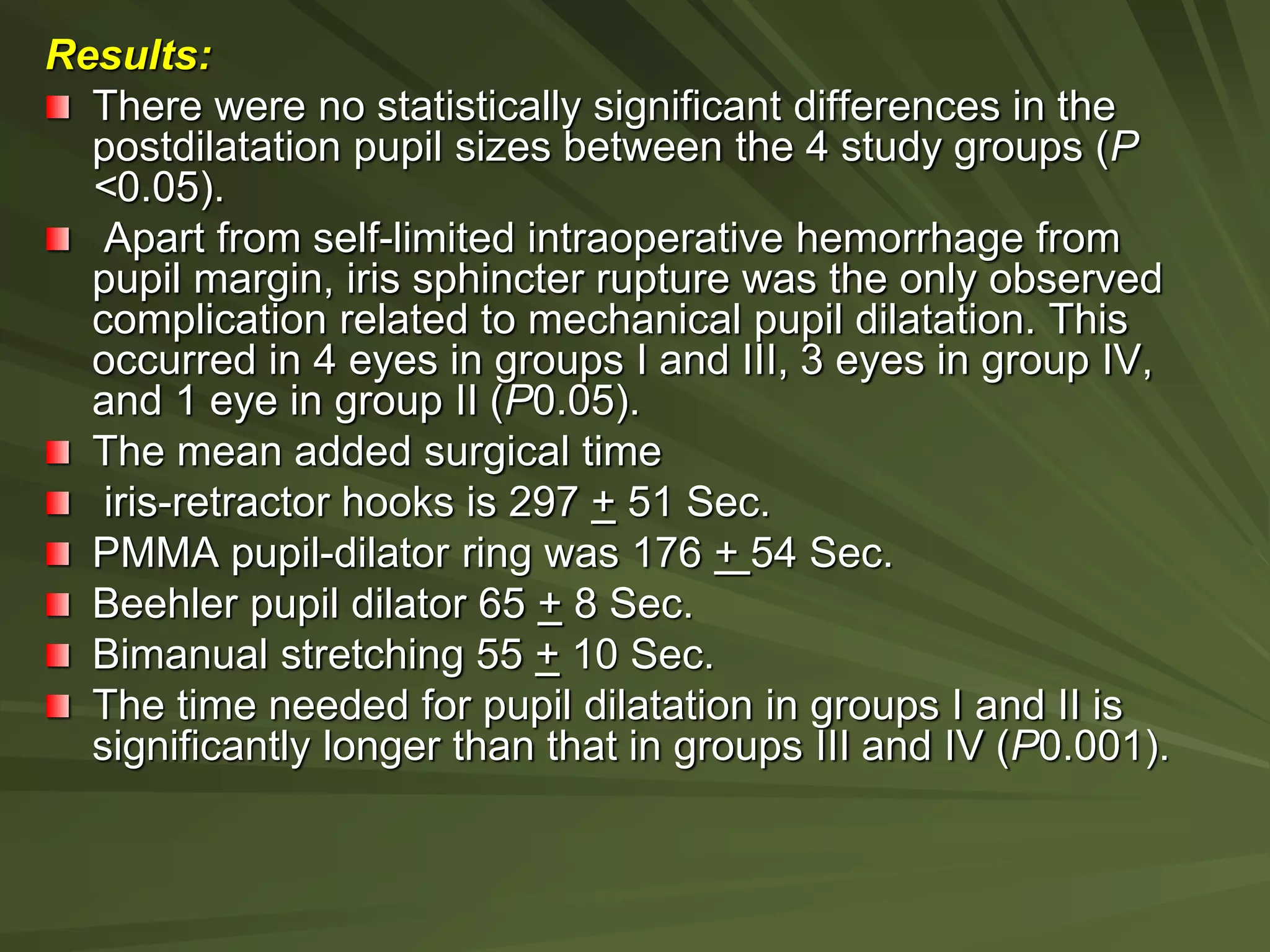
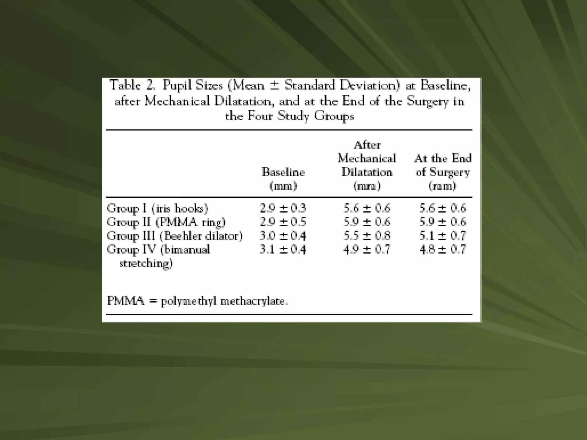
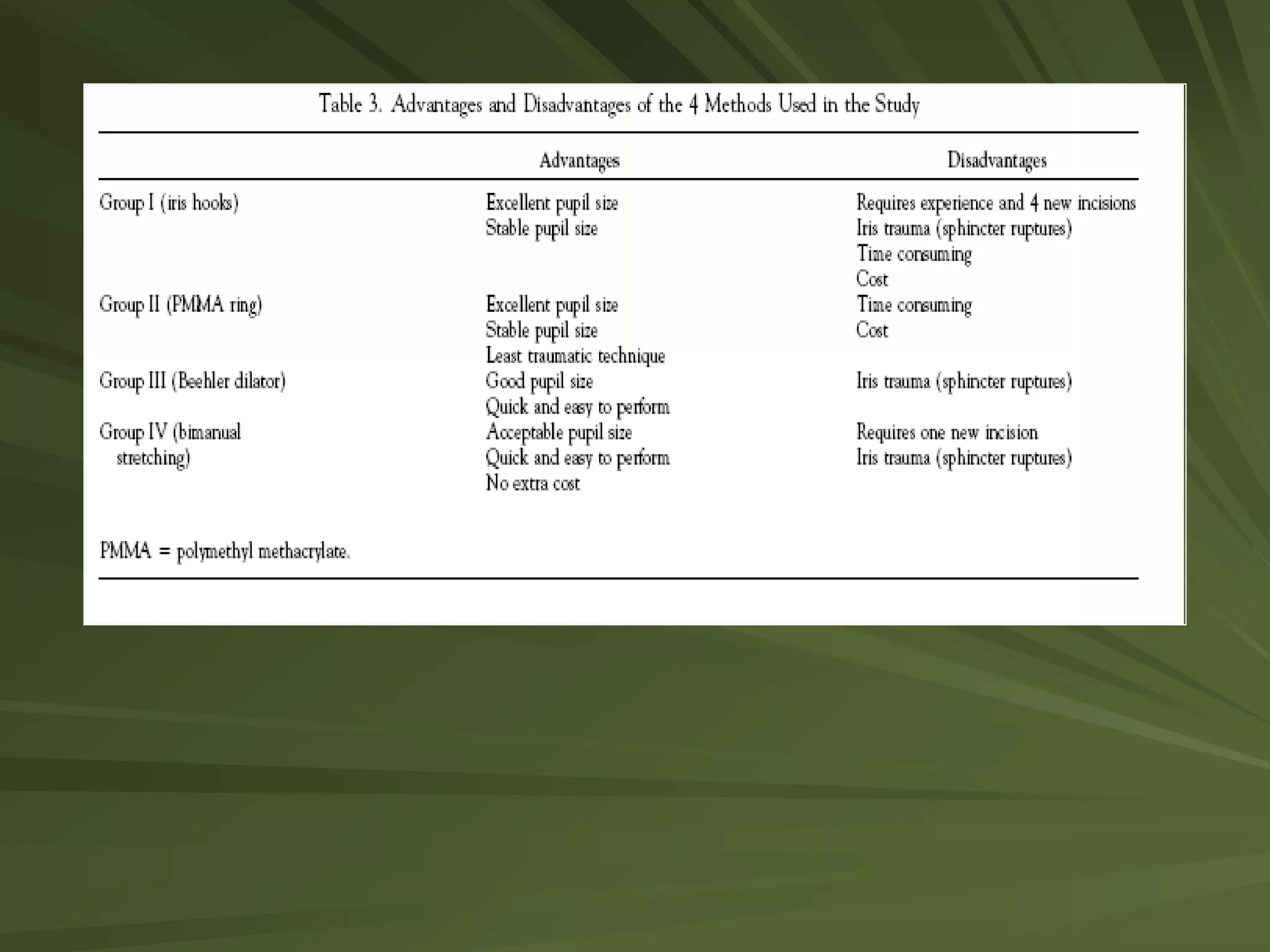
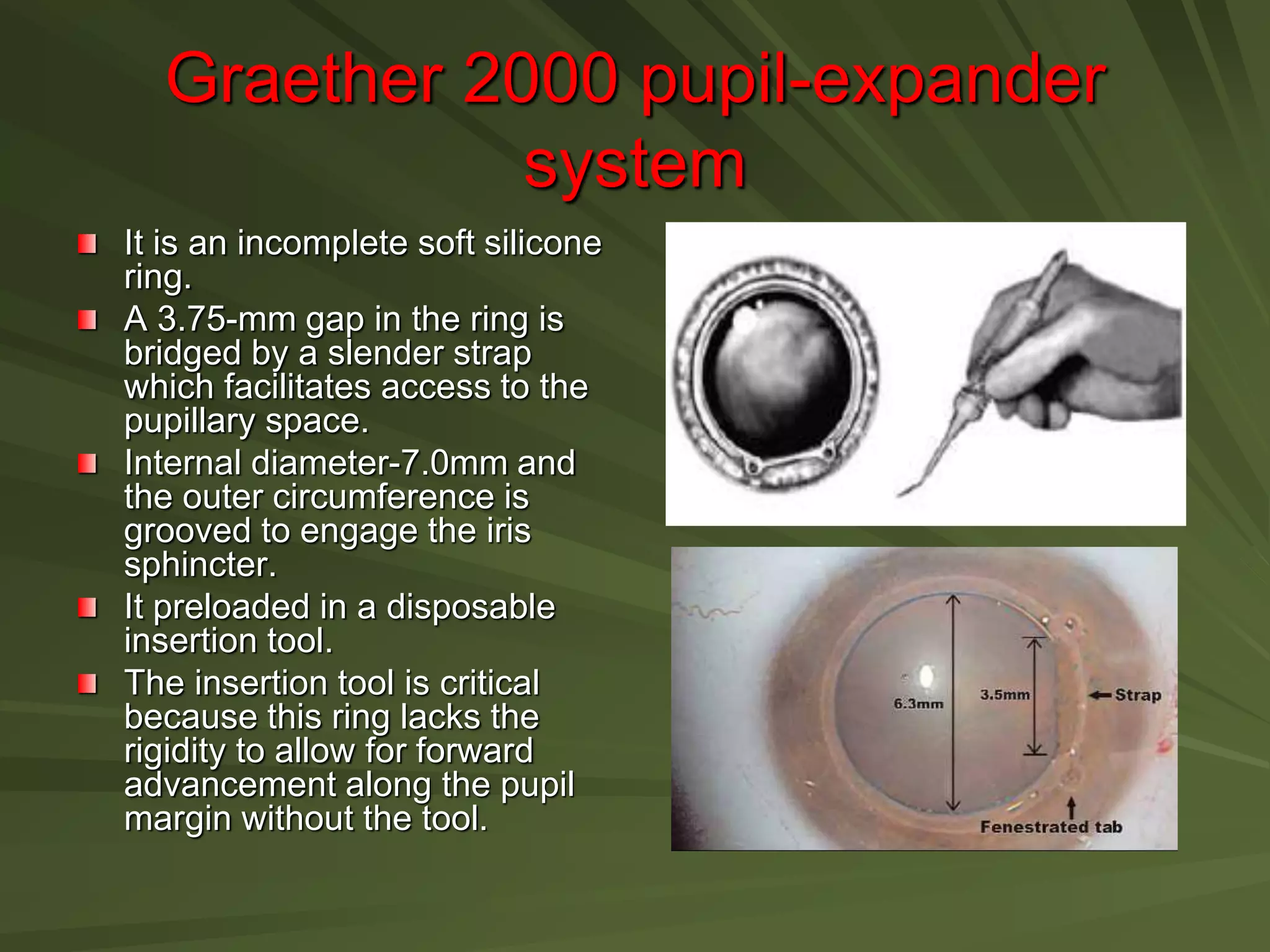
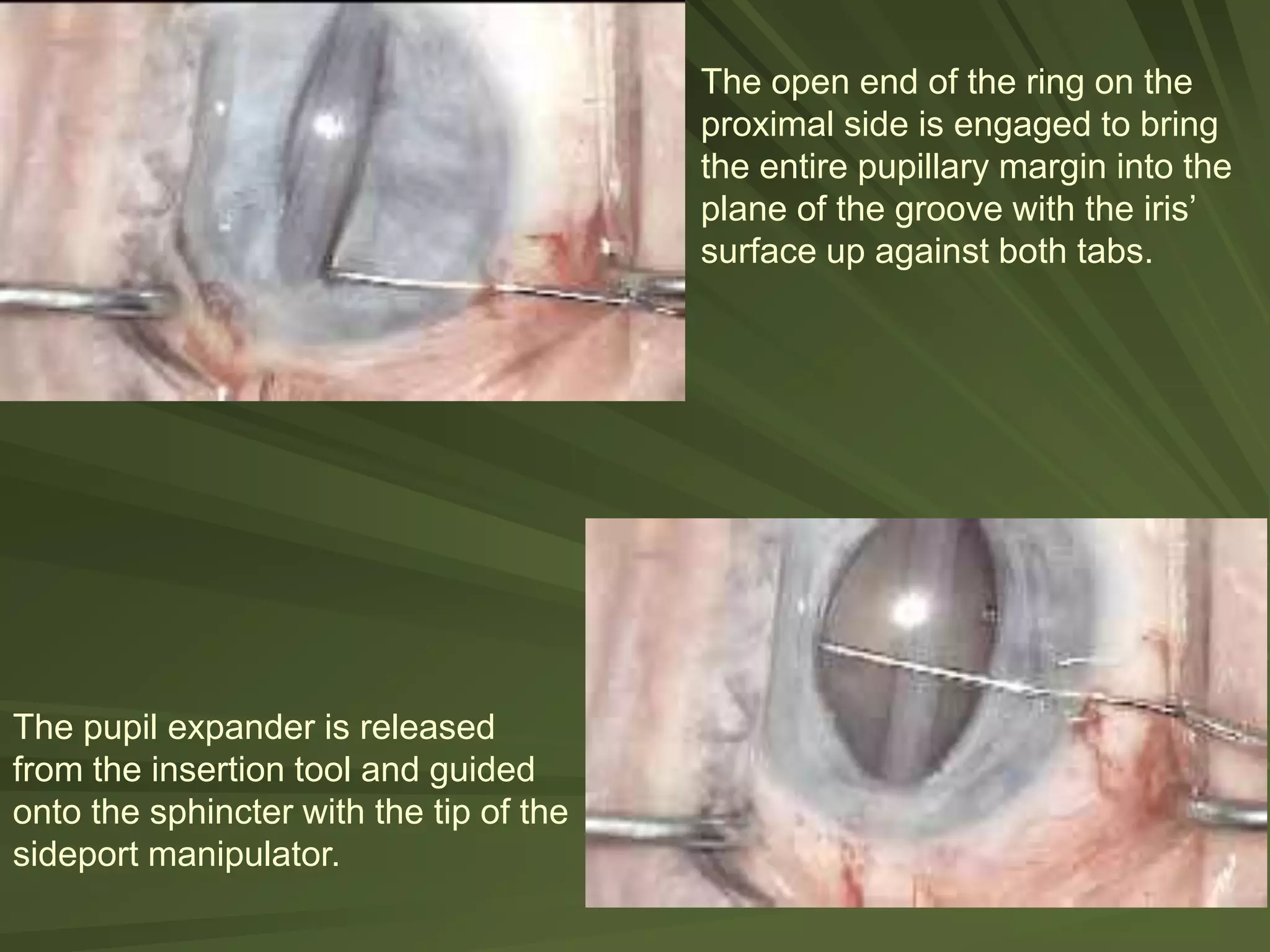
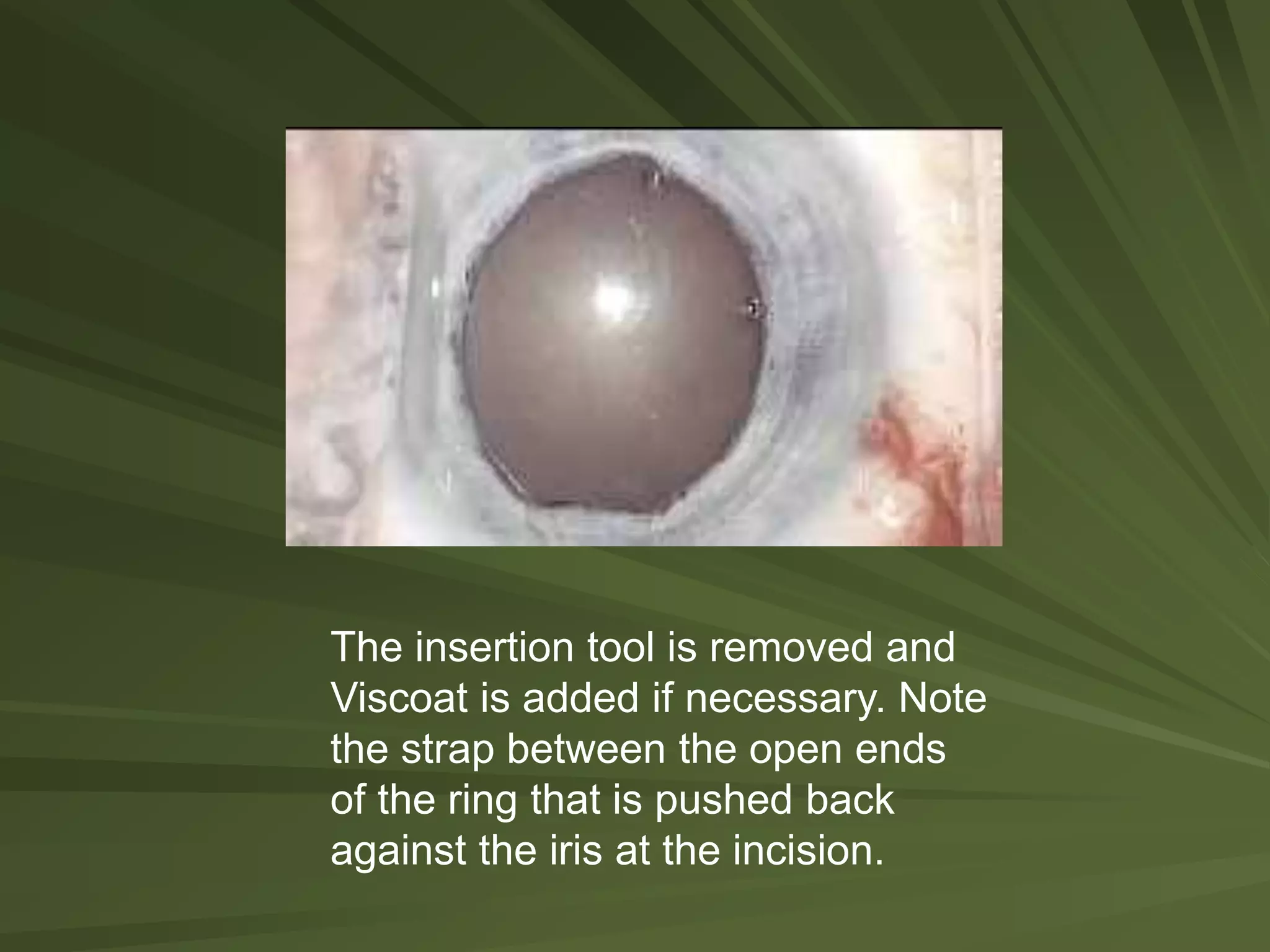
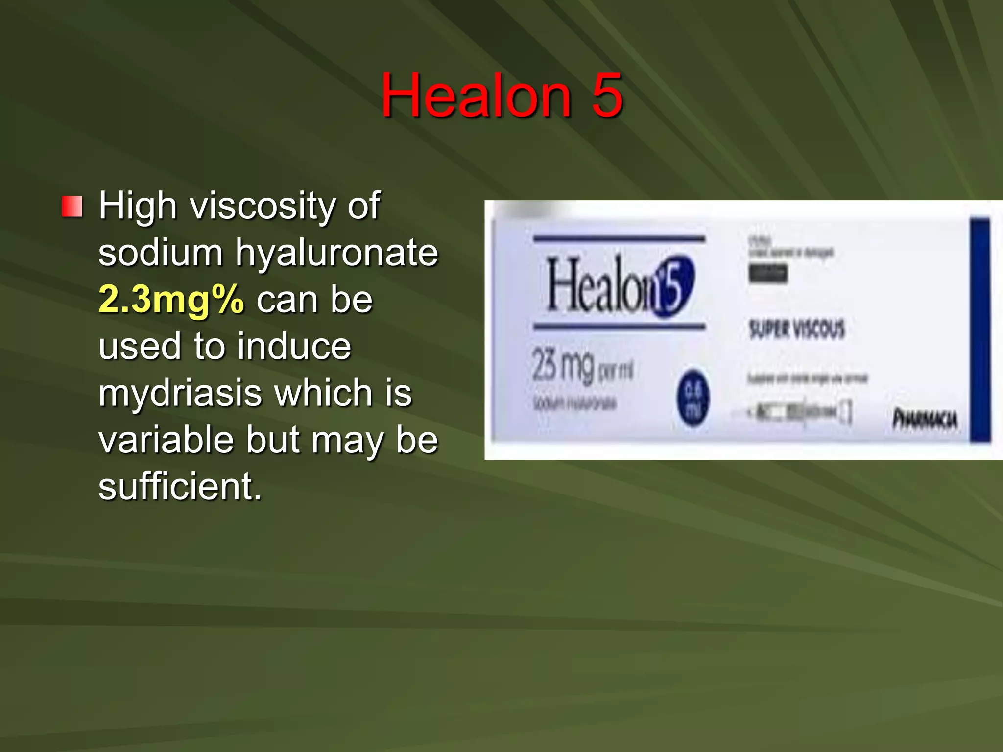
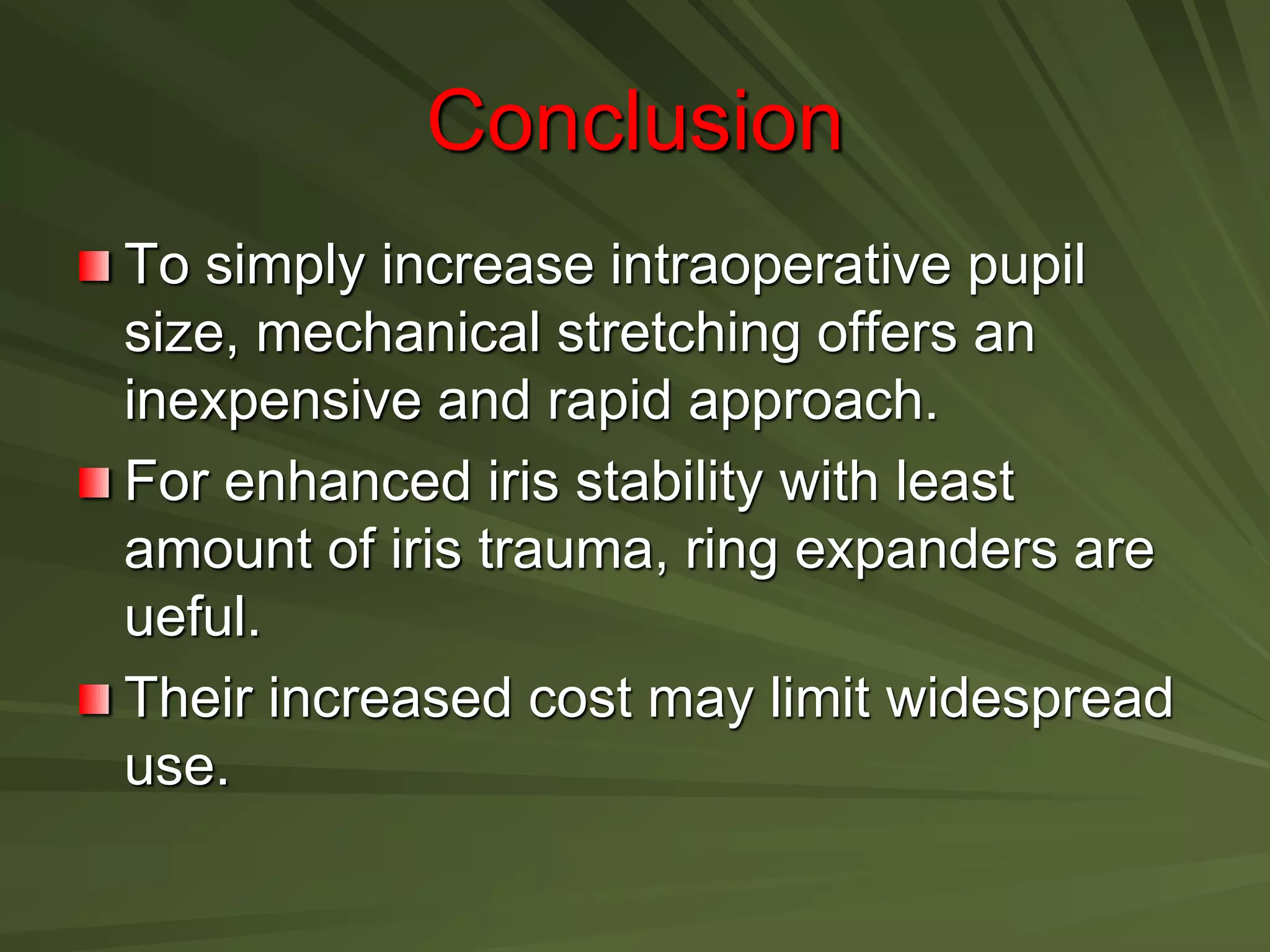
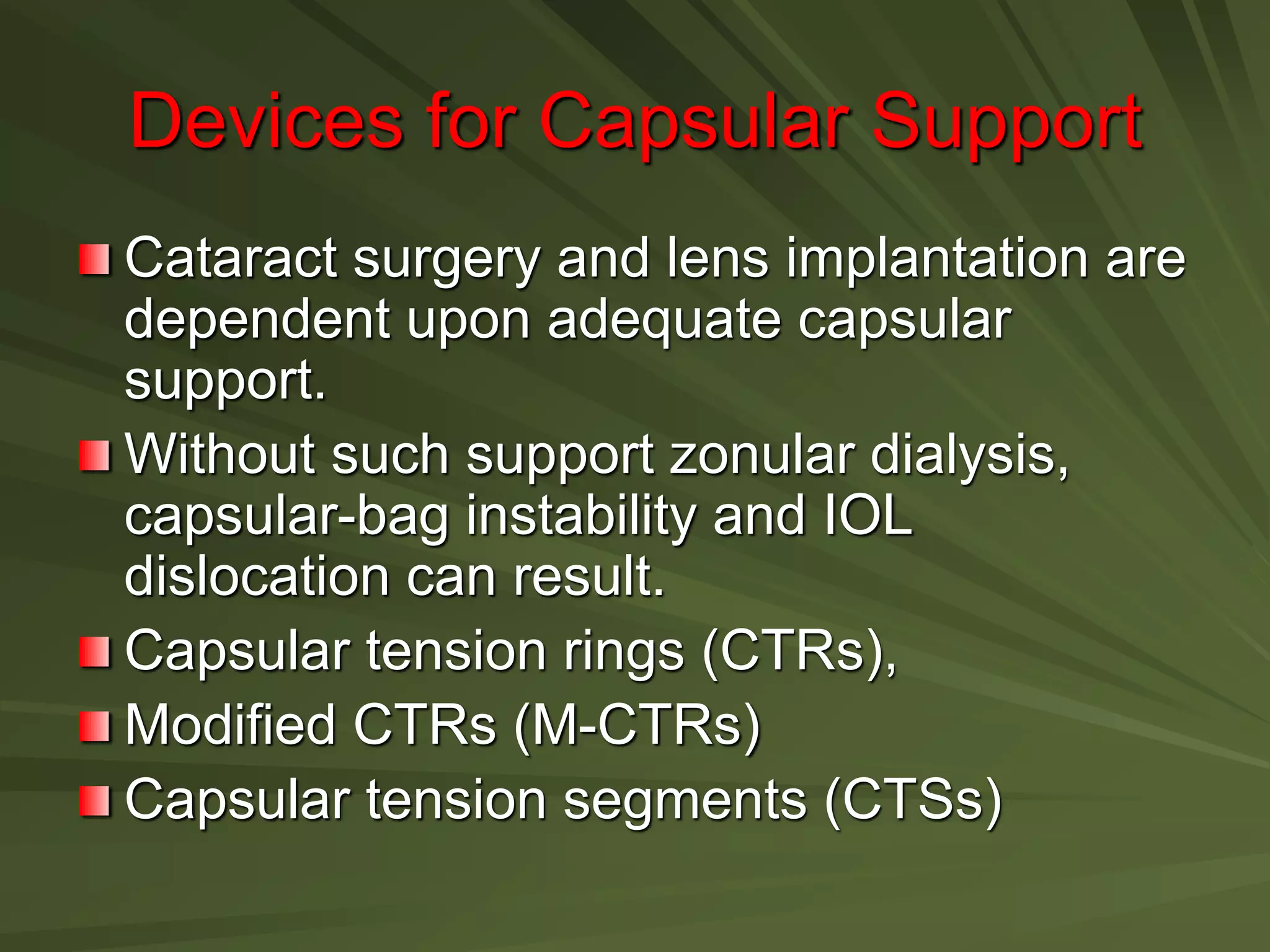
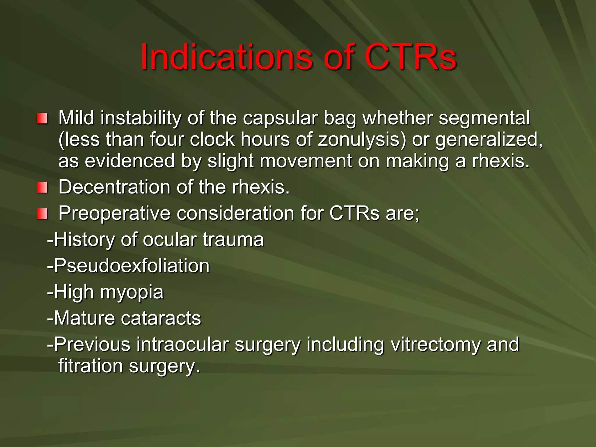
Update on Pupil Stretching and
Capsular Stabilization Devices
Indications
• Inadequate pupil dilation
• Poor capsular support.
• Iris prolapse.
• Intra-operative floppy-iris syndrome
These operative challenges can lead to an
increased risk of
capsular rupture.
zonular dehiscence.
dropped nuclei and vitreous loss.
Adjunct devices for pupil expansion
Iris hooks
Mechanical stretching.
Ring expanders
Sodium hyaluronate 2.3 mg/ml – Healon5.
Iris sutures.
Iris Hooks
In 1992 Mackool described a four point iris retractor
configuration by using small titanium hooks attached to a
square titanium base and an iris repositor.
De Juan introduced a flexible iris retractor made of nylon
with a silastic sleeve to reduce tissue damage due to
titanium.
Reusable nylon hook retractors now used worldwide with
proven utility in small pupils,iris prolapse, intraoperative
floppy iris syndrome, iridoschisis and for capsular
support in cases with zonular dehiscence or subluxed
crystalline lens.
Technique
Placement of side port parallel to
the iris plane and about 1mm
posterior to the sclerolimbal
junction (instead of in clear cornea)
allows for iris retraction in a more
physiological plane.
To minimize trauma to the pupil,the
hooks should be placed
symmetrically and excess iris
retraction should be avoided.
Iris prolapse can be prevented by
placing the four hooks in a diamond
shaped pattern with the fourth hook
in the subincisional position.
This configuration maximizes
visualization for horizontal chopping
and grooving.
The second method is to use a fifth iris hook in
the subincisional location. This may reduce the
need for pupillary stretch by 17%.
Improper positioning and removal of iris hooks
can cause;
- A wide raised iris platform between hooks
-iris prolapse
-iridodialysis
-secondary tears of either the ant capsule or DM
-irregular postoperative pupil.
Mechanical stretching
It’s a simple,
rapid,efficient way to
increase pupil size,
following synechioiysis.
A pair of hooks (second
instrument,’Y’ ) engage
the pupillary margin and
push or pull in opposite
directions.
Beehler’s pupil stretcher
Beehler’s stretcher
uses two or three
retractable
microfingers and one
hook attached to the
tube of the instrument
to stretch the pupil in
an asymmetric four
quadrant pattern.
Ring Expanders
Rings minimize iris-sphincter damage by
inducing circumferential expansion in the
physiologic plane, without elevation or tenting of
the iris. Once in position they stabilize and
protect the pupil margin.
Three commercially available types;
The Perfect Pupil,
The Morcher pupil-dilator ring.
The Graether 2000 pupil-expander system.
Perfect Pupil
It is a grooved polyurethane ring has
an internal diameter of 7.0mm.
The iris is held firmly in position in its
groove.
Attached at one end of the ring is an
integral arm, which is kept to one side
of corneal incision and aids in the
removal of the device.
The ring is open for 45 degree to
accommodate the passage of
instruments.
It is used in cataract patients whose
pupil failed to dilate beyond 4.0mm.
Pupil size after insertion was 7.8mm
and on removal was 1mm larger than
preoperatively.
It is markely reduced iris sphincter
tear,bleeding,ruptured capsule or
irregular pupil following the operation.
Morcher pupil-dilator ring
It is a fixed,rigged 7.5mm
PMMA ring.
Insertion is facilitated with
a Gueder 32970 injector.
It is less malleable than
the Perfect Pupil and
hence more difficult to
insert through a small
incision.
Moreover, lack of an
integral arm makes
insertion and removal
less efficient.
Comparison of various pupil dilatation methods for
phacoemulsification in eyes with small pupil secondary to
pseudoexfoliation. Ophthalmology 2004;111:1693.
Morchers ring was compared with iris hooks,
bimanual stretching, Beehler pupil dilator in a
randomized ,comparative case series of eyes
with PEX syndrome.
Highest mean pupil size was achieved with the
Morcher ring (5.9 +/- 0.6 mm), followed by iris
hooks, Beehler dilation and bimanual stretching.
Rate of sphincter rupture in the Morcher group
was one in ten.
Time for stretch was 5 mins as compared to 1
min for both Beeler and bimanual stretching,and
3 mins for iris hooks.
Comparison of Various Pupil Dilatation Methods for Phacoemulsification
in Eyes
with a Small Pupil Secondary to Pseudoexfoliation
Akman A et al Ophthalmology 2004;111:1693–1698
Purpose: To compare 4 methods for intraoperative pupil dilatation in
eyes with pseudoexfoliation syndrome and insufficient pupil size
during phacoemulsification.
Design: Prospective, randomized, comparative, interventional case
series.
Participants: Forty eyes of 40 patients with pseudoexfoliation and
maximally dilated pupil size smaller than 3.5 mm.
Intervention:
Group I - Mechanical pupil dilatation with iris-retractor hooks
Group II - Polymethyl methacrylate (PMMA) pupil dilator-ring
Group III - Beehler pupil dilator and
Group IV - Bimanual stretching
Main Outcome Measures: Performance (pupil size achieved),
complications, and added surgical time.
Results:
There were no statistically significant differences in the
postdilatation pupil sizes between the 4 study groups (P
<0.05).
Apart from self-limited intraoperative hemorrhage from
pupil margin, iris sphincter rupture was the only observed
complication related to mechanical pupil dilatation. This
occurred in 4 eyes in groups I and III, 3 eyes in group IV,
and 1 eye in group II (P0.05).
The mean added surgical time
iris-retractor hooks is 297 + 51 Sec.
PMMA pupil-dilator ring was 176 + 54 Sec.
Beehler pupil dilator 65 + 8 Sec.
Bimanual stretching 55 + 10 Sec.
The time needed for pupil dilatation in groups I and II is
significantly longer than that in groups III and IV (P0.001).
Iris-retractor hooks and the PMMA pupil-
dilator ring are the most time-consuming
techniques but have the advantage of a
stable pupil size throughout the surgery.
The PMMA pupil-dilator ring causes the
least iris trauma.
The Beehler pupil dilator and bimanual
stretching technique were the least time-
consuming methods for mechanical pupil
dilatation.
Conclusions
Graether 2000 pupil-expander
system
It is an incomplete soft silicone
ring.
A 3.75-mm gap in the ring is
bridged by a slender strap
which facilitates access to the
pupillary space.
Internal diameter-7.0mm and
the outer circumference is
grooved to engage the iris
sphincter.
It preloaded in a disposable
insertion tool.
The insertion tool is critical
because this ring lacks the
rigidity to allow for forward
advancement along the pupil
margin without the tool.
Expander is inserted through the
incision with the insertion tool
turned on its side so that the edges
of the groove form “runners.”
The pupillary margin is engaged
with the tip of the pupil expander
and pushed to the opposite
limbus. The insertion tool is held
stationary from this point during
the insertion.
The sideport manipulator’s tip is
placed in the pupil and moved
toward the incision to stretch the
sphincter.
The sphincter’s margin is lifted to
engage the open end of the
expander ring on the distal side
away from the sideport incision.
Surgeons should lift the sphincter to
bring it up into the
groove. They should not push down
on the insertion tool.
The open end of the ring on the
proximal side is engaged to bring
the entire pupillary margin into the
plane of the groove with the iris’
surface up against both tabs.
The pupil expander is released
from the insertion tool and guided
onto the sphincter with the tip of the
sideport manipulator.
The insertion tool is removed and
Viscoat is added if necessary. Note
the strap between the open ends
of the ring that is pushed back
against the iris at the incision.
Graether pupil expander for managing the small
pupil during surgery. J Cataract Refract Surg,1996;22.
In Graether’s series of 100 patients two patients
developed pupillary block which resulted in iris
prolapse and raised IOP with shallowing of the
AC.
To prevent a watertight seal of ring with the
capsule, Graether recommended to stretch the
strap prior to the capsulotomy or to create a
generous capsulotomy.
Minute sphincter tears may occur.
He recommended the use of VISCOAT (Alcon
Labs, Inc.),DISCOVISC (Alcon Labs, Inc.) or any
other dispersive vscoelastic to manage the pupil.
10-0 Nylon Iris Sutures
Four 1-1.5-mm stab incisions were made at
the limbus,as if placing iris hooks,followed
by capture of the peripheral iris in these
incisions with the aid of a fine forceps. The
captured iris was then sutured to clear
cornea with a 10-0 nylon suture.
Healon 5
High viscosity of
sodium hyaluronate
2.3mg% can be
used to induce
mydriasis which is
variable but may be
sufficient.
Conclusion
To simply increase intraoperative pupil
size, mechanical stretching offers an
inexpensive and rapid approach.
For enhanced iris stability with least
amount of iris trauma, ring expanders are
ueful.
Their increased cost may limit widespread
use.
Devices for Capsular Support
Cataract surgery and lens implantation are
dependent upon adequate capsular
support.
Without such support zonular dialysis,
capsular-bag instability and IOL
dislocation can result.
Capsular tension rings (CTRs),
Modified CTRs (M-CTRs)
Capsular tension segments (CTSs)
Capsular Tension Rings
CTRs stabilize the zonular
apparatus through the centrifugal
force of an open PMMA ring.
The diameter of the CTR slightly
exceed that of the capsular bag.
Once implanted it expands and
centrates the capsular
apparatus, supporting the week
zonules and recruiting tension
from existing stronger zonules.
The first capsular tension
silicone ring implanted into
the rabbit eyes in 1991 by T.
Hara.
After modifications by Nagamoto
and Bissen-Miyajima the first
CTR was implanted during
cataract surgery into a human in
1993.
Three sizes – 12 / 10 mm
13 / 11 mm
14.5 / 13 mm
Indications of CTRs
Mild instability of the capsular bag whether segmental
(less than four clock hours of zonulysis) or generalized,
as evidenced by slight movement on making a rhexis.
Decentration of the rhexis.
Preoperative consideration for CTRs are;
-History of ocular trauma
-Pseudoexfoliation
-High myopia
-Mature cataracts
-Previous intraocular surgery including vitrectomy and
fitration surgery.
Contraindications of CTRs
Greater than mild zonular instability.
Anterior radial or posterior capsular tears.
Safety and efficacy of CTRs
In 2006, Hasance et al. studied safety and
efficacy of CTRs, one retrospective and
one prospective where fellow eye served
as a control.
They concluded that the use of a CTR for
mild instability helped reduce complication
rates and resulted in good IOL centration.
Investigational Device Study of the Optec
Capsular Tension Ring. Ophthalmology 2005;112:460
Purpose: To evaluate safety and efficacy of Optec CTR in providing
capsular support during and/or after cataract extraction in cases of
weak or partially broken ciliary zonule.
12 investigators at 9 sites enrolled 224 subjects and placed 255
CTRs.
CTR’s were placed in patients who were found to have a weakened
or partially broken ciliary zonules. Two CTR models were evaluated,
with diameters of 12 and 13 mm.
Results: 98.8% of IOLs were centered and 1.2% were not.
Prevalence of decentered IOLs was 1.7%,3.8% and 2.3% at 3,6 and
12 months post surgery. Primary complication was PCO which is
unlikely to be due to CTR insertion.
They concluded that OPTEC CTR models 275 and 276 safely
provided capsular support during and after cataract surgery in cases
where the zonules were weak or partially broken.
Modified Capsular Tension Rings
The standard CTR was
modified by Cionni and
Osher in 1998.
M-CTR should only be
used in the setting of an
intact anterior
capsulorhexis and
posterior capsule.
Cionni rings have one or
two fixation eyelets
,which lie in a plane just
above the anterior
capsulotomy. The eyelets
allow for suture(10-0
prolene) fixation of the
capsular bag to the eye
wall.
In 2003 Cionni et al.
studied a larger series of
patients with congenital
loss of zonular support
and crystalline lens
decentration from Weil-
Marchesani syndrome,
idiopathic ectopia lentis
and Marfans syndrome.
Postoperatively most of
patients eyes had well
centered posterior-
chamber IOLs.
Six eyes developed late
symptomatic IOL
decetration at 12 months.
Potential benefits of CTR
(Gimbel HV et al:Ophth Surg and Lasers 1997)
Capsular zonular anatomical barrier partially
reformed –reduce vitreous herniation in AC.
A taut capsular equator offers counter traction
for all tractional maneuvers,making them easier
to perform & decreasing the risk of extending
zonular dialysis.
Maintains circular contour of capsular bag.
Reduce IOL decentration.
Inhibit lens epithelial proliferation on posterior
capsule by compression.
Complications of phaco with CTR
(Dietlin TS et al;J Cataract Refract Surg 2000)
Inadverant tear of anterior capsule during
CCC or insertion of CTR
Nuclear drop
Posterior displacement of CTR
Lens epithelial proliferation with IOL
decentration especially in children.
Capsular Tension Segments
Used when capsular instability is
secondary to an anterior or
posterior capsular tear.
Neither the standard nor the
modified CTR should be used.
CTS designed by Ahmed in 2002
is a partial open ring segment
(120o) with an anteriorly
positioned fixation eyelet.
It need not be dialed like the CTR
or M-CTR.
Can be placed atraumatically in
the setting of a radial anterior or
posterior capsular rupture.
In case of significant tear of
capsular bag, device can be
sutured into position with a fixation
eyelet for intraoperative support or
can be left in place for more
permanent stabilization.
Iris retractors for capsular stabilization
In 1997,Novak was the first to
describe capsular-bag
stabilization in eyes with
traumatic zolular damage using
a modified flexible iris hook
made of 5-0 prolene and a large
ring-shaped handle.
In 1999, Lee and Bloom described
a technique using flexible iris
hooks through four stab incision at
sclerolimbal junction to prevent
anterior tenting of the capsule with
retraction.
In this manner capsular bag was
fixated relative to the sclera at four
equally spaced points in the plane
of he anterior capsule.
Conclusions on capsular
stabilization
When capsular bag instability is in
question, the surgeon may choose the
appropriate technique for reestablishment
of capsualr bag contour from a standard or
modified CTR, one or more CTS’s or an
iris hook variant for support, depending on
the degree of instability.
THANK YOU

Update on pupil stretching and capsular stabilization devices

  • 1.
    Update on PupilStretching and Capsular Stabilization Devices
  • 2.
    Indications • Inadequate pupildilation • Poor capsular support. • Iris prolapse. • Intra-operative floppy-iris syndrome
  • 3.
    These operative challengescan lead to an increased risk of capsular rupture. zonular dehiscence. dropped nuclei and vitreous loss.
  • 4.
    Adjunct devices forpupil expansion Iris hooks Mechanical stretching. Ring expanders Sodium hyaluronate 2.3 mg/ml – Healon5. Iris sutures.
  • 5.
    Iris Hooks In 1992Mackool described a four point iris retractor configuration by using small titanium hooks attached to a square titanium base and an iris repositor. De Juan introduced a flexible iris retractor made of nylon with a silastic sleeve to reduce tissue damage due to titanium. Reusable nylon hook retractors now used worldwide with proven utility in small pupils,iris prolapse, intraoperative floppy iris syndrome, iridoschisis and for capsular support in cases with zonular dehiscence or subluxed crystalline lens.
  • 6.
    Technique Placement of sideport parallel to the iris plane and about 1mm posterior to the sclerolimbal junction (instead of in clear cornea) allows for iris retraction in a more physiological plane. To minimize trauma to the pupil,the hooks should be placed symmetrically and excess iris retraction should be avoided. Iris prolapse can be prevented by placing the four hooks in a diamond shaped pattern with the fourth hook in the subincisional position. This configuration maximizes visualization for horizontal chopping and grooving.
  • 7.
    The second methodis to use a fifth iris hook in the subincisional location. This may reduce the need for pupillary stretch by 17%. Improper positioning and removal of iris hooks can cause; - A wide raised iris platform between hooks -iris prolapse -iridodialysis -secondary tears of either the ant capsule or DM -irregular postoperative pupil.
  • 8.
    Mechanical stretching It’s asimple, rapid,efficient way to increase pupil size, following synechioiysis. A pair of hooks (second instrument,’Y’ ) engage the pupillary margin and push or pull in opposite directions.
  • 9.
    Beehler’s pupil stretcher Beehler’sstretcher uses two or three retractable microfingers and one hook attached to the tube of the instrument to stretch the pupil in an asymmetric four quadrant pattern.
  • 10.
    Ring Expanders Rings minimizeiris-sphincter damage by inducing circumferential expansion in the physiologic plane, without elevation or tenting of the iris. Once in position they stabilize and protect the pupil margin. Three commercially available types; The Perfect Pupil, The Morcher pupil-dilator ring. The Graether 2000 pupil-expander system.
  • 11.
    Perfect Pupil It isa grooved polyurethane ring has an internal diameter of 7.0mm. The iris is held firmly in position in its groove. Attached at one end of the ring is an integral arm, which is kept to one side of corneal incision and aids in the removal of the device. The ring is open for 45 degree to accommodate the passage of instruments. It is used in cataract patients whose pupil failed to dilate beyond 4.0mm. Pupil size after insertion was 7.8mm and on removal was 1mm larger than preoperatively. It is markely reduced iris sphincter tear,bleeding,ruptured capsule or irregular pupil following the operation.
  • 12.
    Morcher pupil-dilator ring Itis a fixed,rigged 7.5mm PMMA ring. Insertion is facilitated with a Gueder 32970 injector. It is less malleable than the Perfect Pupil and hence more difficult to insert through a small incision. Moreover, lack of an integral arm makes insertion and removal less efficient.
  • 13.
    Comparison of variouspupil dilatation methods for phacoemulsification in eyes with small pupil secondary to pseudoexfoliation. Ophthalmology 2004;111:1693. Morchers ring was compared with iris hooks, bimanual stretching, Beehler pupil dilator in a randomized ,comparative case series of eyes with PEX syndrome. Highest mean pupil size was achieved with the Morcher ring (5.9 +/- 0.6 mm), followed by iris hooks, Beehler dilation and bimanual stretching. Rate of sphincter rupture in the Morcher group was one in ten. Time for stretch was 5 mins as compared to 1 min for both Beeler and bimanual stretching,and 3 mins for iris hooks.
  • 14.
    Comparison of VariousPupil Dilatation Methods for Phacoemulsification in Eyes with a Small Pupil Secondary to Pseudoexfoliation Akman A et al Ophthalmology 2004;111:1693–1698 Purpose: To compare 4 methods for intraoperative pupil dilatation in eyes with pseudoexfoliation syndrome and insufficient pupil size during phacoemulsification. Design: Prospective, randomized, comparative, interventional case series. Participants: Forty eyes of 40 patients with pseudoexfoliation and maximally dilated pupil size smaller than 3.5 mm. Intervention: Group I - Mechanical pupil dilatation with iris-retractor hooks Group II - Polymethyl methacrylate (PMMA) pupil dilator-ring Group III - Beehler pupil dilator and Group IV - Bimanual stretching Main Outcome Measures: Performance (pupil size achieved), complications, and added surgical time.
  • 15.
    Results: There were nostatistically significant differences in the postdilatation pupil sizes between the 4 study groups (P <0.05). Apart from self-limited intraoperative hemorrhage from pupil margin, iris sphincter rupture was the only observed complication related to mechanical pupil dilatation. This occurred in 4 eyes in groups I and III, 3 eyes in group IV, and 1 eye in group II (P0.05). The mean added surgical time iris-retractor hooks is 297 + 51 Sec. PMMA pupil-dilator ring was 176 + 54 Sec. Beehler pupil dilator 65 + 8 Sec. Bimanual stretching 55 + 10 Sec. The time needed for pupil dilatation in groups I and II is significantly longer than that in groups III and IV (P0.001).
  • 18.
    Iris-retractor hooks andthe PMMA pupil- dilator ring are the most time-consuming techniques but have the advantage of a stable pupil size throughout the surgery. The PMMA pupil-dilator ring causes the least iris trauma. The Beehler pupil dilator and bimanual stretching technique were the least time- consuming methods for mechanical pupil dilatation. Conclusions
  • 19.
    Graether 2000 pupil-expander system Itis an incomplete soft silicone ring. A 3.75-mm gap in the ring is bridged by a slender strap which facilitates access to the pupillary space. Internal diameter-7.0mm and the outer circumference is grooved to engage the iris sphincter. It preloaded in a disposable insertion tool. The insertion tool is critical because this ring lacks the rigidity to allow for forward advancement along the pupil margin without the tool.
  • 20.
    Expander is insertedthrough the incision with the insertion tool turned on its side so that the edges of the groove form “runners.” The pupillary margin is engaged with the tip of the pupil expander and pushed to the opposite limbus. The insertion tool is held stationary from this point during the insertion.
  • 21.
    The sideport manipulator’stip is placed in the pupil and moved toward the incision to stretch the sphincter. The sphincter’s margin is lifted to engage the open end of the expander ring on the distal side away from the sideport incision. Surgeons should lift the sphincter to bring it up into the groove. They should not push down on the insertion tool.
  • 22.
    The open endof the ring on the proximal side is engaged to bring the entire pupillary margin into the plane of the groove with the iris’ surface up against both tabs. The pupil expander is released from the insertion tool and guided onto the sphincter with the tip of the sideport manipulator.
  • 23.
    The insertion toolis removed and Viscoat is added if necessary. Note the strap between the open ends of the ring that is pushed back against the iris at the incision.
  • 24.
    Graether pupil expanderfor managing the small pupil during surgery. J Cataract Refract Surg,1996;22. In Graether’s series of 100 patients two patients developed pupillary block which resulted in iris prolapse and raised IOP with shallowing of the AC. To prevent a watertight seal of ring with the capsule, Graether recommended to stretch the strap prior to the capsulotomy or to create a generous capsulotomy. Minute sphincter tears may occur. He recommended the use of VISCOAT (Alcon Labs, Inc.),DISCOVISC (Alcon Labs, Inc.) or any other dispersive vscoelastic to manage the pupil.
  • 25.
    10-0 Nylon IrisSutures Four 1-1.5-mm stab incisions were made at the limbus,as if placing iris hooks,followed by capture of the peripheral iris in these incisions with the aid of a fine forceps. The captured iris was then sutured to clear cornea with a 10-0 nylon suture.
  • 26.
    Healon 5 High viscosityof sodium hyaluronate 2.3mg% can be used to induce mydriasis which is variable but may be sufficient.
  • 27.
    Conclusion To simply increaseintraoperative pupil size, mechanical stretching offers an inexpensive and rapid approach. For enhanced iris stability with least amount of iris trauma, ring expanders are ueful. Their increased cost may limit widespread use.
  • 28.
    Devices for CapsularSupport Cataract surgery and lens implantation are dependent upon adequate capsular support. Without such support zonular dialysis, capsular-bag instability and IOL dislocation can result. Capsular tension rings (CTRs), Modified CTRs (M-CTRs) Capsular tension segments (CTSs)
  • 29.
    Capsular Tension Rings CTRsstabilize the zonular apparatus through the centrifugal force of an open PMMA ring. The diameter of the CTR slightly exceed that of the capsular bag. Once implanted it expands and centrates the capsular apparatus, supporting the week zonules and recruiting tension from existing stronger zonules. The first capsular tension silicone ring implanted into the rabbit eyes in 1991 by T. Hara. After modifications by Nagamoto and Bissen-Miyajima the first CTR was implanted during cataract surgery into a human in 1993. Three sizes – 12 / 10 mm 13 / 11 mm 14.5 / 13 mm
  • 30.
    Indications of CTRs Mildinstability of the capsular bag whether segmental (less than four clock hours of zonulysis) or generalized, as evidenced by slight movement on making a rhexis. Decentration of the rhexis. Preoperative consideration for CTRs are; -History of ocular trauma -Pseudoexfoliation -High myopia -Mature cataracts -Previous intraocular surgery including vitrectomy and fitration surgery.
  • 31.
    Contraindications of CTRs Greaterthan mild zonular instability. Anterior radial or posterior capsular tears.
  • 32.
    Safety and efficacyof CTRs In 2006, Hasance et al. studied safety and efficacy of CTRs, one retrospective and one prospective where fellow eye served as a control. They concluded that the use of a CTR for mild instability helped reduce complication rates and resulted in good IOL centration.
  • 33.
    Investigational Device Studyof the Optec Capsular Tension Ring. Ophthalmology 2005;112:460 Purpose: To evaluate safety and efficacy of Optec CTR in providing capsular support during and/or after cataract extraction in cases of weak or partially broken ciliary zonule. 12 investigators at 9 sites enrolled 224 subjects and placed 255 CTRs. CTR’s were placed in patients who were found to have a weakened or partially broken ciliary zonules. Two CTR models were evaluated, with diameters of 12 and 13 mm. Results: 98.8% of IOLs were centered and 1.2% were not. Prevalence of decentered IOLs was 1.7%,3.8% and 2.3% at 3,6 and 12 months post surgery. Primary complication was PCO which is unlikely to be due to CTR insertion. They concluded that OPTEC CTR models 275 and 276 safely provided capsular support during and after cataract surgery in cases where the zonules were weak or partially broken.
  • 34.
    Modified Capsular TensionRings The standard CTR was modified by Cionni and Osher in 1998. M-CTR should only be used in the setting of an intact anterior capsulorhexis and posterior capsule. Cionni rings have one or two fixation eyelets ,which lie in a plane just above the anterior capsulotomy. The eyelets allow for suture(10-0 prolene) fixation of the capsular bag to the eye wall.
  • 35.
    In 2003 Cionniet al. studied a larger series of patients with congenital loss of zonular support and crystalline lens decentration from Weil- Marchesani syndrome, idiopathic ectopia lentis and Marfans syndrome. Postoperatively most of patients eyes had well centered posterior- chamber IOLs. Six eyes developed late symptomatic IOL decetration at 12 months.
  • 36.
    Potential benefits ofCTR (Gimbel HV et al:Ophth Surg and Lasers 1997) Capsular zonular anatomical barrier partially reformed –reduce vitreous herniation in AC. A taut capsular equator offers counter traction for all tractional maneuvers,making them easier to perform & decreasing the risk of extending zonular dialysis. Maintains circular contour of capsular bag. Reduce IOL decentration. Inhibit lens epithelial proliferation on posterior capsule by compression.
  • 37.
    Complications of phacowith CTR (Dietlin TS et al;J Cataract Refract Surg 2000) Inadverant tear of anterior capsule during CCC or insertion of CTR Nuclear drop Posterior displacement of CTR Lens epithelial proliferation with IOL decentration especially in children.
  • 38.
    Capsular Tension Segments Usedwhen capsular instability is secondary to an anterior or posterior capsular tear. Neither the standard nor the modified CTR should be used. CTS designed by Ahmed in 2002 is a partial open ring segment (120o) with an anteriorly positioned fixation eyelet. It need not be dialed like the CTR or M-CTR. Can be placed atraumatically in the setting of a radial anterior or posterior capsular rupture. In case of significant tear of capsular bag, device can be sutured into position with a fixation eyelet for intraoperative support or can be left in place for more permanent stabilization.
  • 39.
    Iris retractors forcapsular stabilization In 1997,Novak was the first to describe capsular-bag stabilization in eyes with traumatic zolular damage using a modified flexible iris hook made of 5-0 prolene and a large ring-shaped handle. In 1999, Lee and Bloom described a technique using flexible iris hooks through four stab incision at sclerolimbal junction to prevent anterior tenting of the capsule with retraction. In this manner capsular bag was fixated relative to the sclera at four equally spaced points in the plane of he anterior capsule.
  • 40.
    Conclusions on capsular stabilization Whencapsular bag instability is in question, the surgeon may choose the appropriate technique for reestablishment of capsualr bag contour from a standard or modified CTR, one or more CTS’s or an iris hook variant for support, depending on the degree of instability.
  • 41.