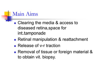
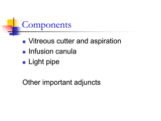
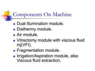
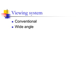
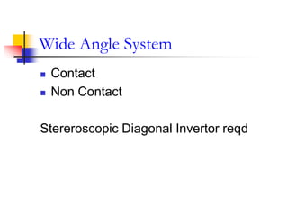
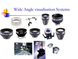
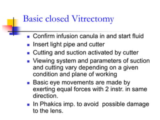
VITRECTOMY PRINCIPLES
DR.SHAH-NOOR HASSAN
FCPS,FRCS(Glasgow)
Introduction
 1968-open sky vitrectomy-KASNER
 1971-closed system vitrectomy-modern
pars plana surgery -MACHEMER
 Bimanual vitrectomy-O’MALLEY
 CHARLES ,SCHEPENS,PEYMAN-
instrumentation&surgical techniques.
Surgical Anatomy
MUSCLE INSERTIONS
 Pars Plicata-2.5 MM
 Pars Plana –3-5MM
 Ora –5-7MM From Limbus
 Vitreous Base-2-6mm wide zone
straddling the ora, extending 2-3mm
posterior to it .
Main Aims
 Clearing the media & access to
diseased retina,space for
int.tamponade
 Retinal manipulation & reattachment
 Release of v-r traction
 Removal of tissue or foreign material &
to obtain vit. biopsy.
Components
 Vitreous cutter and aspiration
 Infusion canula
 Light pipe
Other important adjuncts
Components On Machine
 Dual illumination module.
 Diathermy module.
 Air module.
 Vitrectomy module with viscous fluid
inj(VFI).
 Fragmentation module.
 Irrigation/Aspiration module, also
Viscous fluid extraction.
Viewing system
 Conventional
 Wide angle
Conventional
 Plano concave lenses
 Hand held contact
lenses,placed on a
ring,self stabilizing
 Small
field,nonmagnified
 Additional lenses reqd
 High
resolution,excellent
stereopsis
Wide Angle System
 Contact
 Non Contact
Stereroscopic Diagonal Invertor reqd
Wide Angle visualisation Systems
LENS MAG FIELD USE
PLANO
CONCAVE FLAT
1.0 20* POSTERIOR
POLE
,CENTRAL
VITREOUS
PRISM LENS 1.0 20* MIDPERIPHERA
L FUNDUS AND
VITREOUS
SUPER
MACULA (V)
1.03 64*-77* POSTERIOR
POLE
,CENTRAL
VITREOUS
MINI QUAD 0.71 106*-127* UPTO ORA
LANDERS
WIDEFIELD
0.38 130*-146* UPTO ORA
LANDERS
EQUATORIAL
0.65 101*-131* GOOD UPTO
EQUATOR
Vitreous Cutter
 Presence of outer
fixed tube and an
opening thru’ which
vit. is aspirated.
 Inner tip slides
across inner Port. of
opening thus cutting
the vitreous
Eg.Guillotine,Rotatio
n,Oscillation.
 Safe to use low
suction,high cut rate
Vitreous Cutter
Aspiration
 Device based on venturi principle creates
vacuum used to provide aspiration
 The venturi has inlet for compressed air
& uses the energy from air to produce a
vacuum.
Light Pipe/Endoilluminator
 Halogen Bulb
 Endo illum.
Probe-20 gauge
(0.9mm) Fibre
optic cable
 Yellow light and
White light.
Infusion Canula
Intraocular Irrigating Solution
 CRITERIA–should be sterile,isotonic and
which constitute near those of normal
ocular fluid and with a balanced pH.
 Mixture of GBR(glutathione, bicarbonate
and ringer lactate).
 BSSPlus with similar comb.
 Lens opacification in diabetic pt.
Advocated- Glucose fortified BSS Plus
 Regular BSS Plus for non diabetic pt.
Before You Begin
 Inspection and testing of cutter for
cutting and aspiration
 Flushing of air bubble from system &
infusion canula.
 Testing of fiberoptic probe/light pipe.
 Adjustment of infusion bottle height.
Making Sclerotomies
 Peritomy
 Hemostasis
 Location
 Instruments
 Technique and direction
Sclerotomy for Infusion canula
FIRST TO GO IN ,LAST TO COME OUT
 Length of canula
 Site
 Location
 Anchoring sutures
 Sclerotomy
Checking for canula tip
 APHAKIC/PSEUDOPHAKIC
 PHAKIC
Basic closed Vitrectomy
 Confirm infusion canula in and start fluid
 Insert light pipe and cutter
 Cutting and suction activated by cutter
 Viewing system and parameters of suction
and cutting vary depending on a given
condition and plane of working
 Basic eye movements are made by
exerting equal forces with 2 instr. in same
direction.
 In Phakics imp. to avoid possible damage
to the lens.
Simple Vitrectomy
 Core vitrectomy
 Peripheral vitrectomy
 Removal of posterior hyaloid
 Inspection of peripheral retina
 Closure
Core vitrectomy
 Mid vitreous
 Instruments held steady
 Advance posteriorly
Peripheral Vitrectomy
 Cutter position
 Scleral depression
 High speed cutting
Removal of posterior hyaloid
 Engaged in peripapillary region
 Soft tipped extrusion canula
 Signs of engagement
 Technique
 MVR / Pick
Inspection of peripheral retina
 To ascertain no tears
 Inspect vulnerable areas
 SOS Cryo/Laser
Closure
 Excision of any prolapsed vitreous
 Suturing ports(infusion canula last to
come out)
 Suturing conjunctiva
Other adjunctive procedures
 Lensectomy –opacification /PVR
/trauma /IOFB
 Air fluid exchange
 PFCL/Silicone oil /gases
 Endolaser / endodiathermy
Air fluid exchange
 Air via infusion canula
(pump)
 Active aspiration
 Passive aspiration
 Removal of SRF
Use of PFCL
 Giant retinal tear
 PVR
 Dislocated nucleus / dropped nucleus
Use of silicone oil
 PVR
 Diabetic vitrectomies
For prolonged tamponade
Use of gases C3f8 Sf6
 Pneumoretinopexy
 Tamponade
Recent advances
 25 G vitrectomy –transconjunctival,sutureless
Tried in less complicated cases like
ERM /Diabetic macular edema/ macular hole
/persistent vitreous haem
 4-port vitrectomy(both hands actively
functioning with help of chandelier system)
 23 G
 Endoscope assisted
 OFFICE system
Write the OT notes of the following situations
 Recent Total RRD in a 60 year old
patient
 Recent TRD in a diabetic patient
 Fresh Vit. Hg in a 35 yr old male patient
 Fresh Rd from 2 to 4 o clock in a young
patient
Thank You

Vitrectomy Principles

  • 1.
  • 2.
    Introduction  1968-open skyvitrectomy-KASNER  1971-closed system vitrectomy-modern pars plana surgery -MACHEMER  Bimanual vitrectomy-O’MALLEY  CHARLES ,SCHEPENS,PEYMAN- instrumentation&surgical techniques.
  • 3.
  • 4.
  • 5.
     Pars Plicata-2.5MM  Pars Plana –3-5MM  Ora –5-7MM From Limbus  Vitreous Base-2-6mm wide zone straddling the ora, extending 2-3mm posterior to it .
  • 7.
    Main Aims  Clearingthe media & access to diseased retina,space for int.tamponade  Retinal manipulation & reattachment  Release of v-r traction  Removal of tissue or foreign material & to obtain vit. biopsy.
  • 8.
    Components  Vitreous cutterand aspiration  Infusion canula  Light pipe Other important adjuncts
  • 9.
    Components On Machine Dual illumination module.  Diathermy module.  Air module.  Vitrectomy module with viscous fluid inj(VFI).  Fragmentation module.  Irrigation/Aspiration module, also Viscous fluid extraction.
  • 10.
  • 11.
    Conventional  Plano concavelenses  Hand held contact lenses,placed on a ring,self stabilizing  Small field,nonmagnified  Additional lenses reqd  High resolution,excellent stereopsis
  • 12.
    Wide Angle System Contact  Non Contact Stereroscopic Diagonal Invertor reqd
  • 13.
  • 14.
    LENS MAG FIELDUSE PLANO CONCAVE FLAT 1.0 20* POSTERIOR POLE ,CENTRAL VITREOUS PRISM LENS 1.0 20* MIDPERIPHERA L FUNDUS AND VITREOUS SUPER MACULA (V) 1.03 64*-77* POSTERIOR POLE ,CENTRAL VITREOUS MINI QUAD 0.71 106*-127* UPTO ORA LANDERS WIDEFIELD 0.38 130*-146* UPTO ORA LANDERS EQUATORIAL 0.65 101*-131* GOOD UPTO EQUATOR
  • 15.
    Vitreous Cutter  Presenceof outer fixed tube and an opening thru’ which vit. is aspirated.  Inner tip slides across inner Port. of opening thus cutting the vitreous Eg.Guillotine,Rotatio n,Oscillation.  Safe to use low suction,high cut rate
  • 16.
  • 17.
    Aspiration  Device basedon venturi principle creates vacuum used to provide aspiration  The venturi has inlet for compressed air & uses the energy from air to produce a vacuum.
  • 19.
    Light Pipe/Endoilluminator  HalogenBulb  Endo illum. Probe-20 gauge (0.9mm) Fibre optic cable  Yellow light and White light.
  • 20.
  • 21.
    Intraocular Irrigating Solution CRITERIA–should be sterile,isotonic and which constitute near those of normal ocular fluid and with a balanced pH.  Mixture of GBR(glutathione, bicarbonate and ringer lactate).  BSSPlus with similar comb.  Lens opacification in diabetic pt. Advocated- Glucose fortified BSS Plus  Regular BSS Plus for non diabetic pt.
  • 22.
    Before You Begin Inspection and testing of cutter for cutting and aspiration  Flushing of air bubble from system & infusion canula.  Testing of fiberoptic probe/light pipe.  Adjustment of infusion bottle height.
  • 23.
    Making Sclerotomies  Peritomy Hemostasis  Location  Instruments  Technique and direction
  • 24.
    Sclerotomy for Infusioncanula FIRST TO GO IN ,LAST TO COME OUT  Length of canula  Site  Location  Anchoring sutures  Sclerotomy
  • 26.
    Checking for canulatip  APHAKIC/PSEUDOPHAKIC  PHAKIC
  • 28.
    Basic closed Vitrectomy Confirm infusion canula in and start fluid  Insert light pipe and cutter  Cutting and suction activated by cutter  Viewing system and parameters of suction and cutting vary depending on a given condition and plane of working  Basic eye movements are made by exerting equal forces with 2 instr. in same direction.  In Phakics imp. to avoid possible damage to the lens.
  • 29.
    Simple Vitrectomy  Corevitrectomy  Peripheral vitrectomy  Removal of posterior hyaloid  Inspection of peripheral retina  Closure
  • 30.
    Core vitrectomy  Midvitreous  Instruments held steady  Advance posteriorly
  • 31.
    Peripheral Vitrectomy  Cutterposition  Scleral depression  High speed cutting
  • 32.
    Removal of posteriorhyaloid  Engaged in peripapillary region  Soft tipped extrusion canula  Signs of engagement  Technique  MVR / Pick
  • 33.
    Inspection of peripheralretina  To ascertain no tears  Inspect vulnerable areas  SOS Cryo/Laser
  • 34.
    Closure  Excision ofany prolapsed vitreous  Suturing ports(infusion canula last to come out)  Suturing conjunctiva
  • 35.
    Other adjunctive procedures Lensectomy –opacification /PVR /trauma /IOFB  Air fluid exchange  PFCL/Silicone oil /gases  Endolaser / endodiathermy
  • 36.
    Air fluid exchange Air via infusion canula (pump)  Active aspiration  Passive aspiration  Removal of SRF
  • 37.
    Use of PFCL Giant retinal tear  PVR  Dislocated nucleus / dropped nucleus
  • 38.
    Use of siliconeoil  PVR  Diabetic vitrectomies For prolonged tamponade
  • 39.
    Use of gasesC3f8 Sf6  Pneumoretinopexy  Tamponade
  • 40.
    Recent advances  25G vitrectomy –transconjunctival,sutureless Tried in less complicated cases like ERM /Diabetic macular edema/ macular hole /persistent vitreous haem  4-port vitrectomy(both hands actively functioning with help of chandelier system)  23 G  Endoscope assisted  OFFICE system
  • 41.
    Write the OTnotes of the following situations  Recent Total RRD in a 60 year old patient  Recent TRD in a diabetic patient  Fresh Vit. Hg in a 35 yr old male patient  Fresh Rd from 2 to 4 o clock in a young patient
  • 42.