Downloaded 197 times

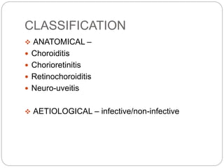


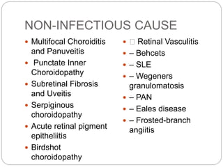
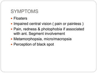


This document discusses choroiditis, an inflammation of the choroid layer of the eye. It begins by introducing choroiditis and classifying it anatomically and by etiology. It then discusses infectious causes such as parasites, bacteria, viruses, and fungi. Non-infectious causes are also listed. The document outlines symptoms, signs, and characteristics of choroiditis lesions. Specific sections provide details on toxoplasmosis and toxocariasis, two common causes of choroiditis. Treatment options including medications, surgery, and procedures are mentioned. In summary, the document provides a comprehensive overview of choroiditis, including causes, presentation, diagnosis, and management.


