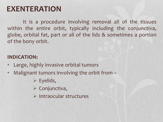
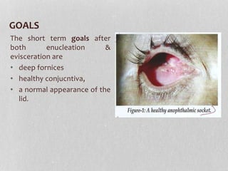
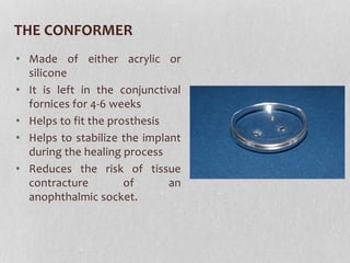
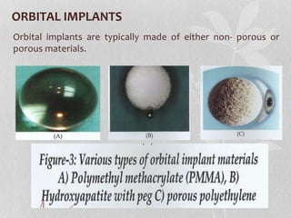
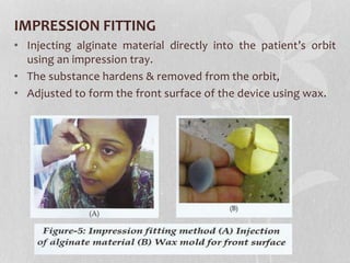
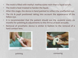
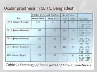
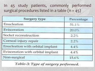
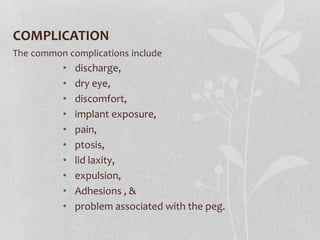
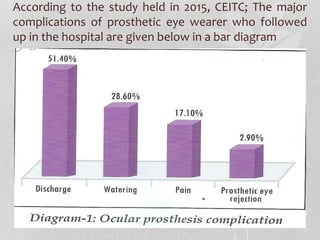
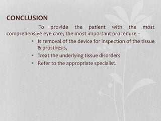
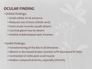
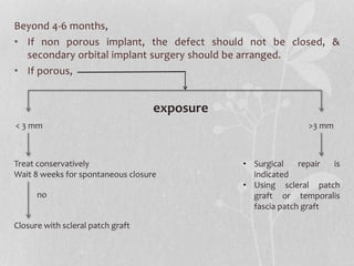
Ophthalmic horizon
Published on September 2016
OCULAR PROSTHESIS:
AN ESSENTIAL
COSMETIC
MANAGEMENT
Prof. Munirujzaman Osmani
BACKGROUND
• The ocular prosthetic device is very important to
provide the best possible functional & cosmetic
results.
• It is important to have a firm understanding of
management of the anophthalmic socket, and when
to make appropriate referrals to the ophthalmic
surgeon.
SURGICAL PROCEDURES
To understand & manage the complications associated
with prosthetic eye wear, eye care professionals first must
understand anophthalmic procedures.
There are three main surgical techniques used in the
partial or complete removal of the eye:
• Enucleation,
• Evisceration, &
• Exenteration.
ENUCLEATION
It is the complete removal of the globe, along with a
portion of the optic nerve, while maintaining the surrounding
orbital tissue.
INDICATION:
 Intraocular malignancy include –
• Retinoblastomas &
• Uveal melanomas
 Phthisical eyes of unknown etiology.
 Severely ruptured globe.
EVISCERATION
It involves the surgical removal of the contents of globe,
while preserving the sclera, extraocular muscles & optic nerve.
INDICATION:
 Painful blind eyes found in end stage of –
• Chronic uveitis or
• Neovascular glaucoma, and
• Corneal perforation.
 Endophthalmitis
EXENTERATION
It is a procedure involving removal all of the tissues
within the entire orbit, typically including the conjunctiva,
globe, orbital fat, part or all of the lids & sometimes a portion
of the bony orbit.
INDICATION:
• Large, highly invasive orbital tumors
• Malignant tumors involving the orbit from –
 Eyelids,
 Conjunctiva,
 Intraocular structures
GOALS
The short term goals after
both enucleation &
evisceration are
• deep fornices
• healthy conjucntiva,
• a normal appearance of the
lid.
THE CONFORMER
• Made of either acrylic or
silicone
• It is left in the conjunctival
fornices for 4-6 weeks
• Helps to fit the prosthesis
• Helps to stabilize the implant
during the healing process
• Reduces the risk of tissue
contracture of an
anophthalmic socket.
ORBITAL IMPLANTS
Orbital implants are typically made of either non- porous or
porous materials.
NONPOROUS ALLOPLASTIC IMPLANTS
 Silicone & polymethyl methacrylate (PMMA) .
 Solid, spherical implant that are well tolerated,
POROUS MATERIAL
 More commonly used
 Material: porous polyethylene, hydroxyapatite & aluminum
oxide.
 Due to the porous nature of these materials, fibrovascular
ingrowth occurs, allowing for improved implant stability &
decreases rejection risk.
TYPES OF OCULAR PROSTHESIS
• BASED ON THICKNESS:
• BASED ON FABRICATION:
Prosthetic eye Prosthetic shell
Thickness more than 1.5 mm Thickness less than 1.5mm
Ready made Custom made
Advantage:
 Inexpensive
 Time limitation exists
Disadvantage:
 Ill fitting
 Improper shade matching
• Increases the adapting with
movement of the eyeball as well as
fitting
• Exactly matches the iris position as
that of the adjacent natural eye
FABRICATION OF OCULAR PROSTHESIS
• Six to eight weeks after surgery, an ocular prosthesis can be
fitted.
• A prosthetic device can be fabricated in two forms,
• A scleral shell -- fit over a phthisical eye
• A full thickness prosthesis -- fit over the anophthalmic
socket.
• Prosthetic devices can be fit either from a stock set of pre-
fabricated eyes or can be custom made.
• Custom made prosthetic eye is preferred to increase stability
& aid in movement.
• The fitting method chosen is upon the ocularist.
• One of the most common fitting techniques is the
impression fitting.
IMPRESSION FITTING
• Injecting alginate material directly into the patient’s orbit
using an impression tray.
• The substance hardens & removed from the orbit,
• Adjusted to form the front surface of the device using wax.
• The mold is filled with methyl- methacrylate resin that is liquid acrylic.
• The mold is heat treated to harden the liquid.
• After this stage, the device is hand painted to reflect the unaffected eye.
• The iris & pupil positioned taking into account the appearance of the
fellow eye.
• It is recommended that the patient should see the ocularist every six
months for polishing & adjustments to the device at least annually.
• Removal of prosthetic device is similar in fashion to the removal of a
hard contact lens.
polishing contouring
Ocular prosthesis in CEITC, Bangladesh
In 45 study patients, commonly performed
surgical procedures listed in a table (n = 45)
COMPLICATION
The common complications include
• discharge,
• dry eye,
• discomfort,
• implant exposure,
• pain,
• ptosis,
• lid laxity,
• expulsion,
• Adhesions , &
• problem associated with the peg.
According to the study held in 2015, CEITC; The major
complications of prosthetic eye wearer who followed
up in the hospital are given below in a bar diagram
CONCLUSION
To provide the patient with the most
comprehensive eye care, the most important procedure –
• Is removal of the device for inspection of the tissue
& prosthesis,
• Treat the underlying tissue disorders
• Refer to the appropriate specialist.
LITERATURE REVIEW
ANOPHTHALMIA
• Anophthalmia is absence of globe
• It may be congenital or acquired.
CONGENITAL ANOPHTHALMIA
• Very rare condition
• Optic vesicle fails to develop
• Causes:
• Idiopathic/ sporadic
• Inherited as dominant, recessive
or sex- linked
• Maternal exposure or
teratogenic infection
OCULAR FINDING
• Orbital findings:
Small orbital rim & entrance
Reduced size of bony orbital cavity
Extra ocular muscles usually absent
Lacrimal gland may be absent
Small & maldeveloped optic foramen
• Eyelid findings:
Foreshortening of the lids in all directions
Absent or decreased levator function with decreased lid folds
Contraction of orbicularis oculi muscle
Shallow conjunctival fornix, especially inferiorly
ACQUIRED ANOPHTHALMIA
 After enucleation, evisceration or exenteration.
IDEAL ANOPHTHALMIC SOCKET
1. Centrally placed, well- covered, buried implant of adequate
volume.
2. Fabricated from a bio- inert material.
3. Socket lined with healthy conjunctiva.
4. Fornices deep enough to retain a prosthesis
5. Eyelids with normal position & appearance, & adequate
tone to support a prosthesis
6. Normal position of the eyelashes & eyelid margin
7. A comfortable ocular prosthesis that looks similar to the
sighted, contralateral globe & in the same horizontal plane.
ADVANTAGES OF EVISCERATION OVER
ENUCLEATION:
• Less disruption of orbital anatomy
• Good motility of prosthesis
• Lower rate of migration, extrusion & reoperation.
Changes associated with anophthalmia
POST ENUCLEATION SOCKET SYNDROME
• Introduced by Tyler's & Collin
• Sequelae of an enucleation are orbital volume deficiency &
changes in the orbital soft tissue architecture leading to the
clinical picture of “POST- ENUCLEATION SOCKET SYNDROME
(PESS)”.
• CLINICAL FEATURES:
 Enophthalmos
 An upper eyelid sulcus deformity
 Ptosis or eyelid retraction
 Laxity of the lower eyelid
 A backward tilt of the ocular prosthesis.
MANAGEMENT
1. Orbital volume replacement
a) Secondary implant (intraconal) if no implant was placed at
the time of primary surgery
b) Orbital floor implant
c) Dermis fat graft to upper sulcus (option in patients with
associated surface contracture)
2. Lower lid tightening
a) Lateral canthal sling
b) Medial canthal reconstruction
c) Fascial sling
3. Correction of shallow lower fornix
Fornix deepening sutures
4. Ptosis correction
IDEAL ORBITAL IMPLANT
• Maintain natural Lid shape
• Light weight
• Porosity
• Natural biocompatibility
• Non toxic & non allergic.
COMPLICATION OF IMPLANT:
Exposure & Extrusion of implant:
• Implant exposure may occur with any type of implant or at
any time may lead to implant extrusion or explantation.
• Porous orbital implants have a lower incidence of implant
exposure than traditional non porous implants.
PREDISPOSING FACTORS OF EXPOSURE OF
IMPLANT
1. Closing the wound under tension
2. Poor wound closure techniques
3. Infection
4. Mechanical or inflammatory irritation from the speculated
surface of the porous implant
5. Delayed ingrowth of fibrovascular tissue with subsequent
tissue breakdown
PREVENTIVE MEASURE FOR IMPLANT
EXPOSURE
• Proper placement of the implant within the orbit followed by a
two- layered closure of anterior Tenon’s capsule & conjunctiva
• The rectus muscles are then attached to the wrapped implant.
TREATMENT:
if few weeks,
• No infection, simple reclosure or with a patch graft (e.g.
sclera, temporalis fascia) is required.
• If infection is suspected & treated vigorously with topical &
systemic antibiotics, an extrusion & removal of the implant
may be avoided.
Beyond 4-6 months,
• If non porous implant, the defect should not be closed, &
secondary orbital implant surgery should be arranged.
• If porous,
exposure
< 3 mm >3 mm
Treat conservatively
Wait 8 weeks for spontaneous closure
no
Closure with scleral patch graft
• Surgical repair is
indicated
• Using scleral patch
graft or temporalis
fascia patch graft
THANK YOU
Next case presentation by DR. BURHAN UDDIN
Topic: THYROID ORBITOPATHY

Ocular Prosthesis

  • 2.
    Ophthalmic horizon Published onSeptember 2016 OCULAR PROSTHESIS: AN ESSENTIAL COSMETIC MANAGEMENT Prof. Munirujzaman Osmani
  • 3.
    BACKGROUND • The ocularprosthetic device is very important to provide the best possible functional & cosmetic results. • It is important to have a firm understanding of management of the anophthalmic socket, and when to make appropriate referrals to the ophthalmic surgeon.
  • 4.
    SURGICAL PROCEDURES To understand& manage the complications associated with prosthetic eye wear, eye care professionals first must understand anophthalmic procedures. There are three main surgical techniques used in the partial or complete removal of the eye: • Enucleation, • Evisceration, & • Exenteration.
  • 5.
    ENUCLEATION It is thecomplete removal of the globe, along with a portion of the optic nerve, while maintaining the surrounding orbital tissue. INDICATION:  Intraocular malignancy include – • Retinoblastomas & • Uveal melanomas  Phthisical eyes of unknown etiology.  Severely ruptured globe.
  • 6.
    EVISCERATION It involves thesurgical removal of the contents of globe, while preserving the sclera, extraocular muscles & optic nerve. INDICATION:  Painful blind eyes found in end stage of – • Chronic uveitis or • Neovascular glaucoma, and • Corneal perforation.  Endophthalmitis
  • 7.
    EXENTERATION It is aprocedure involving removal all of the tissues within the entire orbit, typically including the conjunctiva, globe, orbital fat, part or all of the lids & sometimes a portion of the bony orbit. INDICATION: • Large, highly invasive orbital tumors • Malignant tumors involving the orbit from –  Eyelids,  Conjunctiva,  Intraocular structures
  • 8.
    GOALS The short termgoals after both enucleation & evisceration are • deep fornices • healthy conjucntiva, • a normal appearance of the lid.
  • 9.
    THE CONFORMER • Madeof either acrylic or silicone • It is left in the conjunctival fornices for 4-6 weeks • Helps to fit the prosthesis • Helps to stabilize the implant during the healing process • Reduces the risk of tissue contracture of an anophthalmic socket.
  • 10.
    ORBITAL IMPLANTS Orbital implantsare typically made of either non- porous or porous materials.
  • 11.
    NONPOROUS ALLOPLASTIC IMPLANTS Silicone & polymethyl methacrylate (PMMA) .  Solid, spherical implant that are well tolerated, POROUS MATERIAL  More commonly used  Material: porous polyethylene, hydroxyapatite & aluminum oxide.  Due to the porous nature of these materials, fibrovascular ingrowth occurs, allowing for improved implant stability & decreases rejection risk.
  • 12.
    TYPES OF OCULARPROSTHESIS • BASED ON THICKNESS: • BASED ON FABRICATION: Prosthetic eye Prosthetic shell Thickness more than 1.5 mm Thickness less than 1.5mm Ready made Custom made Advantage:  Inexpensive  Time limitation exists Disadvantage:  Ill fitting  Improper shade matching • Increases the adapting with movement of the eyeball as well as fitting • Exactly matches the iris position as that of the adjacent natural eye
  • 13.
    FABRICATION OF OCULARPROSTHESIS • Six to eight weeks after surgery, an ocular prosthesis can be fitted. • A prosthetic device can be fabricated in two forms, • A scleral shell -- fit over a phthisical eye • A full thickness prosthesis -- fit over the anophthalmic socket.
  • 14.
    • Prosthetic devicescan be fit either from a stock set of pre- fabricated eyes or can be custom made. • Custom made prosthetic eye is preferred to increase stability & aid in movement. • The fitting method chosen is upon the ocularist. • One of the most common fitting techniques is the impression fitting.
  • 15.
    IMPRESSION FITTING • Injectingalginate material directly into the patient’s orbit using an impression tray. • The substance hardens & removed from the orbit, • Adjusted to form the front surface of the device using wax.
  • 16.
    • The moldis filled with methyl- methacrylate resin that is liquid acrylic. • The mold is heat treated to harden the liquid. • After this stage, the device is hand painted to reflect the unaffected eye. • The iris & pupil positioned taking into account the appearance of the fellow eye. • It is recommended that the patient should see the ocularist every six months for polishing & adjustments to the device at least annually. • Removal of prosthetic device is similar in fashion to the removal of a hard contact lens. polishing contouring
  • 17.
    Ocular prosthesis inCEITC, Bangladesh
  • 18.
    In 45 studypatients, commonly performed surgical procedures listed in a table (n = 45)
  • 19.
    COMPLICATION The common complicationsinclude • discharge, • dry eye, • discomfort, • implant exposure, • pain, • ptosis, • lid laxity, • expulsion, • Adhesions , & • problem associated with the peg.
  • 20.
    According to thestudy held in 2015, CEITC; The major complications of prosthetic eye wearer who followed up in the hospital are given below in a bar diagram
  • 21.
    CONCLUSION To provide thepatient with the most comprehensive eye care, the most important procedure – • Is removal of the device for inspection of the tissue & prosthesis, • Treat the underlying tissue disorders • Refer to the appropriate specialist.
  • 22.
  • 23.
    ANOPHTHALMIA • Anophthalmia isabsence of globe • It may be congenital or acquired.
  • 24.
    CONGENITAL ANOPHTHALMIA • Veryrare condition • Optic vesicle fails to develop • Causes: • Idiopathic/ sporadic • Inherited as dominant, recessive or sex- linked • Maternal exposure or teratogenic infection
  • 25.
    OCULAR FINDING • Orbitalfindings: Small orbital rim & entrance Reduced size of bony orbital cavity Extra ocular muscles usually absent Lacrimal gland may be absent Small & maldeveloped optic foramen • Eyelid findings: Foreshortening of the lids in all directions Absent or decreased levator function with decreased lid folds Contraction of orbicularis oculi muscle Shallow conjunctival fornix, especially inferiorly
  • 26.
    ACQUIRED ANOPHTHALMIA  Afterenucleation, evisceration or exenteration.
  • 27.
    IDEAL ANOPHTHALMIC SOCKET 1.Centrally placed, well- covered, buried implant of adequate volume. 2. Fabricated from a bio- inert material. 3. Socket lined with healthy conjunctiva. 4. Fornices deep enough to retain a prosthesis 5. Eyelids with normal position & appearance, & adequate tone to support a prosthesis 6. Normal position of the eyelashes & eyelid margin 7. A comfortable ocular prosthesis that looks similar to the sighted, contralateral globe & in the same horizontal plane.
  • 28.
    ADVANTAGES OF EVISCERATIONOVER ENUCLEATION: • Less disruption of orbital anatomy • Good motility of prosthesis • Lower rate of migration, extrusion & reoperation.
  • 29.
  • 30.
    POST ENUCLEATION SOCKETSYNDROME • Introduced by Tyler's & Collin • Sequelae of an enucleation are orbital volume deficiency & changes in the orbital soft tissue architecture leading to the clinical picture of “POST- ENUCLEATION SOCKET SYNDROME (PESS)”. • CLINICAL FEATURES:  Enophthalmos  An upper eyelid sulcus deformity  Ptosis or eyelid retraction  Laxity of the lower eyelid  A backward tilt of the ocular prosthesis.
  • 33.
    MANAGEMENT 1. Orbital volumereplacement a) Secondary implant (intraconal) if no implant was placed at the time of primary surgery b) Orbital floor implant c) Dermis fat graft to upper sulcus (option in patients with associated surface contracture) 2. Lower lid tightening a) Lateral canthal sling b) Medial canthal reconstruction c) Fascial sling 3. Correction of shallow lower fornix Fornix deepening sutures 4. Ptosis correction
  • 34.
    IDEAL ORBITAL IMPLANT •Maintain natural Lid shape • Light weight • Porosity • Natural biocompatibility • Non toxic & non allergic. COMPLICATION OF IMPLANT: Exposure & Extrusion of implant: • Implant exposure may occur with any type of implant or at any time may lead to implant extrusion or explantation. • Porous orbital implants have a lower incidence of implant exposure than traditional non porous implants.
  • 35.
    PREDISPOSING FACTORS OFEXPOSURE OF IMPLANT 1. Closing the wound under tension 2. Poor wound closure techniques 3. Infection 4. Mechanical or inflammatory irritation from the speculated surface of the porous implant 5. Delayed ingrowth of fibrovascular tissue with subsequent tissue breakdown
  • 36.
    PREVENTIVE MEASURE FORIMPLANT EXPOSURE • Proper placement of the implant within the orbit followed by a two- layered closure of anterior Tenon’s capsule & conjunctiva • The rectus muscles are then attached to the wrapped implant. TREATMENT: if few weeks, • No infection, simple reclosure or with a patch graft (e.g. sclera, temporalis fascia) is required. • If infection is suspected & treated vigorously with topical & systemic antibiotics, an extrusion & removal of the implant may be avoided.
  • 37.
    Beyond 4-6 months, •If non porous implant, the defect should not be closed, & secondary orbital implant surgery should be arranged. • If porous, exposure < 3 mm >3 mm Treat conservatively Wait 8 weeks for spontaneous closure no Closure with scleral patch graft • Surgical repair is indicated • Using scleral patch graft or temporalis fascia patch graft
  • 38.
    THANK YOU Next casepresentation by DR. BURHAN UDDIN Topic: THYROID ORBITOPATHY