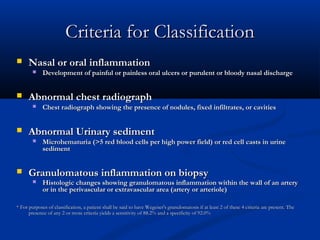
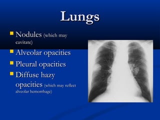
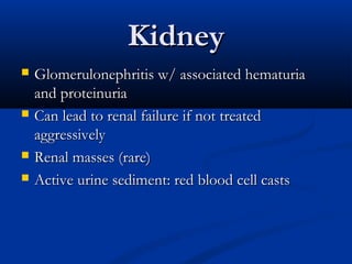
Wegener’s
Granulomatosis
     Kelly Mitchell
     July 5, 2006
    Morning Report
History of Wegener’s
    In 1931, two patients died from prolonged
    sepsis with inflammation of blood vessels
    scattered throughout the body.
   In 1936, Wegener first described a distinct
    syndrome in three patients found to have
    necrotizing granulomas involving the upper and
    lower respiratory tract.
   In 1954, seven more patients described, resulting
    in definate criteria
The Controversy
   Wegener’s vs PR3-ANCA vasculitis
     Lancet, 22 April 2006
     Suggestion that using Wegener’s name “needs
      balanced discussion within the scientific community”

       Reiter's syndrome- reactive arthritis
The Problem with Changing

   Multiple ANCA+ diseases:
       microscopic polyangiitis (MPA)
       "renal-limited" vasculitis (pauci-immune glomerulonephritis without
        evidence of extrarenal disease)
       Churg-Strauss syndrome (CSS)
       Drug-induced vasculitis
       Goodpasture’s
       Rheumatic disorders
       Autoimmune GI disorders
       CF
   Diagnostic Criteria primarily clinical
Criteria for Classification
     Nasal or oral inflammation
            Development of painful or painless oral ulcers or purulent or bloody nasal discharge


     Abnormal chest radiograph
            Chest radiograph showing the presence of nodules, fixed infiltrates, or cavities


     Abnormal Urinary sediment
            Microhematuria (>5 red blood cells per high power field) or red cell casts in urine
             sediment


     Granulomatous inflammation on biopsy
            Histologic changes showing granulomatous inflammation within the wall of an artery
             or in the perivascular or extravascular area (artery or arteriole)

* For purposes of classification, a patient shall be said to have Wegener's granulomatosis if at least 2 of these 4 criteria are present. The
      presence of any 2 or more criteria yields a sensitivity of 88.2% and a specificity of 92.0%
Classic Symptoms
   Upper respiratory tract
     sinuses
     Nose

     ears

     trachea

   Lungs
   Kidneys
Eye
 Scleritis

 Uveitis

 Orbital
  pseudotumor
  /proptosis
Upper Respiratory Tract
                  Ear
 Ear infections that are slow to resolve.

 Recurrent otitis media.

 Decrease in hearing.
Upper Respiratory Tract
                                         Nose
 Nasal crusting
 Frequent
  nosebleeds
 Erosion and
  perforation of the
  nasal septum. The bridge
    of the nose can collapse resulting in a
    “saddle–nose deformity”.
Upper Respiratory Tract
                    Sinuses/Trachea
   Sinuses
       Chronic sinus
        inflammation
   Trachea
       subglottic stenosis
Lungs
   Nodules (which may
    cavitate)
 Alveolar opacities
 Pleural opacities
 Diffuse hazy
  opacities (which may reflect
    alveolar hemorrhage)
Kidney
   Glomerulonephritis w/ associated hematuria
    and proteinuria
   Can lead to renal failure if not treated
    aggressively
   Renal masses (rare)
   Active urine sediment: red blood cell casts
RBC casts
Skin
   “palpable purpura” most
    common

   Raynaud’s phenomenon
    —due to inadequate
    blood flow to fingers and
    toes

   Ulcers
Miscellaneous
   Joints
    Arthritis can occur, with joint swelling and pain
   Nerves
    Peripheral nerve involvement leads to numbness,
    tingling, shooting pains in the extremities, and
    sometimes to weakness in a foot, hand, arm, or leg
   Meninges
   Prostate gland
   Genito–urinary tract
   Constitutional symptoms of fatigue, low–grade fever,
    and weight loss
Incidence of symptoms
    Symptom                  At Onset                         Total
   ENT                      75%                              95%
   Lung                     50                               85
   Joints                   30                               70
   Fever                    25                               50
   Kidney                   20                               75
   Cough                    20                               50
   Eye                      15                               50
   Skin                     15                               45
   Weight Loss              10                               35
   Nervous System (Central/Peripheral) 0            10/15

One-third of patients may be without symptoms at onset of disease
Pathogenesis
                     Risk factors and inciting events

   Exact events obscure
     Infectious—staph?
     Genetic
            single nucleotide polymorphism in a gene encoding a protein tyrosine
             phosphatase (PTPN22)
            AAT deficiency
       Environmental—inhalational?
          Silica
          lead
          mercury
Pathogenesis
                       ANCA
   ANCAs may be not only markers for Wegener's
    granulomatosis and related disorders, but they
    may also be actors in pathogenesis
   Neutrophils exposed to cytokines such as TNF,
    express PR3 & MPO (the targets for ANCAs)
   Adding ANCAs to these cytokine-primed
    neutrophils causes them to generate oxygen
    radicals and release enzymes capable of
    damaging blood vessels.
Pathogenesis
   “Priming” of Neutrophils
       Exposing PR3 and MPO epitopes
   ANCA binding
   Degranulation/ROS production/neutrophil-
    endothelial cell interaction
   Increased ANCA = Increased degranulation rate
Diagnosis
                  Criteria for Classification
   Nasal or oral inflammation
       Development of painful or painless oral ulcers or purulent or bloody nasal
        discharge

   Abnormal chest radiograph
       Chest radiograph showing the presence of nodules, fixed infiltrates, or
        cavities

   Abnormal urinary sediment
       Microhematuria (>5 red blood cells per high power field) or red cell casts
        in urine sediment

   Granulomatous inflammation on biopsy
       Histologic changes showing granulomatous inflammation within the wall
        of an artery or in the perivascular or extravascular area (artery or arteriole)
Diagnosis
   Biopsy specimens showing the triad of vasculitis, granulomata,
    and large areas of necrosis
      Sinuses
      Nose
      Skin--leukocytoclastic vasculitis with little or no complement and
         immunoglobulin on immunofluorescence
        Kidney--segmental necrotizing glomerulonephritis that is usually pauci-
         immune on immunofluorescence / EM
         Lung--vasculitis and granulomatous inflammation
         (Only large sections of lung tissue obtained via thoracoscopic or open
          lung biopsy are likely to show all of the histologic features)
   Seropositivity for C-ANCAs
Antineutrophil cytoplasmic
       antibodies
ANCA
   ~90% of Wegener's cases are ANCA+
       In limited dz, up to 40% may be ANCA neg

   80 - 90 % PR3-ANCA

   Remaining MPO-ANCA
Is ANCA sufficient?
   Concensus is that tissue dx is necessary

   Rarely may initiate tx w/o biopsy

   Should attempt to confirm w/ biopsy when able
Treatment
                   Traditional
   Prednisone (initiated at 1 mg/kg daily for 1 to
    2 months. then tapered)

   Cyclophosphamide (2mg/kg daily for at least
    12 months)

   >90% improve and 75% remit
Treatment
           However, 50% in remission relapse
           AND daily cyclophos is very toxic
   pancytopenia,
   infection,
   hemorrhagic cystitis
   bladder cancer (increased 33-fold)
   lymphoma (increased 11-fold)
Treatment
   Monthly IV cyclophosphamide -- less toxic but less
    effective
   Weekly methotrexate -- maintains remission
   Trimethoprim-sulfamethoxazole -- controversial (?
    effective for disease limited to the respiratory tract), reduces the relapse rate
   Steroids —prednisone vs solumedrol
   Plasmapheresis -unproven, awaiting MEPEX trial
        Recommended for anti-GBM+, pulm hemmorhage, renal failure
   IVIG— recommended in the setting of infection during PLEX
Vasculidities
   Large vessel vasculitis
        Takayasu arteritis
        Giant cell arteritis
   Medium sized vessel vasculitis
        Polyarteritis nodosa
        Isolated central nervous system vasculitis
   Small vessel vasculitis
        Churg-Strauss arteritis
        Wegener's granulomatosis
        Microscopic polyarteritis
        Henoch-Schönlein purpura
        Essential cryoglobulinemic vasculitis
        Hypersensitivity vasculitis
        Vasculitis secondary to connective tissue disorders -- SLE, rheumatoid
         arthritis, relapsing polychondritis, Behcet's disease
        Vasculitis secondary to viral infection —hepatitis B and C, HIV, CMV,
         EBV, Parvo B19
What, then, is the role of ANCA?
   Is a positive test result a "true-positive"?
   Does a negative ANCA assay exclude an "ANCA-
    associated" vasculitis?
   Is the presence of a positive ANCA assay in and of
    itself sufficient to establish the diagnosis (ie, does it
    preclude the need for biopsy?)
   Does an increase in ANCA titer predict a disease flare?
   Does a persistently negative ANCA ensure disease
    quiescence?

9a1c wg mitchell-7-5-06

  • 1.
    Wegener’s Granulomatosis Kelly Mitchell July 5, 2006 Morning Report
  • 2.
    History of Wegener’s  In 1931, two patients died from prolonged sepsis with inflammation of blood vessels scattered throughout the body.  In 1936, Wegener first described a distinct syndrome in three patients found to have necrotizing granulomas involving the upper and lower respiratory tract.  In 1954, seven more patients described, resulting in definate criteria
  • 3.
    The Controversy  Wegener’s vs PR3-ANCA vasculitis  Lancet, 22 April 2006  Suggestion that using Wegener’s name “needs balanced discussion within the scientific community”  Reiter's syndrome- reactive arthritis
  • 4.
    The Problem withChanging  Multiple ANCA+ diseases:  microscopic polyangiitis (MPA)  "renal-limited" vasculitis (pauci-immune glomerulonephritis without evidence of extrarenal disease)  Churg-Strauss syndrome (CSS)  Drug-induced vasculitis  Goodpasture’s  Rheumatic disorders  Autoimmune GI disorders  CF  Diagnostic Criteria primarily clinical
  • 5.
    Criteria for Classification  Nasal or oral inflammation  Development of painful or painless oral ulcers or purulent or bloody nasal discharge  Abnormal chest radiograph  Chest radiograph showing the presence of nodules, fixed infiltrates, or cavities  Abnormal Urinary sediment  Microhematuria (>5 red blood cells per high power field) or red cell casts in urine sediment  Granulomatous inflammation on biopsy  Histologic changes showing granulomatous inflammation within the wall of an artery or in the perivascular or extravascular area (artery or arteriole) * For purposes of classification, a patient shall be said to have Wegener's granulomatosis if at least 2 of these 4 criteria are present. The presence of any 2 or more criteria yields a sensitivity of 88.2% and a specificity of 92.0%
  • 6.
    Classic Symptoms  Upper respiratory tract  sinuses  Nose  ears  trachea  Lungs  Kidneys
  • 7.
    Eye  Scleritis  Uveitis Orbital pseudotumor /proptosis
  • 8.
    Upper Respiratory Tract Ear  Ear infections that are slow to resolve.  Recurrent otitis media.  Decrease in hearing.
  • 9.
    Upper Respiratory Tract Nose  Nasal crusting  Frequent nosebleeds  Erosion and perforation of the nasal septum. The bridge of the nose can collapse resulting in a “saddle–nose deformity”.
  • 10.
    Upper Respiratory Tract Sinuses/Trachea  Sinuses  Chronic sinus inflammation  Trachea  subglottic stenosis
  • 11.
    Lungs  Nodules (which may cavitate)  Alveolar opacities  Pleural opacities  Diffuse hazy opacities (which may reflect alveolar hemorrhage)
  • 12.
    Kidney  Glomerulonephritis w/ associated hematuria and proteinuria  Can lead to renal failure if not treated aggressively  Renal masses (rare)  Active urine sediment: red blood cell casts
  • 13.
  • 14.
    Skin  “palpable purpura” most common  Raynaud’s phenomenon —due to inadequate blood flow to fingers and toes  Ulcers
  • 15.
    Miscellaneous  Joints Arthritis can occur, with joint swelling and pain  Nerves Peripheral nerve involvement leads to numbness, tingling, shooting pains in the extremities, and sometimes to weakness in a foot, hand, arm, or leg  Meninges  Prostate gland  Genito–urinary tract  Constitutional symptoms of fatigue, low–grade fever, and weight loss
  • 16.
    Incidence of symptoms Symptom At Onset Total  ENT 75% 95%  Lung 50 85  Joints 30 70  Fever 25 50  Kidney 20 75  Cough 20 50  Eye 15 50  Skin 15 45  Weight Loss 10 35  Nervous System (Central/Peripheral) 0 10/15 One-third of patients may be without symptoms at onset of disease
  • 17.
    Pathogenesis Risk factors and inciting events  Exact events obscure  Infectious—staph?  Genetic  single nucleotide polymorphism in a gene encoding a protein tyrosine phosphatase (PTPN22)  AAT deficiency  Environmental—inhalational?  Silica  lead  mercury
  • 18.
    Pathogenesis ANCA  ANCAs may be not only markers for Wegener's granulomatosis and related disorders, but they may also be actors in pathogenesis  Neutrophils exposed to cytokines such as TNF, express PR3 & MPO (the targets for ANCAs)  Adding ANCAs to these cytokine-primed neutrophils causes them to generate oxygen radicals and release enzymes capable of damaging blood vessels.
  • 19.
    Pathogenesis  “Priming” of Neutrophils  Exposing PR3 and MPO epitopes  ANCA binding  Degranulation/ROS production/neutrophil- endothelial cell interaction  Increased ANCA = Increased degranulation rate
  • 20.
    Diagnosis Criteria for Classification  Nasal or oral inflammation  Development of painful or painless oral ulcers or purulent or bloody nasal discharge  Abnormal chest radiograph  Chest radiograph showing the presence of nodules, fixed infiltrates, or cavities  Abnormal urinary sediment  Microhematuria (>5 red blood cells per high power field) or red cell casts in urine sediment  Granulomatous inflammation on biopsy  Histologic changes showing granulomatous inflammation within the wall of an artery or in the perivascular or extravascular area (artery or arteriole)
  • 21.
    Diagnosis  Biopsy specimens showing the triad of vasculitis, granulomata, and large areas of necrosis  Sinuses  Nose  Skin--leukocytoclastic vasculitis with little or no complement and immunoglobulin on immunofluorescence  Kidney--segmental necrotizing glomerulonephritis that is usually pauci- immune on immunofluorescence / EM  Lung--vasculitis and granulomatous inflammation (Only large sections of lung tissue obtained via thoracoscopic or open lung biopsy are likely to show all of the histologic features)  Seropositivity for C-ANCAs
  • 23.
  • 24.
    ANCA  ~90% of Wegener's cases are ANCA+  In limited dz, up to 40% may be ANCA neg  80 - 90 % PR3-ANCA  Remaining MPO-ANCA
  • 25.
    Is ANCA sufficient?  Concensus is that tissue dx is necessary  Rarely may initiate tx w/o biopsy  Should attempt to confirm w/ biopsy when able
  • 26.
    Treatment Traditional  Prednisone (initiated at 1 mg/kg daily for 1 to 2 months. then tapered)  Cyclophosphamide (2mg/kg daily for at least 12 months)  >90% improve and 75% remit
  • 27.
    Treatment However, 50% in remission relapse AND daily cyclophos is very toxic  pancytopenia,  infection,  hemorrhagic cystitis  bladder cancer (increased 33-fold)  lymphoma (increased 11-fold)
  • 28.
    Treatment  Monthly IV cyclophosphamide -- less toxic but less effective  Weekly methotrexate -- maintains remission  Trimethoprim-sulfamethoxazole -- controversial (? effective for disease limited to the respiratory tract), reduces the relapse rate  Steroids —prednisone vs solumedrol  Plasmapheresis -unproven, awaiting MEPEX trial  Recommended for anti-GBM+, pulm hemmorhage, renal failure  IVIG— recommended in the setting of infection during PLEX
  • 31.
    Vasculidities  Large vessel vasculitis  Takayasu arteritis  Giant cell arteritis  Medium sized vessel vasculitis  Polyarteritis nodosa  Isolated central nervous system vasculitis  Small vessel vasculitis  Churg-Strauss arteritis  Wegener's granulomatosis  Microscopic polyarteritis  Henoch-Schönlein purpura  Essential cryoglobulinemic vasculitis  Hypersensitivity vasculitis  Vasculitis secondary to connective tissue disorders -- SLE, rheumatoid arthritis, relapsing polychondritis, Behcet's disease  Vasculitis secondary to viral infection —hepatitis B and C, HIV, CMV, EBV, Parvo B19
  • 32.
    What, then, isthe role of ANCA?  Is a positive test result a "true-positive"?  Does a negative ANCA assay exclude an "ANCA- associated" vasculitis?  Is the presence of a positive ANCA assay in and of itself sufficient to establish the diagnosis (ie, does it preclude the need for biopsy?)  Does an increase in ANCA titer predict a disease flare?  Does a persistently negative ANCA ensure disease quiescence?