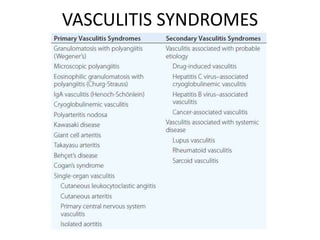
VASCULITIS
TAJUL NISA AJLA
IMS Bangalore, 2016
INTRODUCTION
• DEFINITION:
– Clinicopathologic process characterized by inflammation of and
damage to blood vessels- tissue ischemia of affected vessel
• Have diverse clinical feature and group of syndromes since its
involvement in any type, size, and site of vascular beds. Clinical
feature also include constitutional symptoms.
• Can present as a sole manifestation (primary)
• May be a secondary component of another disease.
• Can be confined to a single organ or involve several organs
CLASSIFICATION
• Overlapping and lack of understanding of the pathogenesis
of vasculitis syndrome has made vasculitis difficult to classify.
• Various classification schemes are attempted; based on
vessel size, role of immune complex, presence of specific
antibody, granuloma formation and organ specificity.
• Chapel Hill nomenclature has been used in organizing this
diverse group of entities
– By dividing the disease based on size of vessel
Chapel Hill
VASCULITIS SYNDROMES
PATHOGENESIS
Immune complex formation
ANCA
Pathogenic T-lymphocyte response
Pathogenic immune-
complex formation
and/or deposition
• IgA vasculitis (Henoch-
Schönlein)
• Lupus vasculitis
• Serum sickness and
cutaneous vasculitis
syndromes
• Hepatitis C virus–
associated
cryoglobulinemic
vasculitis
• Hepatitis B virus–
associated vasculitis
Production of ANCA
• Granulomatosis with
polyangiitis (Wegener’s)
• Microscopic polyangiitis
• Eosinophilic
granulomatosis with
polyangiitis (Churg-
Strauss)
Pathogenic T lymphocyte
responses and
granuloma formation
• Giant cell arteritis
• Takayasu arteritis
• Granulomatosis with
polyangiitis (Wegener’s)
• Eosinophilic
granulomatosis with
polyangiitis (Churg-
Strauss)
Potential Mechanisms of Vessel Damage in
Vasculitis Syndromes
IMMUNE COMPLEX MECHANISM
• Damage to the vessel wall is due to deposition of
immune complex, but the cause of immune complex
formation is not clearly establish
• The actual antigen in the immune complex is rarely
identified
• In Polyarteritis nodosa, hepatitis B antigen is found in
both circulating and deposited immune complexes.
• In cryoglobulinemic vasculitis, hepatitis C has been
identified in the cryoprecipitate of the pts.
• The mechanism of of tissue damage resembles serum
sickness, type 3 hypersensitivity reaction.
ANTINEUTROPHIL CYTOPLASMIC
ANTIBODIES (ANCA)
• ANCA is autoantibodies that is directed against certain
protein in cytoplasmic granules of neutrophils and
monocyte
• ANCA was found in:
– Granulomatous polyangiitis (Wegener’s)
– Microscopic polyangiitis
– Eosinophilic granulomatous polyangiitis (Churg-Strauss)
• These disease are grouped as “ANCA-associated vasculitis”
Two category of ANCA:
cytoplasmic ANCA
(cANCA): proteinase-3
antigen
Perinuclear ANCA
(pANCA): enzyme
myeloperoxidase
PATHOGENIC T-LYMPHOCYTE RESPONSE
AND GRANULOMA FORMATION
• Histopathologic feature of granulomatous
vasculitis shows there is role of pathogenic T
lymphocyte response and cell mediated injury
Endothelial cells express HLA
class 2 molecule following
activation by cytokines; INF ƴ
Act like APC; interact with CD+4
T cells
Endothelial cell secrete IL
Activate the T cell and cause
immunologic process within the
blood vessel
IL-1 and INF-α are potent inducer of:
• endothelial-leucocyte adhesion
molecule 1 (ELAM-1)
• vascular cell adhesion molecule 1
(VCAM-1)
These enhance the adhesion of
leucocytes to endothelial cell
APPROACH TO THE
PATIENT
Diagnosis
• Unexplained systemic illness
• Palpable purpura
• Pulmonary infiltrates
• Microscopic hematuria
• Chronic inflammatory sinusitis
• Mononeuritis multiplex
• Unexplained ischemic event
• Glomerulonephritis with evidence of multisystem
disease
*Exclude other disease that mimic vasculitis
Infectious diseases
• Bacterial
endocarditis
• Disseminated
gonococcal infection
• Pulmonary
histoplasmosis
• Coccidioidomycosis
• Syphilis
• Lyme disease
• Rocky Mountain
spotted fever
• Whipple’s disease
Coagulopathies/thrombotic
microangiopathies
• Antiphospholipid
syndrome
• Thrombotic
thrombocytopenic
purpura
Drug toxicity
• Cocaine
• Levamisole
• Amphetamines
• Ergot alkaloids
• Methysergide
• Arsenic
Antiglomerular basement
membrane disease
(Goodpasture’s syndrome)
Neoplasms
Atrial myxoma
Lymphoma
Carcinomatosis
Atheroembolic disease
Migraine Amyloidosis
CONDITION THAT MIMIC VASCULITIS
LARGE VESSEL ARTERITIS:
GIANT CELL ARTERITIS (TEMPORAL)
TAKAYASU ARTERITIS
GIANT CELL ARTERITIS
• Historically referred to as temporal arteritis
• Chronic granulomatous inflammation of medium and
large arteries
• Involves one or more branches of carotid artery;
temporal artery,
• Systemic disease affect aorta and its branches,
• Can also involves vertebral arteries ophthalmic artery,
aorta
• Closely associated with polymyalgia rheumatica
– Stiffness, aching, and pain in the muscle of neck, shoulder,
lower back, hips, and thighs.
– Polymyalgia usually occurs as an isolation, 40-50% is
associated with giant cell arteritis, 10-20% pt presented
with polymyalgia may develop giant cell arteritis later
GIANT CELL ARTERITIS
• Age > 50 years
• Common more in women, rare in blacks
• High incidence in Scandinavia and region in US with
large population of Scandinavians
• Familial aggregation; association with HLA-DR4.
• Genetic studies shows association of giant cell arteritis
with alleles at HLA-DRB1 locus,
GIANT CELL ARTERITIS; INCIDENCE
AND PREVALENCE
PATHOLOGY
• Panarteritis, multiple arteries
are involved
• Histopathology: inflammatory
mononuclear cell infiltrates
within vessel wall with frequent
giant cell formation
• Proliferation of intima and
fragmentation of internal elastic
lamina
• Often cause ischemia of affected
organs.
PATHOGENESIS
• T-cell mediated immune response against an
unknown vessel wall antigen
• T-lymphocytes, dendritic cells, and
macrophages plays a critical role in disease
pathogenesis.
CLINICAL MANIFESTATION
• Constitutional symptoms:
– Fever, malaise, myalgia, night sweats, weight loss
• Hallmark symptom:
– New onset of localized headache; temporal area,
diffuse, bilateral
• Cranial artery symptoms:
– Headache, tender, thickened or nodular artery,
pulsatile but may occlude later, scalp pain,
claudication of jaw and tongue
CLINICAL MANIFESTATION
• Ophthalmic manifestations: ischemic optic
neuropathy
– Visual blurring, transient blurring, diplopia, eye
pain, sudden loss of vision
• Polymyalgia Rheumatica:
– Fever, malaise, pain and stiffness of neck,
shoulder, hip muscle without any abnormalities in
physical examination
CLINICAL MANIFESTATION
• Large vessel manifestation:
– Subclavian artery stenosis- arm claudication
– Aortic aneurysm; thoracic, abdominal aorta lead
to rupture and dissection
• LAB FINDING:
– Elevated ESR, normochromic or hypochromic
anemia, increased IgG and complement
– Alkaline phosphatase
DIAGNOSIS
• >50 yrs old pt with fever, anemia, high ESR
• With or without symptom of polymyalgia
rheumatica
• Diminish pulse, bruit
• Ocular signs and symptom
• Biopsy of temporal artery, ultrasound, and
arteriogram
TREATMENT OF GAINT CELL ARTERITIS
• Goal of treatment: Reduce symptom, prevent
visual loss
• Glucocorticoid;
– Prednisone: 40-60 mg/d for one month, gradual
tapering
– Methylprednisolone: 1000 mg daily, for 3 days, if
ocular sign occurs
– Level of ESR can be a guide while tapering the dose of drugs
• Aspirin, 81 mg daily to reduce risk of cranial
ischemia
• Tocilizumab (anti IL 6 receptor)- need further
study
COMPLICATION
• Fatality due to:
– Cerebrovascular disease
– Myocardial infarction
– Aortic aneurysm rupture
– dissection
TAKAYASU ARTERITIS
• DEFINITION:
– Inflammation and stenotic disease of medium and
large-sized arteries
– Have strong predilection for the aortic arc and its
branches, may also involve pulmonary artery
• Uncommon disease
• Adolescent girls and young women, more
common in Asia
PATHOLOGY AND PATHOGENESIS
• Large and medium-sized artery; panarteritis
• Immunopathogenic mechanism; demonstration of
circulating immune complex
• Marked intimal proliferation, fibrosis, scarring,
vascularisation of media, disruption and degeneration
of elastic lamina.
• Narrowing of lumen occurs with or without thrombosis
• Involve vasa vasorum
• Compromised blood flow- ischemia
CLINICAL FEATURE
• GENERALISED SYMPTOM: malaise, fever, night
sweats, arthralgia, anorexia, weight loss
• Vascular symptom: absent peripheral pulse
(subclavian artery), organ ischemia
• Hypertension- contributes to renal, cardiac,
cerebral injury
• LAB FINDING: elevated ESR, anemia, elevated
immunoglobulin
DIAGNOSIS
• Young women, with decrease or absence of peripheral
pulse, discrepancies in BP, arterial bruit.
• Arteriography: irregular vessel wall, stenosis,
poststenotic dilation, aneurysm, occlusion, increased
collateral circulation
• MRA, complete aortic arteriography by catheter-
directed dye arteriography- to delineate the
distribution and degree of arterial disesase
• HISTOPATHOLOGIC:
– vessel wall inflammation
– predominant lymphocytic with granuloma
formation
– giant cell at vessel wall
– lymphoplasmacytic infiltrate rich in IgG4-positive
plasma cells, storiform.
– Fibrosis, obliterative phlebitis.
DIAGNOSIS
TREATMENT TAKAYASU ARTERITIS
• Glucocorticoids:
– Prednisone; 40-60 mg per day- alleviate symptoms
• Surgical and arterioplastic approach to
stenosed vessels: decrease risk of stroke,
improving blood flow to ischemic viscera and
limbs
• Methotrexate up to 25 mg per week
• Anti-TNF- need further study
COMPLICATION
• Death occur due to:
– Congestive cardiac failure
– CVA
– MI
– Aneurysm rupture
– Renal failure
• Chronic and relapsing
REFERENCE
• Harrison's Principle of Internal Medicine 19th Edition
• Davidson’s Principles & Practice of Medicine 22nd
Edition
THANK YOU

Vasculitis

  • 1.
  • 2.
    INTRODUCTION • DEFINITION: – Clinicopathologicprocess characterized by inflammation of and damage to blood vessels- tissue ischemia of affected vessel • Have diverse clinical feature and group of syndromes since its involvement in any type, size, and site of vascular beds. Clinical feature also include constitutional symptoms. • Can present as a sole manifestation (primary) • May be a secondary component of another disease. • Can be confined to a single organ or involve several organs
  • 3.
    CLASSIFICATION • Overlapping andlack of understanding of the pathogenesis of vasculitis syndrome has made vasculitis difficult to classify. • Various classification schemes are attempted; based on vessel size, role of immune complex, presence of specific antibody, granuloma formation and organ specificity. • Chapel Hill nomenclature has been used in organizing this diverse group of entities – By dividing the disease based on size of vessel
  • 4.
  • 5.
  • 6.
  • 7.
    Pathogenic immune- complex formation and/ordeposition • IgA vasculitis (Henoch- Schönlein) • Lupus vasculitis • Serum sickness and cutaneous vasculitis syndromes • Hepatitis C virus– associated cryoglobulinemic vasculitis • Hepatitis B virus– associated vasculitis Production of ANCA • Granulomatosis with polyangiitis (Wegener’s) • Microscopic polyangiitis • Eosinophilic granulomatosis with polyangiitis (Churg- Strauss) Pathogenic T lymphocyte responses and granuloma formation • Giant cell arteritis • Takayasu arteritis • Granulomatosis with polyangiitis (Wegener’s) • Eosinophilic granulomatosis with polyangiitis (Churg- Strauss) Potential Mechanisms of Vessel Damage in Vasculitis Syndromes
  • 8.
    IMMUNE COMPLEX MECHANISM •Damage to the vessel wall is due to deposition of immune complex, but the cause of immune complex formation is not clearly establish • The actual antigen in the immune complex is rarely identified • In Polyarteritis nodosa, hepatitis B antigen is found in both circulating and deposited immune complexes. • In cryoglobulinemic vasculitis, hepatitis C has been identified in the cryoprecipitate of the pts. • The mechanism of of tissue damage resembles serum sickness, type 3 hypersensitivity reaction.
  • 10.
    ANTINEUTROPHIL CYTOPLASMIC ANTIBODIES (ANCA) •ANCA is autoantibodies that is directed against certain protein in cytoplasmic granules of neutrophils and monocyte • ANCA was found in: – Granulomatous polyangiitis (Wegener’s) – Microscopic polyangiitis – Eosinophilic granulomatous polyangiitis (Churg-Strauss) • These disease are grouped as “ANCA-associated vasculitis”
  • 11.
    Two category ofANCA: cytoplasmic ANCA (cANCA): proteinase-3 antigen Perinuclear ANCA (pANCA): enzyme myeloperoxidase
  • 13.
    PATHOGENIC T-LYMPHOCYTE RESPONSE ANDGRANULOMA FORMATION • Histopathologic feature of granulomatous vasculitis shows there is role of pathogenic T lymphocyte response and cell mediated injury
  • 14.
    Endothelial cells expressHLA class 2 molecule following activation by cytokines; INF ƴ Act like APC; interact with CD+4 T cells Endothelial cell secrete IL Activate the T cell and cause immunologic process within the blood vessel IL-1 and INF-α are potent inducer of: • endothelial-leucocyte adhesion molecule 1 (ELAM-1) • vascular cell adhesion molecule 1 (VCAM-1) These enhance the adhesion of leucocytes to endothelial cell
  • 15.
  • 16.
    Diagnosis • Unexplained systemicillness • Palpable purpura • Pulmonary infiltrates • Microscopic hematuria • Chronic inflammatory sinusitis • Mononeuritis multiplex • Unexplained ischemic event • Glomerulonephritis with evidence of multisystem disease *Exclude other disease that mimic vasculitis
  • 19.
    Infectious diseases • Bacterial endocarditis •Disseminated gonococcal infection • Pulmonary histoplasmosis • Coccidioidomycosis • Syphilis • Lyme disease • Rocky Mountain spotted fever • Whipple’s disease Coagulopathies/thrombotic microangiopathies • Antiphospholipid syndrome • Thrombotic thrombocytopenic purpura Drug toxicity • Cocaine • Levamisole • Amphetamines • Ergot alkaloids • Methysergide • Arsenic Antiglomerular basement membrane disease (Goodpasture’s syndrome) Neoplasms Atrial myxoma Lymphoma Carcinomatosis Atheroembolic disease Migraine Amyloidosis CONDITION THAT MIMIC VASCULITIS
  • 20.
    LARGE VESSEL ARTERITIS: GIANTCELL ARTERITIS (TEMPORAL) TAKAYASU ARTERITIS
  • 21.
    GIANT CELL ARTERITIS •Historically referred to as temporal arteritis • Chronic granulomatous inflammation of medium and large arteries • Involves one or more branches of carotid artery; temporal artery, • Systemic disease affect aorta and its branches, • Can also involves vertebral arteries ophthalmic artery, aorta
  • 22.
    • Closely associatedwith polymyalgia rheumatica – Stiffness, aching, and pain in the muscle of neck, shoulder, lower back, hips, and thighs. – Polymyalgia usually occurs as an isolation, 40-50% is associated with giant cell arteritis, 10-20% pt presented with polymyalgia may develop giant cell arteritis later GIANT CELL ARTERITIS
  • 23.
    • Age >50 years • Common more in women, rare in blacks • High incidence in Scandinavia and region in US with large population of Scandinavians • Familial aggregation; association with HLA-DR4. • Genetic studies shows association of giant cell arteritis with alleles at HLA-DRB1 locus, GIANT CELL ARTERITIS; INCIDENCE AND PREVALENCE
  • 24.
    PATHOLOGY • Panarteritis, multiplearteries are involved • Histopathology: inflammatory mononuclear cell infiltrates within vessel wall with frequent giant cell formation • Proliferation of intima and fragmentation of internal elastic lamina • Often cause ischemia of affected organs.
  • 25.
    PATHOGENESIS • T-cell mediatedimmune response against an unknown vessel wall antigen • T-lymphocytes, dendritic cells, and macrophages plays a critical role in disease pathogenesis.
  • 26.
    CLINICAL MANIFESTATION • Constitutionalsymptoms: – Fever, malaise, myalgia, night sweats, weight loss • Hallmark symptom: – New onset of localized headache; temporal area, diffuse, bilateral • Cranial artery symptoms: – Headache, tender, thickened or nodular artery, pulsatile but may occlude later, scalp pain, claudication of jaw and tongue
  • 27.
    CLINICAL MANIFESTATION • Ophthalmicmanifestations: ischemic optic neuropathy – Visual blurring, transient blurring, diplopia, eye pain, sudden loss of vision • Polymyalgia Rheumatica: – Fever, malaise, pain and stiffness of neck, shoulder, hip muscle without any abnormalities in physical examination
  • 28.
    CLINICAL MANIFESTATION • Largevessel manifestation: – Subclavian artery stenosis- arm claudication – Aortic aneurysm; thoracic, abdominal aorta lead to rupture and dissection • LAB FINDING: – Elevated ESR, normochromic or hypochromic anemia, increased IgG and complement – Alkaline phosphatase
  • 29.
    DIAGNOSIS • >50 yrsold pt with fever, anemia, high ESR • With or without symptom of polymyalgia rheumatica • Diminish pulse, bruit • Ocular signs and symptom • Biopsy of temporal artery, ultrasound, and arteriogram
  • 30.
    TREATMENT OF GAINTCELL ARTERITIS • Goal of treatment: Reduce symptom, prevent visual loss • Glucocorticoid; – Prednisone: 40-60 mg/d for one month, gradual tapering – Methylprednisolone: 1000 mg daily, for 3 days, if ocular sign occurs – Level of ESR can be a guide while tapering the dose of drugs • Aspirin, 81 mg daily to reduce risk of cranial ischemia • Tocilizumab (anti IL 6 receptor)- need further study
  • 31.
    COMPLICATION • Fatality dueto: – Cerebrovascular disease – Myocardial infarction – Aortic aneurysm rupture – dissection
  • 32.
    TAKAYASU ARTERITIS • DEFINITION: –Inflammation and stenotic disease of medium and large-sized arteries – Have strong predilection for the aortic arc and its branches, may also involve pulmonary artery • Uncommon disease • Adolescent girls and young women, more common in Asia
  • 33.
    PATHOLOGY AND PATHOGENESIS •Large and medium-sized artery; panarteritis • Immunopathogenic mechanism; demonstration of circulating immune complex • Marked intimal proliferation, fibrosis, scarring, vascularisation of media, disruption and degeneration of elastic lamina. • Narrowing of lumen occurs with or without thrombosis • Involve vasa vasorum • Compromised blood flow- ischemia
  • 34.
    CLINICAL FEATURE • GENERALISEDSYMPTOM: malaise, fever, night sweats, arthralgia, anorexia, weight loss • Vascular symptom: absent peripheral pulse (subclavian artery), organ ischemia • Hypertension- contributes to renal, cardiac, cerebral injury • LAB FINDING: elevated ESR, anemia, elevated immunoglobulin
  • 35.
    DIAGNOSIS • Young women,with decrease or absence of peripheral pulse, discrepancies in BP, arterial bruit. • Arteriography: irregular vessel wall, stenosis, poststenotic dilation, aneurysm, occlusion, increased collateral circulation • MRA, complete aortic arteriography by catheter- directed dye arteriography- to delineate the distribution and degree of arterial disesase
  • 36.
    • HISTOPATHOLOGIC: – vesselwall inflammation – predominant lymphocytic with granuloma formation – giant cell at vessel wall – lymphoplasmacytic infiltrate rich in IgG4-positive plasma cells, storiform. – Fibrosis, obliterative phlebitis. DIAGNOSIS
  • 37.
    TREATMENT TAKAYASU ARTERITIS •Glucocorticoids: – Prednisone; 40-60 mg per day- alleviate symptoms • Surgical and arterioplastic approach to stenosed vessels: decrease risk of stroke, improving blood flow to ischemic viscera and limbs • Methotrexate up to 25 mg per week • Anti-TNF- need further study
  • 38.
    COMPLICATION • Death occurdue to: – Congestive cardiac failure – CVA – MI – Aneurysm rupture – Renal failure • Chronic and relapsing
  • 39.
    REFERENCE • Harrison's Principleof Internal Medicine 19th Edition • Davidson’s Principles & Practice of Medicine 22nd Edition
  • 40.