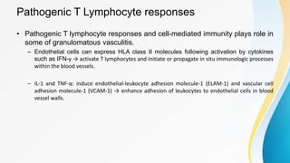
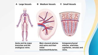
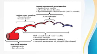
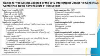
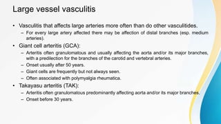
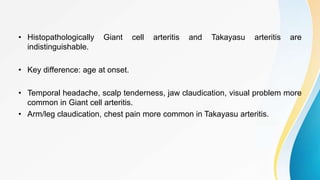
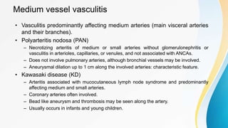
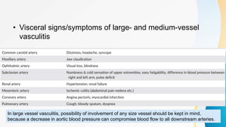
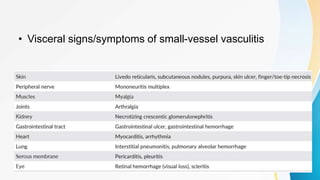
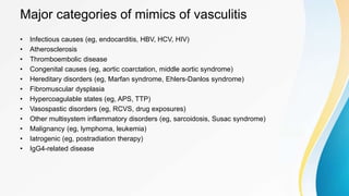
Vasculitis refers to inflammation of blood vessels. This document discusses the pathophysiology, classification, clinical presentation, diagnosis, and treatment of various types of vasculitis. The main types include large vessel vasculitis (e.g. giant cell arteritis, Takayasu arteritis), medium vessel vasculitis (e.g. polyarteritis nodosa, Kawasaki disease), small vessel vasculitis (ANCA-associated vasculitis like granulomatosis with polyangiitis, microscopic polyangiitis, eosinophilic granulomatosis with polyangiitis), and immune complex small vessel vasculitis (e.g. IgA vasculitis, antiglomer
2. INTRODUCTION
• The vasculitides are defined by the presence of inflammatory leukocytes in
vessel walls with reactive damage to mural structures.
• Clinicopathological process characterized by inflammation of and damage to
blood vessels.
• Both loss of vessel integrity leading to bleeding, and compromise of the
lumen may result in downstream tissue ischemia and necrosis.
3. INTRODUCTION
• In general, affected vessels vary in size, type, and location in association with
the specific type of vasculitis.
• Often associated with ischemia of tissues supplied.
• May be confined to single organ or simultaneously involve multiple organ or
systems.
4. PATHOPHYSIOLOGY
• Most: mediated at least in part by immunogenic mechanisms that occur in
response to certain antigenic stimuli.
• A number of factors contribute to this mechanism:
– Genetic predisposition
– Environmental exposures
– Regulatory mechanisms associated with immune response to certain antigens.
5. • Prominent hypothesized mechanisms:
– Pathogenic immune complex formation
• IgA vasculitis (Henoch-Schonlein)
• Lupus vasculitis
• Serum sickness and cutaneous vasculitis syndromes
• Hep B/Hep C virus associated vasculitis
– Anti-neutrophil cytoplasmic antigen (ANCA)
• Granulomatosis with polyangiitis (Wegener’s)
• Microsopic polyangiitis
• Eosinophilic granulomatosis with polyangiitis (Churg-Strauss)
– Pathogenic T lymphocyte responses and granuloma formation
• Giant cell arteritis
• Takayasu arteritis
• Granulomatosis with polyangiitis (Wegener’s)
• Eosinophilic granulomatosis with polyangiitis (Churg-Strauss)
6. Pathogenic immune complex formation
• First and most widely accepted mechanism.
– Causal role not clearly established.
Antigen
Antigen-Antibody complex
Complement activation (particularly C5a)
Deposited in vessel wall
Chemotactic for Neutrophil
Inflammation and damage of vessel wall
Compromise of vessel lumen.
Ischemic changes in tissue supplied.
IgE triggered mechanism
Mast cells
Platelets
Histamine, Bradykinin, Leukotriene
Increased permeability of vessel wall
7. Antineutrophil cytoplasmic antibodies (ANCA)
• Antibodies directed against certain proteins in cytoplasmic granules of
neutrophils and monocytes.
cANCA (cytoplasmic ANCA) pANCA (perinuclear ANCA)
•Gives diffuse granular cytoplasmic staining
pattern.
• More localized perinuclear staining pattern
•Against Proteinase-3 •Against Myeloperoxidase
•Granulomatosis with polyangitis (Wegener’s) •Microscopic polyangiitis
•Eosinophilic granulomatosis with polyangitis
(Churg-Strauss)
•Isolated necrotizing crescentic
glomerulonephritis
A perinuclear pattern of staining that is not due to anti-MPO antibodies has been associated with
nonvasculitic entities such as rheumatoid and nonrheumatoid autoimmune diseases, inflammatory
bowel disease, certain drugs and infections such as endocarditis and bacterial airway infections in
patients with cystic fibrosis.
8. How do ANCAs cause disease?
• PR3 and MPO reside in the azurophilic granules and lysosomes of neutrophils
– apparently inaccessible to serum antibodies.
• Primed by TNF-α and IL-1: come to cell membrane, interact with extracellular ANCA.
ANCA ↔ PR3/MPO
ANCA activated neutrophil
Neutrophil degranulation
Reactive oxygen
intermediates
Tissue
damage
Adhere to and kill
endothelial cells
Release of proinflammatory
cytokines (e.g. IL-1, IL-6)
Complement activation
9. Pathogenic T Lymphocyte responses
• Pathogenic T lymphocyte responses and cell-mediated immunity plays role in
some of granulomatous vasculitis.
– Endothelial cells can express HLA class II molecules following activation by cytokines
such as IFN-γ → activate T lymphocytes and initiate or propagate in situ immunologic processes
within the blood vessels.
– IL-1 and TNF-α: induce endothelial-leukocyte adhesion molecule-1 (ELAM-1) and vascular cell
adhesion molecule-1 (VCAM-1) → enhance adhesion of leukocytes to endothelial cells in blood
vessel walls.
11. Aorta and its major
branches and the
analogous veins.
Main visceral arteries
and veins and their
initial
branches/tributaries.
Intraparenchymal
arteries, arterioles,
capillaries, venules and
veins.
12.
13. Names for vasculitides adopted by the 2012 International Chapel Hill Consensus
Conference on the nomenclature of vasculitides
14. Large vessel vasculitis
• Vasculitis that affects large arteries more often than do other vasculitides.
– For every large artery affected there may be affection of distal branches (esp. medium
arteries).
• Giant cell arteritis (GCA):
– Arteritis often granulomatous and usually affecting the aorta and/or its major branches,
with a predilection for the branches of the carotid and vertebral arteries.
– Onset usually after 50 years.
– Giant cells are frequently but not always seen.
– Often associated with polymyalgia rheumatica.
• Takayasu arteritis (TAK):
– Arteritis often granulomatous predominantly affecting aorta and/or its major branches.
– Onset before 30 years.
15. • Histopathologically Giant cell arteritis and Takayasu arteritis are
indistinguishable.
• Key difference: age at onset.
• Temporal headache, scalp tenderness, jaw claudication, visual problem more
common in Giant cell arteritis.
• Arm/leg claudication, chest pain more common in Takayasu arteritis.
16. Medium vessel vasculitis
• Vasculitis predominantly affecting medium arteries (main visceral arteries
and their branches).
• Polyarteritis nodosa (PAN)
– Necrotizing arteritis of medium or small arteries without glomerulonephritis or
vasculitis in arterioles, capillaries, or venules, and not associated with ANCAs.
– Does not involve pulmonary arteries, although bronchial vessels may be involved.
– Aneurysmal dilation up to 1 cm along the involved arteries: characteristic feature.
• Kawasaki disease (KD)
– Arteritis associated with mucocutaneous lymph node syndrome and predominantly
affecting medium and small arteries.
– Coronary arteries often involved.
– Bead like aneurysm and thrombosis may be seen along the artery.
– Usually occurs in infants and young children.
17. Small vessel vasculitis
• Vasculitis predominantly affecting small vessels, defined as small
intraparenchymal arteries, arterioles, capillaries and venules.
• Two categories
– ANCA associated vasculitis: necrotizing vasculitis, with few or no immune deposits,
predominantly affecting small vessels, associated with ANCA.
– Immune complex small vessel vasculitis: vasculitis with moderate to marked vessel wall
deposits of immunoglobulin or complement, predominantly affecting small vessels.
Arterial involvement much less common in immune complex small vessel vasculitis than in
ANCA associated vasculitis.
18. ANCA associated vasculitis
• Granulomatosis with polyangiitis (Wegener’s granulomatosis)
– Necrotizing granulomatous inflammation usually involving the upper and lower respiratory
tract, and necrotizing vasculitis affecting predominantly small to medium vessels.
– Necrotizing glomerulonephritis common.
– Inflammation, necrosis, granuloma formation with or without vasculitis common in upper
airway.
– Ocular vasculitis and pulmonary capillaritis with hemorrhage are frequent.
– Granulomatous and non-granulomatous extravascular inflammation are common.
– Associated with cANCA (90%).
– Diagnosis by biopsy. cANCA: adjunctive.
19. ANCA associated vasculitis
• Microscopic polyangiitis
– Necrotizing vasculitis with few or no immune deposits, predominantly affecting small
vessels.
– Necrotizing glomerulonephritis very common. Renal lesion similar to that of
Granulomatosis with polyangiitis.
– Pulmonary capillaritis often occurs.
– Granulomatous inflammation is not seen.
– Associated with pANCA (75%).
• Eosinophilic granulomatosis with polyangiitis (Churg-Strauss)
– Eosinophil rich and necrotizing granulomatous inflammation often involving respiratory
tract, and necrotizing vasculitis predominantly involving small vessels and associated
with asthma and eosinophilia.
– Nasal polyps: common association.
– Eosinophilia in blood and tissues: essential feature.
– Associated with pANCA (25% of those without renal disease and 75% of those with renal
disease).
20. Immune complex small vessel vasculitis
• Antiglomerular basement membrane disease
– Vasculitis affecting glomerular capillaries, pulmonary capillaries, or both, with
basement membrane deposition of anti-basement membrane autoantibodies.
– Lung involvement causes pulmonary hemorrhage.
– Renal involvement causes glomerulonephritis with necrosis and crescents.
– Two age groups: young men in late 20’s and men and women in 60’s to 70’s.
– Younger age group: more explosive disease with hemoptysis, hematuria, fever,
dyspnea.
– Older population: prolonged asymptomatic renal injury. Poorer prognosis.
• IgA vasculitis (Henoch-Schonlein)
– Vasculitis with IgA1-dominant immune deposits, affecting small vessels
– Often involves the skin and GI tract and frequently causes arthritis.
– Glomerulonephritis indistinguishable from IgA nephropathy may occur.
21. Secondary vasculitis
• Rheumatoid vasculitis
– Typically occurs in patients with long-standing RA, positive RF, and
hypocomplementemia.
– <1% of the patients.
– Cutaneous signs: petechiae, purpura, digital infarcts, gangrene, livedo reticularis, and
in severe cases large, painful lower extremity ulcerations.
– Sensorymotor polyneuropathies may occur.
• SLE vasculitis
– Reported to involve 11-36%
– Cutaneous lesions representing small vessel involvement: most frequent.
– Medium and large vessel vasculitis may present with visceral affection, with life-
threatening manifestations such as mesenteric vasculitis, pulmonary hemorrhage, or
mononeuritis multiplex.
22. GENERAL APPROACH TO VASCULITIS
• Should be considered in any patient
with unexplained systemic illness.
• Clinical abnormalities that when
present alone or in combination
suggest a diagnosis of vasculitis:
– Palpable purpura
– Pulmonary infiltrates and microscopic
hematuria
– Chronic inflammatory sinusitis
– Mononeuritis multiplex
– Unexplained ischemic events
– Glomerulonephritis with evidence of
multisystem disease.
23. • A number of nonvasculitic diseases may also produce one or more of
these features → First step in work up: exclude mimickers.
24. General Principles of Diagnosis
• Clinical features in general can be categorized into:
– Systemic signs/symptoms caused by inflammation
– Visceral signs/symptoms specific to affected organ
• Systemic manifestations
– Fever
– Weight loss
– Weakness, general malaise
– Arthralgia, muscle pain
• Local/Visceral manifestations: simultaneous or sequential appearance of
symptoms from different affected organs.
25. • Visceral signs/symptoms of large- and medium-vessel
vasculitis
In large vessel vasculitis, possibility of involvement of any size vessel should be kept in mind,
because a decrease in aortic blood pressure can compromise blood flow to all downstream arteries.
27. • Laboratory findings
• General laboratory findings:
– Acute inflammatory markers: ESR, CRP, WBC count may be raised.
– EGPA: significant eosinophilia.
– PAN: high level of plasma renin activity or HBV antigen positivity may be seen.
– Renal dysfunction may be seen
• Urinalysis:
– Proteinuria, Hematuria, Leukocyturia may be seen
28. • ANCA
– GPA: cANCA positive in 90%.
– MPA, EGPA: pANCA positive in 50-80% of cases.
• Anti-GBM antibody
– Especially in cases exhibiting RPGN and/or alveolar hemorrhage.
– Specific to anti-GBM disease.
29. • Imaging Study
• Plain X-ray, ultrasonography, CT, MRI: help to confirm abnormal vascular wall structures
and blood flow dysfunction.
• Takayasu arteritis: aortic wall thickening densely stained with Gadolinium on contrast
MRI.
• Microscopic polyangiitis: X-ray, CT, MRI may show interstitial infiltrative shadows in lung
fields.
• Granulomatosis with polyangiitis, Eosinophilic granulomatosis with polyangiitis:
granulomatous lesions accompanied infiltrative shadows.
• Polyarteritis nodosa: multiple microaneurysms and/or contractions in association with
inflammation of medium to small sized arteries. Aneurysms in branches of abdominal
aorta
30. Renal arteriogram in large-vessel polyarteritis
nodosa showing characteristic
microaneurysms (small arrows) and abrupt
cutoffs of small arteries (large arrows).
31. • Tissue Biopsy
– Definitive method of diagnosis of vasculitis.
– May not be feasible in some forms like Takayasu arteritis, Polyarteritis nodosa, primary
CNS vasculitis: arteriogram of suspected organs.
– Giant cell arteritis: Biopsy of temporal artery essential.
– Microscopic polyangiitis: necrotizing angiitis in arterioles and capillaries in the kidneys
indicating necrotizing crescentic glomerulonephritis.
32. – Granulomatosis with polyangiitis: Necrotizing granulomatous lesion with
giant cells in lesion sites of upper respiratory tract. Various pattern of
glomerulonephritis in kidney biopsy.
– Eosinophilic Granulomatosis with polyangiitis: Peripheral nerves, muscles
and lungs: vasculitis and granulomas with prominent eosinophil infiltration.
– Anti-GBM disease: diffuse crescentic glomerulonephritis. Deposits of IgG
and C3 along glomerular capillary walls.
– IgA vasculitis: necrotizing angiitis in the area from papillary to reticular
layer of skin. Deposition of IgA in vascular endothelium.
35. General Principles of Treatment
• If offending antigen recognized, the antigen must be removed where
possible.
• If associated with underlying disease (infection, malignancy, connective
tissue disease): underlying disease should be treated.
• If the syndrome represents a primary vasculitic disease, treatment should be
initiated according to category of the vasculitis syndrome.
36. • Glucocorticoids: in those systemic vasculitides that cannot be specifically
categorized or for which there is no established standard therapy.
– Other immunosuppressive therapy added only if an adequate response does not result or
if remission can only be achieved and maintained with an unacceptably toxic regimen of
glucocorticoids.
• When remission is achieved, one should continually attempt to taper
glucocorticoids and discontinue when possible.
• When using other immunosuppressive regimens, one should base the choice
of agent upon the available therapeutic data supporting efficacy in that
disease, the site and severity of organ involvement, and the toxicity profile of
the drug.
37. • Glucocorticoids and/or other immunosuppressive agents should be instituted
immediately in diseases where irreversible organ system dysfunction and
high morbidity and mortality rates have been clearly established (e.g. GPA).
• On the other hand, aggressive therapy should be avoided for vasculitic
manifestations that rarely result in irreversible organ dysfunction and that
usually do not respond to such therapy (e.g. isolated idiopathic cutaneous
vasculitis).
38. • Treatment of large-vessel vasculitis
– Corticosteroid effective for most patients with TAK and GCA.
• 40-60 mg/day for one month followed by gradual tapering.
• If ocular symptoms in Giant cell arteritis, Methylprednisolone 1 g/day for 3 days followed by oral steroid.
– Methotrexate, anti TNF antibodyn( (Infliximab), Cyclophosphamide, Cyclosporine:
variable response. Can be used in those who do not respond/cannot tolerate steroid
therapy.
39. • Treatment for medium-vessel vasculitis
– Good response with corticosteroids.
– In patients with organ failure, combination therapy with immunosuppressive agents is
desirable.
• Treatment for small-vessel vasculitis
– Corticosteroids plus immunosuppressive therapy (usually with cyclophosphamide) to be
initiated.
– Additional immunosuppressants/plasmapheresis may be needed based on severity/initial
response.
40. Infection prophylaxis
• Infections with Pneumocystis jiroveci and certain fungi can be seen in
patients receiving glucocorticoids.
• Criteria for primary prophylaxis against Pneumocystis pneumonia:
– Age ≥50 years
– For those receiving glucocorticoids:
• Prednisolone ≥1.2 mg/kg/day or ≥0.8 mg/kg/day in combination with immunosuppressive drugs.
• Discontinue when: Prednisolone dose is ≤0.4 mg/kg/day.
– For those receiving immunosuppressive drugs
• Prednisolone ≥0.8 mg/kg/day or with a peripheral leukocyte count of ≤500/mm3.
• Discontinue when: Prednisolone dose is ≤0.4 mg/kg/day or peripheral lymphocyte count ≥500/mm3.
41. • Criteria for secondary prophylaxis
– All patients who have experienced Pneumocystis pneumonia.
– Discontinuation criteria: same as for primary prophylaxis.
• Regimen:
– TMP/SMX one double strength tablet per day.
– Inhaled pentamidine 300 mg 2-4 weekly.
A unique feature of neutrophils is their ability to generate Neutrophil Extracellular Traps (NETs), a ‘sticky mesh’ of extruded chromatin, nucleic acid and a range of cytoplasmic proteins. These serve antibacterial functions, but also represent a means by which cytoplasmic proteins may be exposed to the immune system.
NETs represent an important means for autoantigen uptake by antigen presenting cells (APCs) in AAV.
NETs specifically interact with myeloid dendritic cells (mDCs) to facilitate the uptake of MPO and PR3.
Infectious vasculitis: known to be caused by direct invasion and proliferation of pathogens in vessel walls with resultant inflammation.
Examples of infectious vasculitis include rickettsial vasculitis, syphilitic aortitis, and Aspergillus arteritis.
For example, imaging and fluorescein angiography studies have shown that ocular involvement in giant cell arteritis may affect not only ophthalmic arteries, but also retinal arteries and multiple ciliary arteries (medium arteries), and even smaller branches of the ciliary and retinal arteries (small arteries).
The term “temporal arteritis” is not a suitable alternative for GCA because not all patients have temporal artery involvement, and other categories of vasculitis can affect the temporal arteries.
KD: Characterized by non-suppurative cervical adenitis and changes in skin and mucous membranes such as
Edema
Congested conjunctiva
Erythema of oral cavity, lips, and palms
Desquamation of skin of the fingertips
These systemic manifestations also frequently seen in elderly people with malignancy. Also screen for malignancy.