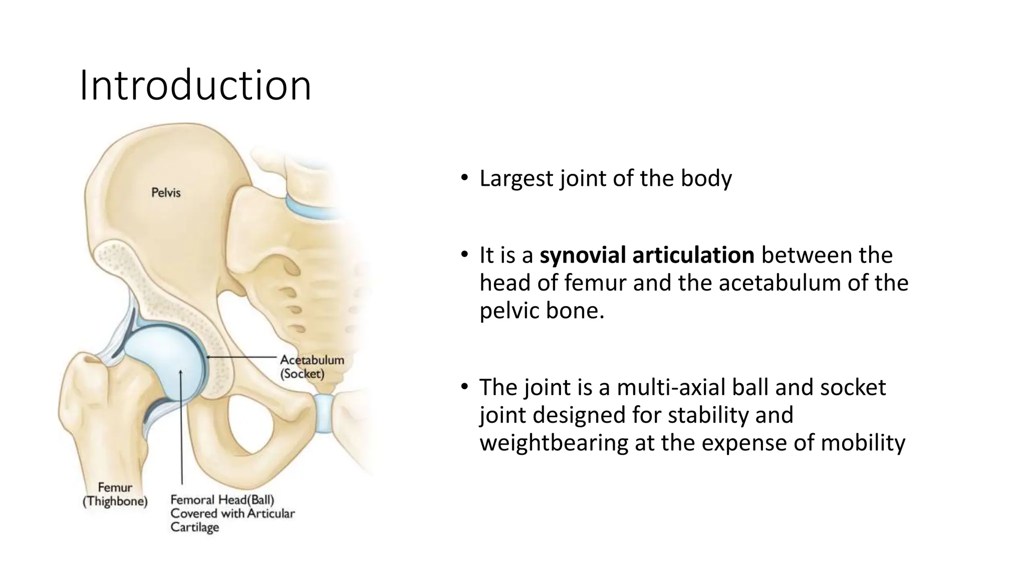
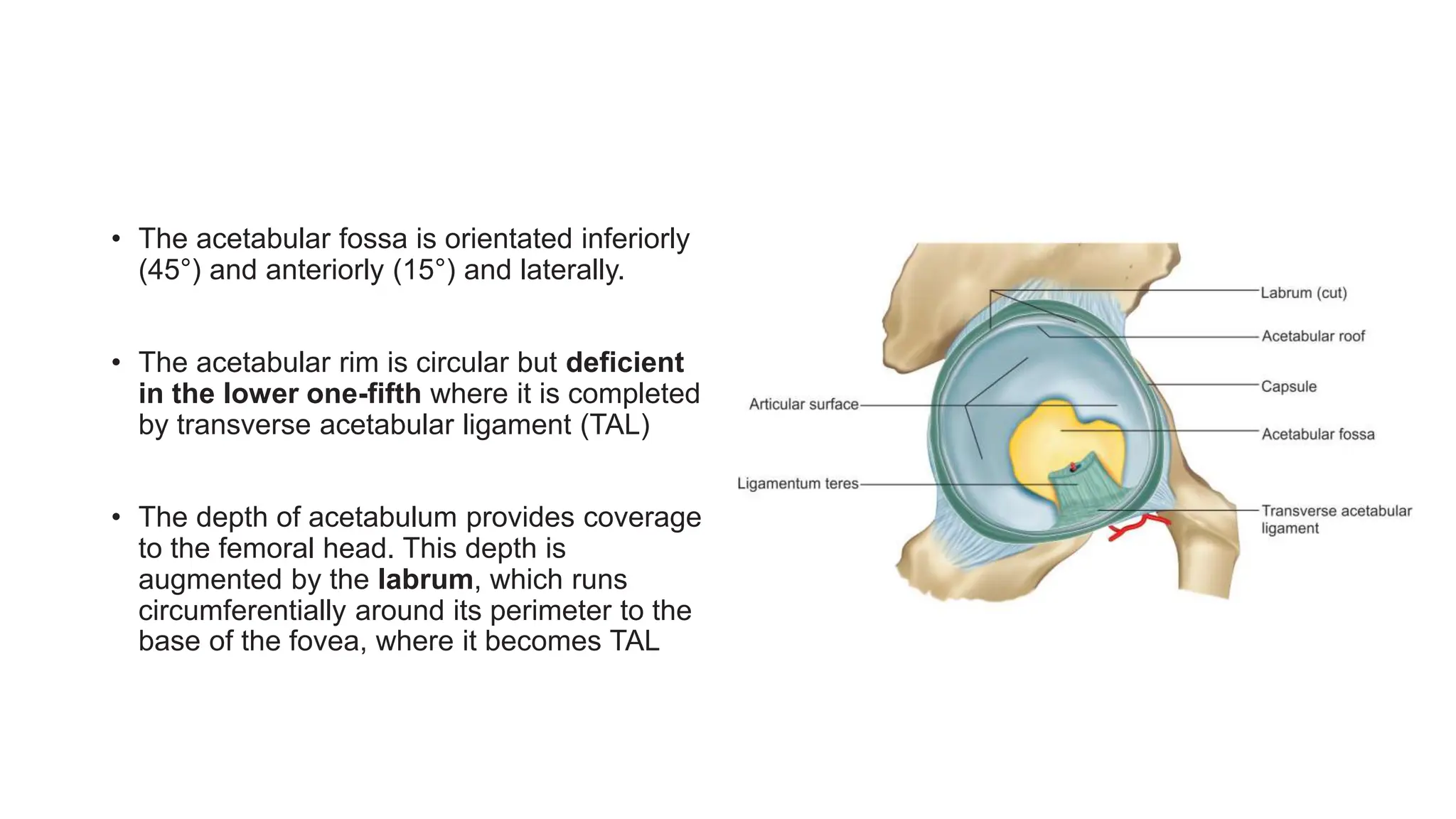
SlideShare a Scribd company logo
1 of 91
Topic- Surgical hip
approaches
Presenter – Dr. Karthik M V
Moderator – Dr. Jayant Jain
Introduction
• Largest joint of the body
• It is a synovial articulation between the
head of femur and the acetabulum of the
pelvic bone.
• The joint is a multi-axial ball and socket
joint designed for stability and
weightbearing at the expense of mobility
• The acetabular fossa is orientated inferiorly
(45°) and anteriorly (15°) and laterally.
• The acetabular rim is circular but deficient
in the lower one-fifth where it is completed
by transverse acetabular ligament (TAL)
• The depth of acetabulum provides coverage
to the femoral head. This depth is
augmented by the labrum, which runs
circumferentially around its perimeter to the
base of the fovea, where it becomes TAL
Femur
• The femur is the longest and strongest bone in the
human body. It is mostly cylindrical throughout its
length, and it is anterolaterally bowed in its
midportion
• Head is covered by articular hyaline cartilage in
approximately 60–70% of a sphere also the fovea
centralis is devoid of cartilage.
• The neck of the femur is approximately
5 cm long. The proximal metaphysis and neck are
anteverted by approximately 14°. The angle
between the
femoral shaft and the neck is approximately 125°
Capsule
• The capsule is attached along the anterior and posterior periphery of the acetabulum
just outside the acetabular labrum.
• Inferiorly, the capsule is attached to the transverse acetabular ligament.
• The capsule is attached to the femur anteriorly along the intertrochanteric line, but
posteriorly it attaches to middle of the neck so that the Basi cervical portion and
intertrochanteric crest are extracapsular posteriorly
Ligaments
• Ileofemoral ligament (Bigelow):
This is fan-shaped modified “λ”
structure that resembles inverted
letter Y
• It is the strongest ligament in the
body
• Attached to the lower portion of the
anterior inferior iliac spine and the
diverging fibers of the Y fan out to
attach along the intertrochanteric
line.
• Taut in: full extension
• Pubofemoral ligament: It arises
from pubic portion of the
acetabular rim and the
obturator aspect of the superior
pubic ramus and blends with
the inferior-most fibers of the
iliofemoral ligament.
• Taut in: Hip extension and
abduction
• Ischio-femoral Ligament : It arises from
the ischial portion of the acetabular
rim and spirals laterally and upward to
blend in the capsule
• It Blends with the posterior surface of
the capsule and zona orbicularis
• Taut in: Extension
• Ligamentum teres—it is intra-
articular but actually
extra-synovial as it is surrounded by
synovial membrane.
• The ligamentum attaches to the
fovea of the femoral head and
acetabular central fovea
• Zona orbicularis —“annular
ligament” encircles the femoral neck
like a button hole and again plays
little role in stability
Muscles
Movements
• Flexion of 120°
• Extension 20°
• Abduction 40–45°
• Adduction of 20–25°
• Internal rotation 35°
• External rotation 45°
Surgical approaches
The characteristic of a good approach should:
• Provide adequate approach to both femoral head and acetabulum.
• Involve minimal dissection of soft tissues.
• Reduce operative time and blood loss.
• Should be between intervascular and interneural plane to minimize
morbidity.
• Provide least postoperative pain and early mobilization.
• Be associated with minimal chance of damage to neurovascular bundles
and other vital structures and with minimal risk of infection or thrombosis.
• Should reliably restore the anatomy after procedure so that functional
deterioration is minimal and stability of joint is maintained
• Anterior approach – interval b/w
sartorius and tensor fascia lata.
• Anterolateral approach – interval
b/w tensor fascia lata and gluteus
medius.
• Posterior approach – interval b/w
gluteus medius and maximus or
gluteus maximus splitting.
• Medial approach – interval b/w
adductor longus and Gracilis.
Anterior Approach
(Smith-Petersen, Iliofemoral)
It exploits internervous plane b/w Sartorius (femoral nerve) & Tensor
Fascia Lata ( Superior gluteal nerve).
Indications:
• Open reduction of congenital dislocations of the hip when the dislocated femoral
head lies anterosuperior to the true acetabulum
• Synovial biopsies
• Intra-articular fusions
• Total hip replacement
• Hemiarthroplasty
• Excision of tumors, especially of the pelvis
• The patient is placed supine with a small sandbag under the affected buttock.
• Curvilinear Incision starting from the ant. half of iliac crest to ASIS and from there curve it
vertically down for 8-10cm
Internervous Plane:
The superficial plane Sartorius (femoral nerve) and the tensor fasciae latae (superior gluteal nerve)
Identify the gap between the tensor fasciae latae and the sartorius by palpation
The lateral femoral cutaneous nerve (lateral cutaneous nerve of the thigh) pierces the deep fascia close to the
intermuscular interval between the tensor fasciae Iatae and the sartorius
Incise the deep fascia on the medial side of the tensor fascia latae. Retract the sartorius upward and
medially and the tensor fascia downward and laterally.
The deeper internervous plane lies between the rectus femoris (femoral nerve) and the gluteus medius
(superior gluteal nerve)
The deep layer of musculature, consisting of the rectus femoris and the gluteus medius, is now visible.
The ascending branch of the lateral femoral circumflex artery must be ligated.
• Detach the rectus femoris from both its origins, the anterior inferior iliac spine and the superior lip of
the acetabulum, and retract it medially. Retract the gluteus medius laterally.
The hip joint capsule is now partly exposed. Retract the iliopsoas tendon medially
• The hip joint capsule is fully exposed. Detach the muscles of the ilium if further exposure is needed
• Adduct and external rotate to stretch capsule, incise capsule by longitudinal or T-
shaped incision, dislocate hip after capsulotomy by External rotation.
After incising the joint capsule; Proximal extension of the wound exposes the ilium. Distal extension of the
incision exposes the anterior aspect of the femur
Sommerville anterior approach
• Indication: For irreducible congenital dislocation of the hip in a young child
• Incision: Transverse “Bikini” incision – anteriorly- Inferior and medial to ASIS and
coursing obliquely superiorly and posteriorly to the middle of the iliac crest.
• For reduction of CDH following sequential steps must be performed: Psoas
tenotomy > Complete medial capsulotomy > excision of hypertrophied lig. Teres >
reduction of femoral head into true acetabulum
Minimally invasive Anterior approach to hip
• Reduction of displaced femoral neck fractures
• Drainage of hip infections
• Elective joint replacement surgery
Make an 8-cm longitudinal incision beginning 1 cm below and 1 cm lateral to the anterior superior iliac spine. Aim
the incision toward the head of the fibula. The center of the incision should be at the level of the tip of the greater
trochanter.
Identify the interval between sartorius and tensor fasciae latae and incise the deep fascia covering the tensor fasciae
latae muscle at its medial edge.
Retract the tensor fasciae latae laterally to expose the rectus muscle covered by a fascial layer
Incise the fascial sheath covering the anterior aspect of the rectus to expose the muscle.
Retract the rectus muscle medially to expose the fascia covering the posterior aspect of the muscle. Incise
this fascial sheath longitudinally to reveal the lateral circumflex vessels
Ligate the lateral circumflex vessels. Expose the anterior hip joint capsule covered by fatty tissue. Some fibers of psoas—
the iliocapsularis muscle will need to be separated from the capsule by sharp dissection. Incise the anterior capsule of the
hip joint longitudinally.
Anterolateral approach (Watson-jones
approach)
• Excellent exposure of acetabulum with safety during reaming of
femoral shaft.
• It exploits intermuscular plane b/w Tensor fascia latae & Gluteus
medius.
• It involves partial or complete detachment of abductors so that hip
can be adducted during reaming.
• Abductor mechanism released by either trochanteric osteotomy or
detaching anterior part of gluteus medius and whole of minimus.
• There is no true internervous plane for this approach, since the
gluteus medius and the tensor fasciae latae have a common nerve
supply, the superior gluteal nerve.
• Indications:
• Total hip replacement
• Hemiarthroplasty
• Open reduction and internal fixation of femoral neck fractures
• Synovial biopsy of the hip
• Biopsy of the femoral neck
Position of the patient on the operating table for the anterolateral approach to the hip.
Bring the greater trochanter to the edge of the table, and allow the buttocks, skin, and fat to fall
posteriorly, away from the operative plane
Incision for the anterolateral approach to the hip- 8-15cm straight longitudinal incision centered
over tip of greater trochanter
Incise the fascia lata posterior to the tensor fasciae latae.
Retract the fascia lata and the tensor fasciae latae muscle, which it envelopes,
anteriorly, revealing the gluteus medius and a series of vessels that cross the interval between the tensor fasciae
latae and the gluteus medius.
Retract the gluteus medius posteriorly and the tensor fasciae latae anteriorly, uncovering the fatty layer
directly over the joint capsule
Bluntly dissect the fat pad off the anterior portion of the joint capsule to expose it and the rectus femoris
tendon
Osteotomize the greater trochanter
Reflect the osteotomized portion of the trochanter superiorly (with the attached gluteus medius) to reveal the joint
capsule
The joint capsule may also be exposed by partial resection of the gluteus medius tendon from the
anterior portion of the trochanter.
Reflect the head of the rectus femoris from the anterior portion of the joint
capsule.
Incise the anterior joint capsule to reveal the femoral head and neck and the acetabular rim. If further
proximal exposure is needed, incise the fascia lata proximally toward the iliac crest and along the iliac crest
anteriorly.
To expose the acetabulum, dislocate and resect the femoral head. Placing three or four
Hohmann-type retractors around the lip of the acetabulum provides excellent exposure
Extend the incision down the lateral aspect of the thigh, incising the deep fascia and splitting the vastus
lateralis in line with its musculature to reach the lateral aspect of the femur
Lateral approach
• Indications: Total hip replacement surgeries
• Good approach to femur head
• Avoids need for trochanteric osteotomy
• Early mobilization ( bulk of gluteus medius preserved)
Position of the patient on the operating table for the anterolateral approach to the hip.
Bring the greater trochanter to the edge of the table, and allow the buttocks, skin, and fat to fall posteriorly, away
from the operative plane.
Make a longitudinal incision centered over the tip of the greater trochanter in the line of the femoral shaft
• Internervous plane:
• There is no true internervous plane.
• The fibres of the gluteus medius muscle are split in their own line distal to the
point where the superior gluteal nerve supplies the muscle.
• The vastus lateralis muscle is also split in it’s own line lateral to the point where it
is supplied by the femoral nerve.
Divide the deep fascia in the line of the skin incision, retracting the fascial edges to pull the tensor fasciae latae
anteriorly.
Split the fibers of gluteus medius above the tip of the greater trochanter and extend this incision distally
on the lateral aspect of the trochanter until 2 cm of the vastus lateralis is also split
Develop this anterior flap and divide the tendon of the gluteus minimus muscle to reveal the
anterior aspect of the hip joint capsule
Enter the capsule using a longitudinal T-shaped incision.
Osteotomize the femoral neck using an oscillating saw.
Extract the femoral head. Insert appropriate retractors to reveal the acetabulum.
Hardinge modification: (Direct Lateral
approach)
• Position: Patient supine with greater trochanter at the edge of the table
• Incision: Make a posteriorly directed lazy “J” incision centered over greater
trochanter
• The only difference in this step: Instead of osteotomizing GT, incise the tendon of
gluteus medius obliquely across the GT leaving the posterior half still attached to
the trochanter. Carry the incision proximally in line with the fibres of Gluteus
medius
• Distally carry the incision anteriorly in line with the fibers of vastus lateralis.
• Advantages : GT and bulk of gluteus medius preserved allowing rapid
rehabilitation.
Posterior approach (Moore’s approach-
Southern exposure)
• Most common approach used to expose hip joint
• Easy, safe, and quick access to joint
• No interference with abductor mechanism
• Excellent visualisation of femoral shaft
• Popular for revision joint replacement
• Division of posterior capsule, higher rates of dislocation
Indications:
• Hemiarthroplasty
• Total hip replacement, including revision surgery
• Open reduction and internal fixation of posterior acetabular fractures
• Dependent drainage of hip sepsis
• Removal of loose bodies from the hip joint
• Pedicle bone grafting
• Open reduction of posterior hip dislocations
Position of the patient on the operating table for the posterior approach to the hip joint
True lateral position, affected limb uppermost, padding of bony prominences
There is no true internervous plane. Split the fibers of the gluteus maximus, a procedure that does not
cause significant denervation of the muscle.
A: Skin incision for the posterior approach to the hip joint. B: Incise the fascia lata
• 10-15cm curved incision centered
on the posterior aspect of greater
trochanter
• Begin incision 6-8cm above and
posterior to posterior aspect of
trochanter
• Runs along fibers of gluteus
maximus
• Curve the incision across the
buttock, cutting posterior aspect of
trochanter, continue along shaft of
femur
• Flex hip to 90 degree– longitudinal
incision over posterior aspect of
trochanter– ‘ Moore-style’ incision
Retract the gluteus maximus to reveal the fatty layer over the short external rotators of
the hip
• Remember that the sciatic nerve
leaves the pelvis and run down
the back of thigh on the short
external rotators, encased in fatty
layer.
• Don’t dissect to see the nerve,
unnecessary bleeding from the
vessels lying in the fat around it.
Push the fat posteromedially to expose the insertions of the short rotators. Note that the sciatic nerve is not
visible; it lies within the substance of the fatty tissue. Place your retractors within the substance of the gluteus
maximus superficial to the fatty tissue.
A, B: Internally rotate the femur to bring the insertion of the short rotators of the hip as
far lateral to the sciatic nerve as possible.
C: Detach the short rotator muscles close to their femoral insertion and reflect them backward, laying them
over the sciatic nerve to protect it.
• Insert stay sutures into the
piriformis and obturator internus
tendons just before they insert
to GT
• Detach the muscles 1cm from
their femoral insertion and
reflect backward
Incise the posterior joint capsule to expose the femoral head and
neck.
To gain additional exposure, cut the quadratus femoris and the tendinous insertion of the gluteus
maximus.
Medial approach (Ludloff’s Approach)
Indications:
• Open reduction of congenital dislocation of the hip. The approach gives an excellent
exposure of the psoas tendon, which can block reduction of the hip.
• Biopsy and treatment of tumors of the inferior portion of the femoral neck and
medial aspect of proximal shaft
• Psoas release
• Obturator neurectomy
Make a longitudinal incision on the
medial side of the thigh, starting at a
point 3 cm
below the pubic tubercle. The incision
runs down over the adductor longus. Its
length
is determined by the amount of femur
that must be exposed.
Place the patient supine on the operating
table with the affected hip flexed,
abducted,
and externally rotated
The intermuscular interval between the adductor
longus and the gracilis is not an internervous plane
because both muscles are innervated by the anterior
division of the obturator nerve. The plane is safe,
however, because the muscles receive their nerve
supplies proximal to the dissection.
A: Develop the plane between the gracilis and the adductor longus.
B: Retract the adductor longus and the gracilis to reveal the adductor brevis with the overlying
anterior division of the obturator nerve.
C: Retract the adductor brevis from the muscle belly of the adductor magnus to uncover the
posterior division of the obturator nerve. Note the lesser trochanter in the depths of the wound.
The deep muscular layer of the medial approach to
the hip. The dissection lies between the adductor
brevis and the adductor magnus. The gracilis,
adductor longus, and sartorius have been resected
to reveal the deeper structures of the medial
aspect of the thigh.
Anterior and posterior divisions of the obturator
nerve to the adductor longus and adductor brevis
will be visible.
References
• Surgical exposures in orthopaedics, stanley hoppenfeld, 5th edition
• Campbell’s operative Orthopaedics, 14th edition
• Netter’s Atlas
• Essential Orthopaedics, Manish Kumar Varshney
• Orthopaedics principles and its applications, Turek 8th edition
THANK YOU

More Related Content

Similar to Surgical approaches to Hip.pptx

Anterior approach to hip ppt new,
Anterior approach to hip ppt new,Anterior approach to hip ppt new,
Anterior approach to hip ppt new,BipulBorthakur
 
Seminar on applied anatomy and surgical approaches to shoulder
Seminar on applied anatomy and surgical approaches to shoulderSeminar on applied anatomy and surgical approaches to shoulder
Seminar on applied anatomy and surgical approaches to shoulderDr.Hari krishna Bachu
 
Surgical Approach to Hip and Acetabulum
Surgical Approach to Hip and AcetabulumSurgical Approach to Hip and Acetabulum
Surgical Approach to Hip and AcetabulumSijan Bhattachan
 
Posteromedial and posterolateral approach to knee
Posteromedial and posterolateral approach to kneePosteromedial and posterolateral approach to knee
Posteromedial and posterolateral approach to kneeBipulBorthakur
 
Comparetive anatomy & kinesiology of large important synovial joint of h...
Comparetive  anatomy & kinesiology of large important synovial joint of h...Comparetive  anatomy & kinesiology of large important synovial joint of h...
Comparetive anatomy & kinesiology of large important synovial joint of h...Dr Lovely Jain
 
The shoulder joint
The shoulder jointThe shoulder joint
The shoulder jointQuan Fu Gan
 
the gross antomy of the hip hoint and applied anatomy focused for undergradua...
the gross antomy of the hip hoint and applied anatomy focused for undergradua...the gross antomy of the hip hoint and applied anatomy focused for undergradua...
the gross antomy of the hip hoint and applied anatomy focused for undergradua...Shaifaly madan rustagi
 
Surgical approaches tibia fibula
Surgical approaches tibia fibulaSurgical approaches tibia fibula
Surgical approaches tibia fibulaMirant Dave
 
applied aspects of shoulder joint
applied aspects of shoulder jointapplied aspects of shoulder joint
applied aspects of shoulder jointmrinal joshi
 
Posterior approach to the hip
Posterior approach to the hipPosterior approach to the hip
Posterior approach to the hipBipulBorthakur
 
HIP JOINTS-1.pptx.........................
HIP JOINTS-1.pptx.........................HIP JOINTS-1.pptx.........................
HIP JOINTS-1.pptx.........................IshaKanojiya1
 
HEMIARTHROPLASTY.pptx
HEMIARTHROPLASTY.pptxHEMIARTHROPLASTY.pptx
HEMIARTHROPLASTY.pptxMisStrom
 
HipJoint attachment and action of muscles
HipJoint attachment and action of musclesHipJoint attachment and action of muscles
HipJoint attachment and action of musclesHarmanSaini54
 

Similar to Surgical approaches to Hip.pptx (20)

Anterior approach to hip ppt new,
Anterior approach to hip ppt new,Anterior approach to hip ppt new,
Anterior approach to hip ppt new,
 
Seminar on applied anatomy and surgical approaches to shoulder
Seminar on applied anatomy and surgical approaches to shoulderSeminar on applied anatomy and surgical approaches to shoulder
Seminar on applied anatomy and surgical approaches to shoulder
 
Polio 2
Polio 2Polio 2
Polio 2
 
hipjointanatomy
hipjointanatomyhipjointanatomy
hipjointanatomy
 
Hip joint anatomy
Hip joint anatomyHip joint anatomy
Hip joint anatomy
 
Surgical Approach to Hip and Acetabulum
Surgical Approach to Hip and AcetabulumSurgical Approach to Hip and Acetabulum
Surgical Approach to Hip and Acetabulum
 
Posteromedial and posterolateral approach to knee
Posteromedial and posterolateral approach to kneePosteromedial and posterolateral approach to knee
Posteromedial and posterolateral approach to knee
 
Comparetive anatomy & kinesiology of large important synovial joint of h...
Comparetive  anatomy & kinesiology of large important synovial joint of h...Comparetive  anatomy & kinesiology of large important synovial joint of h...
Comparetive anatomy & kinesiology of large important synovial joint of h...
 
The shoulder joint
The shoulder jointThe shoulder joint
The shoulder joint
 
Joints of lower limb
Joints of lower limbJoints of lower limb
Joints of lower limb
 
Hip joint
Hip jointHip joint
Hip joint
 
the gross antomy of the hip hoint and applied anatomy focused for undergradua...
the gross antomy of the hip hoint and applied anatomy focused for undergradua...the gross antomy of the hip hoint and applied anatomy focused for undergradua...
the gross antomy of the hip hoint and applied anatomy focused for undergradua...
 
Surgical approaches tibia fibula
Surgical approaches tibia fibulaSurgical approaches tibia fibula
Surgical approaches tibia fibula
 
applied aspects of shoulder joint
applied aspects of shoulder jointapplied aspects of shoulder joint
applied aspects of shoulder joint
 
Posterior approach to the hip
Posterior approach to the hipPosterior approach to the hip
Posterior approach to the hip
 
HIP JOINTS-1.pptx.........................
HIP JOINTS-1.pptx.........................HIP JOINTS-1.pptx.........................
HIP JOINTS-1.pptx.........................
 
Hip joint part I
Hip joint part IHip joint part I
Hip joint part I
 
HEMIARTHROPLASTY.pptx
HEMIARTHROPLASTY.pptxHEMIARTHROPLASTY.pptx
HEMIARTHROPLASTY.pptx
 
HipJoint attachment and action of muscles
HipJoint attachment and action of musclesHipJoint attachment and action of muscles
HipJoint attachment and action of muscles
 
Shoulder anatomy
Shoulder anatomyShoulder anatomy
Shoulder anatomy
 

More from Karthik MV

pop and bone cement orthopaedics ppt.pptx
pop and bone cement orthopaedics ppt.pptxpop and bone cement orthopaedics ppt.pptx
pop and bone cement orthopaedics ppt.pptxKarthik MV
 
Osteosarcoma and GCT.pptx
Osteosarcoma and GCT.pptxOsteosarcoma and GCT.pptx
Osteosarcoma and GCT.pptxKarthik MV
 
epiphyseal injuries.pptx
epiphyseal injuries.pptxepiphyseal injuries.pptx
epiphyseal injuries.pptxKarthik MV
 
compartment syndrome.pptx
compartment syndrome.pptxcompartment syndrome.pptx
compartment syndrome.pptxKarthik MV
 
BONE ANATOMY.pptx
BONE ANATOMY.pptxBONE ANATOMY.pptx
BONE ANATOMY.pptxKarthik MV
 

More from Karthik MV (6)

pop and bone cement orthopaedics ppt.pptx
pop and bone cement orthopaedics ppt.pptxpop and bone cement orthopaedics ppt.pptx
pop and bone cement orthopaedics ppt.pptx
 
Osteosarcoma and GCT.pptx
Osteosarcoma and GCT.pptxOsteosarcoma and GCT.pptx
Osteosarcoma and GCT.pptx
 
epiphyseal injuries.pptx
epiphyseal injuries.pptxepiphyseal injuries.pptx
epiphyseal injuries.pptx
 
MALUNION.pptx
MALUNION.pptxMALUNION.pptx
MALUNION.pptx
 
compartment syndrome.pptx
compartment syndrome.pptxcompartment syndrome.pptx
compartment syndrome.pptx
 
BONE ANATOMY.pptx
BONE ANATOMY.pptxBONE ANATOMY.pptx
BONE ANATOMY.pptx
 

Recently uploaded

Call Girls Service In Shyam Nagar Whatsapp 8445551418 Independent Escort Service
Call Girls Service In Shyam Nagar Whatsapp 8445551418 Independent Escort ServiceCall Girls Service In Shyam Nagar Whatsapp 8445551418 Independent Escort Service
Call Girls Service In Shyam Nagar Whatsapp 8445551418 Independent Escort Serviceparulsinha
 
Call Girls Service in Bommanahalli - 7001305949 with real photos and phone nu...
Call Girls Service in Bommanahalli - 7001305949 with real photos and phone nu...Call Girls Service in Bommanahalli - 7001305949 with real photos and phone nu...
Call Girls Service in Bommanahalli - 7001305949 with real photos and phone nu...narwatsonia7
 
Housewife Call Girls Hoskote | 7001305949 At Low Cost Cash Payment Booking
Housewife Call Girls Hoskote | 7001305949 At Low Cost Cash Payment BookingHousewife Call Girls Hoskote | 7001305949 At Low Cost Cash Payment Booking
Housewife Call Girls Hoskote | 7001305949 At Low Cost Cash Payment Bookingnarwatsonia7
 
Call Girls Whitefield Just Call 7001305949 Top Class Call Girl Service Available
Call Girls Whitefield Just Call 7001305949 Top Class Call Girl Service AvailableCall Girls Whitefield Just Call 7001305949 Top Class Call Girl Service Available
Call Girls Whitefield Just Call 7001305949 Top Class Call Girl Service Availablenarwatsonia7
 
Call Girl Service Bidadi - For 7001305949 Cheap & Best with original Photos
Call Girl Service Bidadi - For 7001305949 Cheap & Best with original PhotosCall Girl Service Bidadi - For 7001305949 Cheap & Best with original Photos
Call Girl Service Bidadi - For 7001305949 Cheap & Best with original Photosnarwatsonia7
 
Aspirin presentation slides by Dr. Rewas Ali
Aspirin presentation slides by Dr. Rewas AliAspirin presentation slides by Dr. Rewas Ali
Aspirin presentation slides by Dr. Rewas AliRewAs ALI
 
Kesar Bagh Call Girl Price 9548273370 , Lucknow Call Girls Service
Kesar Bagh Call Girl Price 9548273370 , Lucknow Call Girls ServiceKesar Bagh Call Girl Price 9548273370 , Lucknow Call Girls Service
Kesar Bagh Call Girl Price 9548273370 , Lucknow Call Girls Servicemakika9823
 
Vip Call Girls Anna Salai Chennai 👉 8250192130 ❣️💯 Top Class Girls Available
Vip Call Girls Anna Salai Chennai 👉 8250192130 ❣️💯 Top Class Girls AvailableVip Call Girls Anna Salai Chennai 👉 8250192130 ❣️💯 Top Class Girls Available
Vip Call Girls Anna Salai Chennai 👉 8250192130 ❣️💯 Top Class Girls AvailableNehru place Escorts
 
VIP Call Girls Tirunelveli Aaradhya 8250192130 Independent Escort Service Tir...
VIP Call Girls Tirunelveli Aaradhya 8250192130 Independent Escort Service Tir...VIP Call Girls Tirunelveli Aaradhya 8250192130 Independent Escort Service Tir...
VIP Call Girls Tirunelveli Aaradhya 8250192130 Independent Escort Service Tir...narwatsonia7
 
VIP Call Girls Pune Vani 9907093804 Short 1500 Night 6000 Best call girls Ser...
VIP Call Girls Pune Vani 9907093804 Short 1500 Night 6000 Best call girls Ser...VIP Call Girls Pune Vani 9907093804 Short 1500 Night 6000 Best call girls Ser...
VIP Call Girls Pune Vani 9907093804 Short 1500 Night 6000 Best call girls Ser...Miss joya
 
Sonagachi Call Girls Services 9907093804 @24x7 High Class Babes Here Call Now
Sonagachi Call Girls Services 9907093804 @24x7 High Class Babes Here Call NowSonagachi Call Girls Services 9907093804 @24x7 High Class Babes Here Call Now
Sonagachi Call Girls Services 9907093804 @24x7 High Class Babes Here Call NowRiya Pathan
 
Call Girls Service Surat Samaira ❤️🍑 8250192130 👄 Independent Escort Service ...
Call Girls Service Surat Samaira ❤️🍑 8250192130 👄 Independent Escort Service ...Call Girls Service Surat Samaira ❤️🍑 8250192130 👄 Independent Escort Service ...
Call Girls Service Surat Samaira ❤️🍑 8250192130 👄 Independent Escort Service ...CALL GIRLS
 
Call Girl Coimbatore Prisha☎️ 8250192130 Independent Escort Service Coimbatore
Call Girl Coimbatore Prisha☎️  8250192130 Independent Escort Service CoimbatoreCall Girl Coimbatore Prisha☎️  8250192130 Independent Escort Service Coimbatore
Call Girl Coimbatore Prisha☎️ 8250192130 Independent Escort Service Coimbatorenarwatsonia7
 
Artifacts in Nuclear Medicine with Identifying and resolving artifacts.
Artifacts in Nuclear Medicine with Identifying and resolving artifacts.Artifacts in Nuclear Medicine with Identifying and resolving artifacts.
Artifacts in Nuclear Medicine with Identifying and resolving artifacts.MiadAlsulami
 
Call Girl Chennai Indira 9907093804 Independent Call Girls Service Chennai
Call Girl Chennai Indira 9907093804 Independent Call Girls Service ChennaiCall Girl Chennai Indira 9907093804 Independent Call Girls Service Chennai
Call Girl Chennai Indira 9907093804 Independent Call Girls Service ChennaiNehru place Escorts
 
CALL ON ➥9907093804 🔝 Call Girls Hadapsar ( Pune) Girls Service
CALL ON ➥9907093804 🔝 Call Girls Hadapsar ( Pune)  Girls ServiceCALL ON ➥9907093804 🔝 Call Girls Hadapsar ( Pune)  Girls Service
CALL ON ➥9907093804 🔝 Call Girls Hadapsar ( Pune) Girls ServiceMiss joya
 
Call Girls In Andheri East Call 9920874524 Book Hot And Sexy Girls
Call Girls In Andheri East Call 9920874524 Book Hot And Sexy GirlsCall Girls In Andheri East Call 9920874524 Book Hot And Sexy Girls
Call Girls In Andheri East Call 9920874524 Book Hot And Sexy Girlsnehamumbai
 
Call Girls Doddaballapur Road Just Call 7001305949 Top Class Call Girl Servic...
Call Girls Doddaballapur Road Just Call 7001305949 Top Class Call Girl Servic...Call Girls Doddaballapur Road Just Call 7001305949 Top Class Call Girl Servic...
Call Girls Doddaballapur Road Just Call 7001305949 Top Class Call Girl Servic...narwatsonia7
 

Recently uploaded (20)

Call Girls Service In Shyam Nagar Whatsapp 8445551418 Independent Escort Service
Call Girls Service In Shyam Nagar Whatsapp 8445551418 Independent Escort ServiceCall Girls Service In Shyam Nagar Whatsapp 8445551418 Independent Escort Service
Call Girls Service In Shyam Nagar Whatsapp 8445551418 Independent Escort Service
 
Call Girls Service in Bommanahalli - 7001305949 with real photos and phone nu...
Call Girls Service in Bommanahalli - 7001305949 with real photos and phone nu...Call Girls Service in Bommanahalli - 7001305949 with real photos and phone nu...
Call Girls Service in Bommanahalli - 7001305949 with real photos and phone nu...
 
Russian Call Girls in Delhi Tanvi ➡️ 9711199012 💋📞 Independent Escort Service...
Russian Call Girls in Delhi Tanvi ➡️ 9711199012 💋📞 Independent Escort Service...Russian Call Girls in Delhi Tanvi ➡️ 9711199012 💋📞 Independent Escort Service...
Russian Call Girls in Delhi Tanvi ➡️ 9711199012 💋📞 Independent Escort Service...
 
Housewife Call Girls Hoskote | 7001305949 At Low Cost Cash Payment Booking
Housewife Call Girls Hoskote | 7001305949 At Low Cost Cash Payment BookingHousewife Call Girls Hoskote | 7001305949 At Low Cost Cash Payment Booking
Housewife Call Girls Hoskote | 7001305949 At Low Cost Cash Payment Booking
 
Call Girls Whitefield Just Call 7001305949 Top Class Call Girl Service Available
Call Girls Whitefield Just Call 7001305949 Top Class Call Girl Service AvailableCall Girls Whitefield Just Call 7001305949 Top Class Call Girl Service Available
Call Girls Whitefield Just Call 7001305949 Top Class Call Girl Service Available
 
Call Girl Service Bidadi - For 7001305949 Cheap & Best with original Photos
Call Girl Service Bidadi - For 7001305949 Cheap & Best with original PhotosCall Girl Service Bidadi - For 7001305949 Cheap & Best with original Photos
Call Girl Service Bidadi - For 7001305949 Cheap & Best with original Photos
 
Aspirin presentation slides by Dr. Rewas Ali
Aspirin presentation slides by Dr. Rewas AliAspirin presentation slides by Dr. Rewas Ali
Aspirin presentation slides by Dr. Rewas Ali
 
Kesar Bagh Call Girl Price 9548273370 , Lucknow Call Girls Service
Kesar Bagh Call Girl Price 9548273370 , Lucknow Call Girls ServiceKesar Bagh Call Girl Price 9548273370 , Lucknow Call Girls Service
Kesar Bagh Call Girl Price 9548273370 , Lucknow Call Girls Service
 
Vip Call Girls Anna Salai Chennai 👉 8250192130 ❣️💯 Top Class Girls Available
Vip Call Girls Anna Salai Chennai 👉 8250192130 ❣️💯 Top Class Girls AvailableVip Call Girls Anna Salai Chennai 👉 8250192130 ❣️💯 Top Class Girls Available
Vip Call Girls Anna Salai Chennai 👉 8250192130 ❣️💯 Top Class Girls Available
 
Escort Service Call Girls In Sarita Vihar,, 99530°56974 Delhi NCR
Escort Service Call Girls In Sarita Vihar,, 99530°56974 Delhi NCREscort Service Call Girls In Sarita Vihar,, 99530°56974 Delhi NCR
Escort Service Call Girls In Sarita Vihar,, 99530°56974 Delhi NCR
 
VIP Call Girls Tirunelveli Aaradhya 8250192130 Independent Escort Service Tir...
VIP Call Girls Tirunelveli Aaradhya 8250192130 Independent Escort Service Tir...VIP Call Girls Tirunelveli Aaradhya 8250192130 Independent Escort Service Tir...
VIP Call Girls Tirunelveli Aaradhya 8250192130 Independent Escort Service Tir...
 
VIP Call Girls Pune Vani 9907093804 Short 1500 Night 6000 Best call girls Ser...
VIP Call Girls Pune Vani 9907093804 Short 1500 Night 6000 Best call girls Ser...VIP Call Girls Pune Vani 9907093804 Short 1500 Night 6000 Best call girls Ser...
VIP Call Girls Pune Vani 9907093804 Short 1500 Night 6000 Best call girls Ser...
 
Sonagachi Call Girls Services 9907093804 @24x7 High Class Babes Here Call Now
Sonagachi Call Girls Services 9907093804 @24x7 High Class Babes Here Call NowSonagachi Call Girls Services 9907093804 @24x7 High Class Babes Here Call Now
Sonagachi Call Girls Services 9907093804 @24x7 High Class Babes Here Call Now
 
Call Girls Service Surat Samaira ❤️🍑 8250192130 👄 Independent Escort Service ...
Call Girls Service Surat Samaira ❤️🍑 8250192130 👄 Independent Escort Service ...Call Girls Service Surat Samaira ❤️🍑 8250192130 👄 Independent Escort Service ...
Call Girls Service Surat Samaira ❤️🍑 8250192130 👄 Independent Escort Service ...
 
Call Girl Coimbatore Prisha☎️ 8250192130 Independent Escort Service Coimbatore
Call Girl Coimbatore Prisha☎️  8250192130 Independent Escort Service CoimbatoreCall Girl Coimbatore Prisha☎️  8250192130 Independent Escort Service Coimbatore
Call Girl Coimbatore Prisha☎️ 8250192130 Independent Escort Service Coimbatore
 
Artifacts in Nuclear Medicine with Identifying and resolving artifacts.
Artifacts in Nuclear Medicine with Identifying and resolving artifacts.Artifacts in Nuclear Medicine with Identifying and resolving artifacts.
Artifacts in Nuclear Medicine with Identifying and resolving artifacts.
 
Call Girl Chennai Indira 9907093804 Independent Call Girls Service Chennai
Call Girl Chennai Indira 9907093804 Independent Call Girls Service ChennaiCall Girl Chennai Indira 9907093804 Independent Call Girls Service Chennai
Call Girl Chennai Indira 9907093804 Independent Call Girls Service Chennai
 
CALL ON ➥9907093804 🔝 Call Girls Hadapsar ( Pune) Girls Service
CALL ON ➥9907093804 🔝 Call Girls Hadapsar ( Pune)  Girls ServiceCALL ON ➥9907093804 🔝 Call Girls Hadapsar ( Pune)  Girls Service
CALL ON ➥9907093804 🔝 Call Girls Hadapsar ( Pune) Girls Service
 
Call Girls In Andheri East Call 9920874524 Book Hot And Sexy Girls
Call Girls In Andheri East Call 9920874524 Book Hot And Sexy GirlsCall Girls In Andheri East Call 9920874524 Book Hot And Sexy Girls
Call Girls In Andheri East Call 9920874524 Book Hot And Sexy Girls
 
Call Girls Doddaballapur Road Just Call 7001305949 Top Class Call Girl Servic...
Call Girls Doddaballapur Road Just Call 7001305949 Top Class Call Girl Servic...Call Girls Doddaballapur Road Just Call 7001305949 Top Class Call Girl Servic...
Call Girls Doddaballapur Road Just Call 7001305949 Top Class Call Girl Servic...
 

Surgical approaches to Hip.pptx

  • 1. Topic- Surgical hip approaches Presenter – Dr. Karthik M V Moderator – Dr. Jayant Jain
  • 2. Introduction • Largest joint of the body • It is a synovial articulation between the head of femur and the acetabulum of the pelvic bone. • The joint is a multi-axial ball and socket joint designed for stability and weightbearing at the expense of mobility
  • 3. • The acetabular fossa is orientated inferiorly (45°) and anteriorly (15°) and laterally. • The acetabular rim is circular but deficient in the lower one-fifth where it is completed by transverse acetabular ligament (TAL) • The depth of acetabulum provides coverage to the femoral head. This depth is augmented by the labrum, which runs circumferentially around its perimeter to the base of the fovea, where it becomes TAL
  • 4. Femur • The femur is the longest and strongest bone in the human body. It is mostly cylindrical throughout its length, and it is anterolaterally bowed in its midportion • Head is covered by articular hyaline cartilage in approximately 60–70% of a sphere also the fovea centralis is devoid of cartilage. • The neck of the femur is approximately 5 cm long. The proximal metaphysis and neck are anteverted by approximately 14°. The angle between the femoral shaft and the neck is approximately 125°
  • 5.
  • 6. Capsule • The capsule is attached along the anterior and posterior periphery of the acetabulum just outside the acetabular labrum. • Inferiorly, the capsule is attached to the transverse acetabular ligament. • The capsule is attached to the femur anteriorly along the intertrochanteric line, but posteriorly it attaches to middle of the neck so that the Basi cervical portion and intertrochanteric crest are extracapsular posteriorly
  • 7. Ligaments • Ileofemoral ligament (Bigelow): This is fan-shaped modified “λ” structure that resembles inverted letter Y • It is the strongest ligament in the body • Attached to the lower portion of the anterior inferior iliac spine and the diverging fibers of the Y fan out to attach along the intertrochanteric line. • Taut in: full extension
  • 8. • Pubofemoral ligament: It arises from pubic portion of the acetabular rim and the obturator aspect of the superior pubic ramus and blends with the inferior-most fibers of the iliofemoral ligament. • Taut in: Hip extension and abduction
  • 9. • Ischio-femoral Ligament : It arises from the ischial portion of the acetabular rim and spirals laterally and upward to blend in the capsule • It Blends with the posterior surface of the capsule and zona orbicularis • Taut in: Extension
  • 10. • Ligamentum teres—it is intra- articular but actually extra-synovial as it is surrounded by synovial membrane. • The ligamentum attaches to the fovea of the femoral head and acetabular central fovea • Zona orbicularis —“annular ligament” encircles the femoral neck like a button hole and again plays little role in stability
  • 12.
  • 13.
  • 14. Movements • Flexion of 120° • Extension 20° • Abduction 40–45° • Adduction of 20–25° • Internal rotation 35° • External rotation 45°
  • 16. The characteristic of a good approach should: • Provide adequate approach to both femoral head and acetabulum. • Involve minimal dissection of soft tissues. • Reduce operative time and blood loss. • Should be between intervascular and interneural plane to minimize morbidity. • Provide least postoperative pain and early mobilization. • Be associated with minimal chance of damage to neurovascular bundles and other vital structures and with minimal risk of infection or thrombosis. • Should reliably restore the anatomy after procedure so that functional deterioration is minimal and stability of joint is maintained
  • 17. • Anterior approach – interval b/w sartorius and tensor fascia lata. • Anterolateral approach – interval b/w tensor fascia lata and gluteus medius. • Posterior approach – interval b/w gluteus medius and maximus or gluteus maximus splitting. • Medial approach – interval b/w adductor longus and Gracilis.
  • 18. Anterior Approach (Smith-Petersen, Iliofemoral) It exploits internervous plane b/w Sartorius (femoral nerve) & Tensor Fascia Lata ( Superior gluteal nerve). Indications: • Open reduction of congenital dislocations of the hip when the dislocated femoral head lies anterosuperior to the true acetabulum • Synovial biopsies • Intra-articular fusions • Total hip replacement • Hemiarthroplasty • Excision of tumors, especially of the pelvis
  • 19. • The patient is placed supine with a small sandbag under the affected buttock.
  • 20. • Curvilinear Incision starting from the ant. half of iliac crest to ASIS and from there curve it vertically down for 8-10cm
  • 21. Internervous Plane: The superficial plane Sartorius (femoral nerve) and the tensor fasciae latae (superior gluteal nerve)
  • 22. Identify the gap between the tensor fasciae latae and the sartorius by palpation
  • 23. The lateral femoral cutaneous nerve (lateral cutaneous nerve of the thigh) pierces the deep fascia close to the intermuscular interval between the tensor fasciae Iatae and the sartorius
  • 24. Incise the deep fascia on the medial side of the tensor fascia latae. Retract the sartorius upward and medially and the tensor fascia downward and laterally.
  • 25. The deeper internervous plane lies between the rectus femoris (femoral nerve) and the gluteus medius (superior gluteal nerve)
  • 26. The deep layer of musculature, consisting of the rectus femoris and the gluteus medius, is now visible. The ascending branch of the lateral femoral circumflex artery must be ligated.
  • 27. • Detach the rectus femoris from both its origins, the anterior inferior iliac spine and the superior lip of the acetabulum, and retract it medially. Retract the gluteus medius laterally.
  • 28. The hip joint capsule is now partly exposed. Retract the iliopsoas tendon medially
  • 29. • The hip joint capsule is fully exposed. Detach the muscles of the ilium if further exposure is needed
  • 30. • Adduct and external rotate to stretch capsule, incise capsule by longitudinal or T- shaped incision, dislocate hip after capsulotomy by External rotation.
  • 31. After incising the joint capsule; Proximal extension of the wound exposes the ilium. Distal extension of the incision exposes the anterior aspect of the femur
  • 32.
  • 33. Sommerville anterior approach • Indication: For irreducible congenital dislocation of the hip in a young child • Incision: Transverse “Bikini” incision – anteriorly- Inferior and medial to ASIS and coursing obliquely superiorly and posteriorly to the middle of the iliac crest. • For reduction of CDH following sequential steps must be performed: Psoas tenotomy > Complete medial capsulotomy > excision of hypertrophied lig. Teres > reduction of femoral head into true acetabulum
  • 34.
  • 35. Minimally invasive Anterior approach to hip • Reduction of displaced femoral neck fractures • Drainage of hip infections • Elective joint replacement surgery
  • 36. Make an 8-cm longitudinal incision beginning 1 cm below and 1 cm lateral to the anterior superior iliac spine. Aim the incision toward the head of the fibula. The center of the incision should be at the level of the tip of the greater trochanter.
  • 37. Identify the interval between sartorius and tensor fasciae latae and incise the deep fascia covering the tensor fasciae latae muscle at its medial edge.
  • 38. Retract the tensor fasciae latae laterally to expose the rectus muscle covered by a fascial layer
  • 39. Incise the fascial sheath covering the anterior aspect of the rectus to expose the muscle.
  • 40. Retract the rectus muscle medially to expose the fascia covering the posterior aspect of the muscle. Incise this fascial sheath longitudinally to reveal the lateral circumflex vessels
  • 41. Ligate the lateral circumflex vessels. Expose the anterior hip joint capsule covered by fatty tissue. Some fibers of psoas— the iliocapsularis muscle will need to be separated from the capsule by sharp dissection. Incise the anterior capsule of the hip joint longitudinally.
  • 42. Anterolateral approach (Watson-jones approach) • Excellent exposure of acetabulum with safety during reaming of femoral shaft. • It exploits intermuscular plane b/w Tensor fascia latae & Gluteus medius. • It involves partial or complete detachment of abductors so that hip can be adducted during reaming.
  • 43. • Abductor mechanism released by either trochanteric osteotomy or detaching anterior part of gluteus medius and whole of minimus. • There is no true internervous plane for this approach, since the gluteus medius and the tensor fasciae latae have a common nerve supply, the superior gluteal nerve.
  • 44. • Indications: • Total hip replacement • Hemiarthroplasty • Open reduction and internal fixation of femoral neck fractures • Synovial biopsy of the hip • Biopsy of the femoral neck
  • 45. Position of the patient on the operating table for the anterolateral approach to the hip. Bring the greater trochanter to the edge of the table, and allow the buttocks, skin, and fat to fall posteriorly, away from the operative plane
  • 46. Incision for the anterolateral approach to the hip- 8-15cm straight longitudinal incision centered over tip of greater trochanter
  • 47. Incise the fascia lata posterior to the tensor fasciae latae.
  • 48. Retract the fascia lata and the tensor fasciae latae muscle, which it envelopes, anteriorly, revealing the gluteus medius and a series of vessels that cross the interval between the tensor fasciae latae and the gluteus medius.
  • 49. Retract the gluteus medius posteriorly and the tensor fasciae latae anteriorly, uncovering the fatty layer directly over the joint capsule
  • 50. Bluntly dissect the fat pad off the anterior portion of the joint capsule to expose it and the rectus femoris tendon
  • 52. Reflect the osteotomized portion of the trochanter superiorly (with the attached gluteus medius) to reveal the joint capsule
  • 53. The joint capsule may also be exposed by partial resection of the gluteus medius tendon from the anterior portion of the trochanter.
  • 54. Reflect the head of the rectus femoris from the anterior portion of the joint capsule.
  • 55. Incise the anterior joint capsule to reveal the femoral head and neck and the acetabular rim. If further proximal exposure is needed, incise the fascia lata proximally toward the iliac crest and along the iliac crest anteriorly.
  • 56. To expose the acetabulum, dislocate and resect the femoral head. Placing three or four Hohmann-type retractors around the lip of the acetabulum provides excellent exposure
  • 57. Extend the incision down the lateral aspect of the thigh, incising the deep fascia and splitting the vastus lateralis in line with its musculature to reach the lateral aspect of the femur
  • 58.
  • 59. Lateral approach • Indications: Total hip replacement surgeries • Good approach to femur head • Avoids need for trochanteric osteotomy • Early mobilization ( bulk of gluteus medius preserved)
  • 60. Position of the patient on the operating table for the anterolateral approach to the hip. Bring the greater trochanter to the edge of the table, and allow the buttocks, skin, and fat to fall posteriorly, away from the operative plane.
  • 61. Make a longitudinal incision centered over the tip of the greater trochanter in the line of the femoral shaft
  • 62. • Internervous plane: • There is no true internervous plane. • The fibres of the gluteus medius muscle are split in their own line distal to the point where the superior gluteal nerve supplies the muscle. • The vastus lateralis muscle is also split in it’s own line lateral to the point where it is supplied by the femoral nerve.
  • 63. Divide the deep fascia in the line of the skin incision, retracting the fascial edges to pull the tensor fasciae latae anteriorly.
  • 64. Split the fibers of gluteus medius above the tip of the greater trochanter and extend this incision distally on the lateral aspect of the trochanter until 2 cm of the vastus lateralis is also split
  • 65. Develop this anterior flap and divide the tendon of the gluteus minimus muscle to reveal the anterior aspect of the hip joint capsule
  • 66. Enter the capsule using a longitudinal T-shaped incision.
  • 67. Osteotomize the femoral neck using an oscillating saw.
  • 68. Extract the femoral head. Insert appropriate retractors to reveal the acetabulum.
  • 69.
  • 70. Hardinge modification: (Direct Lateral approach) • Position: Patient supine with greater trochanter at the edge of the table • Incision: Make a posteriorly directed lazy “J” incision centered over greater trochanter • The only difference in this step: Instead of osteotomizing GT, incise the tendon of gluteus medius obliquely across the GT leaving the posterior half still attached to the trochanter. Carry the incision proximally in line with the fibres of Gluteus medius • Distally carry the incision anteriorly in line with the fibers of vastus lateralis. • Advantages : GT and bulk of gluteus medius preserved allowing rapid rehabilitation.
  • 71.
  • 72.
  • 73. Posterior approach (Moore’s approach- Southern exposure) • Most common approach used to expose hip joint • Easy, safe, and quick access to joint • No interference with abductor mechanism • Excellent visualisation of femoral shaft • Popular for revision joint replacement • Division of posterior capsule, higher rates of dislocation
  • 74. Indications: • Hemiarthroplasty • Total hip replacement, including revision surgery • Open reduction and internal fixation of posterior acetabular fractures • Dependent drainage of hip sepsis • Removal of loose bodies from the hip joint • Pedicle bone grafting • Open reduction of posterior hip dislocations
  • 75. Position of the patient on the operating table for the posterior approach to the hip joint True lateral position, affected limb uppermost, padding of bony prominences
  • 76. There is no true internervous plane. Split the fibers of the gluteus maximus, a procedure that does not cause significant denervation of the muscle.
  • 77. A: Skin incision for the posterior approach to the hip joint. B: Incise the fascia lata • 10-15cm curved incision centered on the posterior aspect of greater trochanter • Begin incision 6-8cm above and posterior to posterior aspect of trochanter • Runs along fibers of gluteus maximus • Curve the incision across the buttock, cutting posterior aspect of trochanter, continue along shaft of femur • Flex hip to 90 degree– longitudinal incision over posterior aspect of trochanter– ‘ Moore-style’ incision
  • 78. Retract the gluteus maximus to reveal the fatty layer over the short external rotators of the hip • Remember that the sciatic nerve leaves the pelvis and run down the back of thigh on the short external rotators, encased in fatty layer. • Don’t dissect to see the nerve, unnecessary bleeding from the vessels lying in the fat around it.
  • 79. Push the fat posteromedially to expose the insertions of the short rotators. Note that the sciatic nerve is not visible; it lies within the substance of the fatty tissue. Place your retractors within the substance of the gluteus maximus superficial to the fatty tissue.
  • 80. A, B: Internally rotate the femur to bring the insertion of the short rotators of the hip as far lateral to the sciatic nerve as possible. C: Detach the short rotator muscles close to their femoral insertion and reflect them backward, laying them over the sciatic nerve to protect it. • Insert stay sutures into the piriformis and obturator internus tendons just before they insert to GT • Detach the muscles 1cm from their femoral insertion and reflect backward
  • 81. Incise the posterior joint capsule to expose the femoral head and neck.
  • 82. To gain additional exposure, cut the quadratus femoris and the tendinous insertion of the gluteus maximus.
  • 83.
  • 84. Medial approach (Ludloff’s Approach) Indications: • Open reduction of congenital dislocation of the hip. The approach gives an excellent exposure of the psoas tendon, which can block reduction of the hip. • Biopsy and treatment of tumors of the inferior portion of the femoral neck and medial aspect of proximal shaft • Psoas release • Obturator neurectomy
  • 85. Make a longitudinal incision on the medial side of the thigh, starting at a point 3 cm below the pubic tubercle. The incision runs down over the adductor longus. Its length is determined by the amount of femur that must be exposed. Place the patient supine on the operating table with the affected hip flexed, abducted, and externally rotated
  • 86. The intermuscular interval between the adductor longus and the gracilis is not an internervous plane because both muscles are innervated by the anterior division of the obturator nerve. The plane is safe, however, because the muscles receive their nerve supplies proximal to the dissection.
  • 87. A: Develop the plane between the gracilis and the adductor longus.
  • 88. B: Retract the adductor longus and the gracilis to reveal the adductor brevis with the overlying anterior division of the obturator nerve. C: Retract the adductor brevis from the muscle belly of the adductor magnus to uncover the posterior division of the obturator nerve. Note the lesser trochanter in the depths of the wound.
  • 89. The deep muscular layer of the medial approach to the hip. The dissection lies between the adductor brevis and the adductor magnus. The gracilis, adductor longus, and sartorius have been resected to reveal the deeper structures of the medial aspect of the thigh. Anterior and posterior divisions of the obturator nerve to the adductor longus and adductor brevis will be visible.
  • 90. References • Surgical exposures in orthopaedics, stanley hoppenfeld, 5th edition • Campbell’s operative Orthopaedics, 14th edition • Netter’s Atlas • Essential Orthopaedics, Manish Kumar Varshney • Orthopaedics principles and its applications, Turek 8th edition