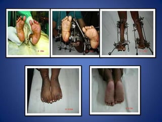
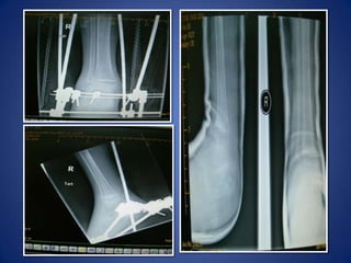
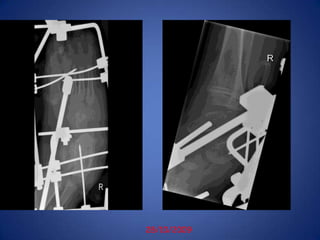
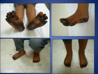
JOSHI’S EXTERNAL STABLISATION SYSTEM-(JESS)INCTEVDR. K.S.V. RaoMBBS, D.Orth, DNB( Ortho)
Causes Of Relapse In Rx Of CTEVErrors in  ctev correction  methods in PonsetiImproper surgical intervension without adequate conservative treatmentInadequate post operative careNon-compliant parents in post correction regime
Causes Of Relapse In Rx Of CTEV-------Lack of rehabilitation exercisesRigid club foot associated with- arthrogryposis, aminiotic band syndrome, Menigomyelocele, spina bifida, spinal cord defects  Unequal growth of muscles during growth spurtsDefective or inadequate orthotic fittings
Relapsed clubfoot is nothing more than an incompletely corrected feet.-(Beatson and Pearson 1966, Evans 1961, Fripp and Shaw 1967, Kite 1972, Turco 1971)Spurious correction later manifests as relapse.
Residual Deformities Adduction & inversion of forefootEquinus at ankle.Cavus & heel varusIn-toeing ±Problem – compounded by secondary changes in skin/bone & joints fibrosis/stiffness
Basic Anatomic Derangement In ClubfootCongenital subluxation of talo-calcaneonavicular joint
Navicular & calcaneus displaced medially in relation to  talus.Club foot- abnormal intertarsal relationshipThe shape of the tarsal bones is altered in accordance with the wolf’s law. Soft tissue contracture  acquired in accordance with the law of Davis “When ligaments and soft tissue are in lax state they will gradually shorten”
Clinical Assessment- (Caroll)Calf atrophyPosterior displacement of the fibulaCreases medial or posteriorCurved lateral borderCavusFixed equinusNavicular fixed to medial malleolusOs cacis fixed to tibiaNo mid tarsal mobilityFixed forefoot supination**Each feature scores 1 point Worst feet would score 10 and a Normal well corrected foot score 0
Radiological AssessmentTalo-calcaneal angle(AP) 15°-40
Talo-calcaneal angle (lat stress) 25-40 °
Talo-calcaneal index > 40 °
Tibio-calcaneal angle (stress lat) 5-15 °nTC25-40N 60-90<15 AbnnTC15-40
OVERCORRECTED FEET
 -to tide over the period till the child reaches  age of 14 before triple arthodesis
Problems  -RevisionRepeat surgical procedure –ChallengingPreexisting fibrosisStiffness of the joints of the foot Hypoplastic anterior tibial vesselsWound closure difficulties with skin necrosis.
Prof. BrijBhushan Joshi  (1928 –  2009)
JESSJoshi External Stablisation SystemDeveloped by  DR. B.B.JOSHI in Mumbai, IndiaFirst Patient - operated  in 1988Today - evolved into a verastile system with application in trauma, defects &  deformities  in upper and lower limb.JESS has a special  application in the correction of resistant clubfoot .
Principle Of JessBasis of  deformity correction - principle Of FRACTIONAL DISTRACTION OF ILIZAROV (1980)Dr Joshi  added the concept of DIFFERENTIAL DISTRACTION  (1988)In differential distraction - concave side  of deformity  is distracted twice the rate of the convex sidePrevents crushing of the tissues on the convex  side, lengthens the limb and effectively corrects the deformity at the same time.
IndicationsDrop out of  conservative treatmentRecurrence after earlier surgical releaseKnown resistant cases- severely contracted foot, AMC, Congenital band syndrome.Late presentation to treatmentAdjunct to surgical treatment -for realignment of skeleton to minimise bone resection and shortening of the foot
The Goal Of TreatmentFoot that is – Cosmetically acceptable Pliable Functional PainlessPlantigrade Fits into standard footwear Spares the parent and the child from the ordeal of frequent hospitalisation and years of treatment with casts and braces.
Components of JESS Fixator
Distractor DevicesThe double holeThe fish mouth The split block The biaxial hinge Connecting  rods- standard connecting rods in the small and medium set is 3 mm rod.
LINK JOINTS Link joints- different sizes-Medium size accommodates a                               -connecting rod upto 3 mm diameter in lower hole                                                                         - a k wire of 1.2 to 3 mm diameter in upper hole.Universal  link joint-independent locking system for each connecting rod and k wire Can hold rods up to 4 mm diameter
Operative TechniqueGA-SupinePneumatic tourniquet is applied- not inflatedNeurovascular markingsHand drill to pass k wires/power drill in older children3 MAIN STEPS:1.The insertion of k-wires2.The creation of holds3.The connection between the holds
Creations Of HoldsThe tibial holdThe Metatarsal holdThe Calcaneal holdTHE CONNECTION BETWEEN HOLDSThe Tibio-metatarsal connection  The Calcaneo-Metatarsal connection  The Tibio-Calcaneal connectionTOE  SLING ATTACHMENT-provides dynamic traction to prevent flexion of the toes as deformity gradually corrects
Application Of Tibial Wires
Application Of Transverse Calcaneal Wires
Application Of Metatarsal Wires
Application Of Axial Calcaneal Wire
Calaneo –Metatarsal DistractionCorrects forefoot adduction at mid tarsal & tarsometarsal joints
Realigns the head of talus with the navicular
Derotates the calcaneumEnd point-Clinical and radiological correction of forefoot deformities(approx 2-4 weeks)Medial- 0.25 mm every 6 hours Lateral- 0.25 mm every 12 hours
The Tibio-calcaneal DistractionTC is carried out in 2 positionsDistractors are mounted  between the inferior limbs of the tibial Z rods and post limb of the calcaneal-L  rodDistractors lie parallel to the leg and just posterior to the transfixing  calcaneal wires. This  corrects varus of the hind foot and equinus
Once the varus is corrected-Tibiocalcanealdistractors are shifted posteriorly-Distraction in this position provides thrust to  stretch the posterior structures and corrects hind foot equinus at the ankle and subtalar jointsEnd point –judged clinically (approx 4 weeks)Medial- 0.25 mm every 6 hoursLateral- 0.25 mm every 12 hours
Tibio-metatarsal ConnectionTibio-metatrsal connection is static.Keeps anterior part of the ankle and subtalar joint open while  the heel equinus is being corrected Weekly adjustment  needed to reduce excessive tension by loosening the clamps.Dorsiflexion of the ankle joint  achieved gradually after correction of the other components of the deformityRocker bottom –pseudo correction occurs if force dorsiflexion
Post Operative ManagementDISTRACTION SCHEDULE—3 rd day onwards360  clock wise in 4 fractions/180 in 2 fractions
Corrective period: 3-6 weeks.
Static period: 3-6 weeks
Casting after complete correction not only protects the osteopenic bones while the pin-tracts heal, but also maintains correction and allows gradual weightbearing.Care of the assemblyCover the pin sites with a dry dressingEncash the whole frame with a thin layer of soft foam or cardboardChange dressing  of pin tracks regularly
The Static Phase20 ° of dorsiflexion  necessary to avoid recurrence and to permit squatting.Following correction - assembly  held in a static position for  3 to 6 wks to allow soft tissue maturation in the elongated position.Static phase  should be twice the period of distraction
Cases
10/5/2009Post STR rt-3/M
28/10/2009
STR-dec2007(Sohar)JESS-28/10/2009KHTib AT-12/5/2010KHEXCELLENT RESULT18/4/2011
RESULTS
In 2003 S. Suresh et all treated 26 children with ctev 44 Joshi's external stabilization system procedure at the Safdarjung Hospital, New Delhi between Jan 1998 and Dec 1999. Three dimensional corrections were achieved by use of the distracter device. Excellent results were obtained in 77% of cases, good results in 13% and poor results in 9% of the cases.S.SURESH et al – Role Of  JESS In The Management Of Idiopathic Club feet, journal Of Orthopaedic Surgery. 2003: 11(2):194-200
Khoula Experience1992-1998  Khoula hospital, paedortho unit treated 112 feet using JESS fixator to correct foot deformities.20 were excluded from study-polio, meningomyelocele, muscular dystrophy92 feet were recurrent/neglected club feet--72 feet (56 patients) were available for study14(19.4%) were neglected-no surgery42(80.6%) were recurrent clubfoot3 (8.3%) had limited soft tissue surgery at time of JESS application. (Heel cord lengthening, plantar fasciotomy, and tibialis post z plasty)
ResultsGOOD result- 58 feet(80.5%)FAIR result- 10 feet(13.9%)POOR result-4 feet(5.6%)—needed reapplication of JESS to correct the deformity prior to triple arthrodesis.None of our patients showed correction to a normal range of talocalcaneal angle radiologically.
Complications
Orthotic DevicesSplints are fitted to maintain the corrected position over prolonged periodsThermoplastic splints are used-allows minor individual  variations.Denis–browne splint with abduction bar –in non ambulatory childChild refered to physiotherapist for gait training and to strengthen weaker muscles to keep foot supple and aligned
Older Children/Adults
Advantages Of JessUse in Small foot

Jess

  • 1.
    JOSHI’S EXTERNAL STABLISATIONSYSTEM-(JESS)INCTEVDR. K.S.V. RaoMBBS, D.Orth, DNB( Ortho)
  • 2.
    Causes Of RelapseIn Rx Of CTEVErrors in ctev correction methods in PonsetiImproper surgical intervension without adequate conservative treatmentInadequate post operative careNon-compliant parents in post correction regime
  • 3.
    Causes Of RelapseIn Rx Of CTEV-------Lack of rehabilitation exercisesRigid club foot associated with- arthrogryposis, aminiotic band syndrome, Menigomyelocele, spina bifida, spinal cord defects Unequal growth of muscles during growth spurtsDefective or inadequate orthotic fittings
  • 4.
    Relapsed clubfoot isnothing more than an incompletely corrected feet.-(Beatson and Pearson 1966, Evans 1961, Fripp and Shaw 1967, Kite 1972, Turco 1971)Spurious correction later manifests as relapse.
  • 5.
    Residual Deformities Adduction& inversion of forefootEquinus at ankle.Cavus & heel varusIn-toeing ±Problem – compounded by secondary changes in skin/bone & joints fibrosis/stiffness
  • 6.
    Basic Anatomic DerangementIn ClubfootCongenital subluxation of talo-calcaneonavicular joint
  • 7.
    Navicular & calcaneusdisplaced medially in relation to talus.Club foot- abnormal intertarsal relationshipThe shape of the tarsal bones is altered in accordance with the wolf’s law. Soft tissue contracture acquired in accordance with the law of Davis “When ligaments and soft tissue are in lax state they will gradually shorten”
  • 8.
    Clinical Assessment- (Caroll)CalfatrophyPosterior displacement of the fibulaCreases medial or posteriorCurved lateral borderCavusFixed equinusNavicular fixed to medial malleolusOs cacis fixed to tibiaNo mid tarsal mobilityFixed forefoot supination**Each feature scores 1 point Worst feet would score 10 and a Normal well corrected foot score 0
  • 9.
  • 10.
  • 11.
  • 12.
    Tibio-calcaneal angle (stresslat) 5-15 °nTC25-40N 60-90<15 AbnnTC15-40
  • 13.
  • 14.
    -to tideover the period till the child reaches age of 14 before triple arthodesis
  • 15.
    Problems -RevisionRepeatsurgical procedure –ChallengingPreexisting fibrosisStiffness of the joints of the foot Hypoplastic anterior tibial vesselsWound closure difficulties with skin necrosis.
  • 16.
    Prof. BrijBhushan Joshi (1928 – 2009)
  • 17.
    JESSJoshi External StablisationSystemDeveloped by DR. B.B.JOSHI in Mumbai, IndiaFirst Patient - operated in 1988Today - evolved into a verastile system with application in trauma, defects & deformities in upper and lower limb.JESS has a special application in the correction of resistant clubfoot .
  • 18.
    Principle Of JessBasisof deformity correction - principle Of FRACTIONAL DISTRACTION OF ILIZAROV (1980)Dr Joshi added the concept of DIFFERENTIAL DISTRACTION (1988)In differential distraction - concave side of deformity is distracted twice the rate of the convex sidePrevents crushing of the tissues on the convex side, lengthens the limb and effectively corrects the deformity at the same time.
  • 19.
    IndicationsDrop out of conservative treatmentRecurrence after earlier surgical releaseKnown resistant cases- severely contracted foot, AMC, Congenital band syndrome.Late presentation to treatmentAdjunct to surgical treatment -for realignment of skeleton to minimise bone resection and shortening of the foot
  • 20.
    The Goal OfTreatmentFoot that is – Cosmetically acceptable Pliable Functional PainlessPlantigrade Fits into standard footwear Spares the parent and the child from the ordeal of frequent hospitalisation and years of treatment with casts and braces.
  • 21.
  • 22.
    Distractor DevicesThe doubleholeThe fish mouth The split block The biaxial hinge Connecting rods- standard connecting rods in the small and medium set is 3 mm rod.
  • 23.
    LINK JOINTS Linkjoints- different sizes-Medium size accommodates a -connecting rod upto 3 mm diameter in lower hole - a k wire of 1.2 to 3 mm diameter in upper hole.Universal link joint-independent locking system for each connecting rod and k wire Can hold rods up to 4 mm diameter
  • 24.
    Operative TechniqueGA-SupinePneumatic tourniquetis applied- not inflatedNeurovascular markingsHand drill to pass k wires/power drill in older children3 MAIN STEPS:1.The insertion of k-wires2.The creation of holds3.The connection between the holds
  • 25.
    Creations Of HoldsThetibial holdThe Metatarsal holdThe Calcaneal holdTHE CONNECTION BETWEEN HOLDSThe Tibio-metatarsal connection The Calcaneo-Metatarsal connection The Tibio-Calcaneal connectionTOE SLING ATTACHMENT-provides dynamic traction to prevent flexion of the toes as deformity gradually corrects
  • 26.
  • 27.
  • 28.
  • 29.
    Application Of AxialCalcaneal Wire
  • 30.
    Calaneo –Metatarsal DistractionCorrectsforefoot adduction at mid tarsal & tarsometarsal joints
  • 31.
    Realigns the headof talus with the navicular
  • 32.
    Derotates the calcaneumEndpoint-Clinical and radiological correction of forefoot deformities(approx 2-4 weeks)Medial- 0.25 mm every 6 hours Lateral- 0.25 mm every 12 hours
  • 33.
    The Tibio-calcaneal DistractionTCis carried out in 2 positionsDistractors are mounted between the inferior limbs of the tibial Z rods and post limb of the calcaneal-L rodDistractors lie parallel to the leg and just posterior to the transfixing calcaneal wires. This corrects varus of the hind foot and equinus
  • 34.
    Once the varusis corrected-Tibiocalcanealdistractors are shifted posteriorly-Distraction in this position provides thrust to stretch the posterior structures and corrects hind foot equinus at the ankle and subtalar jointsEnd point –judged clinically (approx 4 weeks)Medial- 0.25 mm every 6 hoursLateral- 0.25 mm every 12 hours
  • 35.
    Tibio-metatarsal ConnectionTibio-metatrsal connectionis static.Keeps anterior part of the ankle and subtalar joint open while the heel equinus is being corrected Weekly adjustment needed to reduce excessive tension by loosening the clamps.Dorsiflexion of the ankle joint achieved gradually after correction of the other components of the deformityRocker bottom –pseudo correction occurs if force dorsiflexion
  • 36.
    Post Operative ManagementDISTRACTIONSCHEDULE—3 rd day onwards360 clock wise in 4 fractions/180 in 2 fractions
  • 37.
  • 38.
  • 39.
    Casting after completecorrection not only protects the osteopenic bones while the pin-tracts heal, but also maintains correction and allows gradual weightbearing.Care of the assemblyCover the pin sites with a dry dressingEncash the whole frame with a thin layer of soft foam or cardboardChange dressing of pin tracks regularly
  • 40.
    The Static Phase20° of dorsiflexion necessary to avoid recurrence and to permit squatting.Following correction - assembly held in a static position for 3 to 6 wks to allow soft tissue maturation in the elongated position.Static phase should be twice the period of distraction
  • 41.
  • 56.
  • 57.
  • 58.
  • 61.
  • 62.
    In 2003 S.Suresh et all treated 26 children with ctev 44 Joshi's external stabilization system procedure at the Safdarjung Hospital, New Delhi between Jan 1998 and Dec 1999. Three dimensional corrections were achieved by use of the distracter device. Excellent results were obtained in 77% of cases, good results in 13% and poor results in 9% of the cases.S.SURESH et al – Role Of JESS In The Management Of Idiopathic Club feet, journal Of Orthopaedic Surgery. 2003: 11(2):194-200
  • 63.
    Khoula Experience1992-1998 Khoula hospital, paedortho unit treated 112 feet using JESS fixator to correct foot deformities.20 were excluded from study-polio, meningomyelocele, muscular dystrophy92 feet were recurrent/neglected club feet--72 feet (56 patients) were available for study14(19.4%) were neglected-no surgery42(80.6%) were recurrent clubfoot3 (8.3%) had limited soft tissue surgery at time of JESS application. (Heel cord lengthening, plantar fasciotomy, and tibialis post z plasty)
  • 64.
    ResultsGOOD result- 58feet(80.5%)FAIR result- 10 feet(13.9%)POOR result-4 feet(5.6%)—needed reapplication of JESS to correct the deformity prior to triple arthrodesis.None of our patients showed correction to a normal range of talocalcaneal angle radiologically.
  • 66.
  • 67.
    Orthotic DevicesSplints arefitted to maintain the corrected position over prolonged periodsThermoplastic splints are used-allows minor individual variations.Denis–browne splint with abduction bar –in non ambulatory childChild refered to physiotherapist for gait training and to strengthen weaker muscles to keep foot supple and aligned
  • 68.
  • 71.
    Advantages Of JessUsein Small foot

Editor's Notes

  • #5 There appears to be increasing support to the view that the so-called relapsed clubfoot is nothing more than an incompletely corrected clubfoot It is the spurious correction later manifests itself as a relapse.
  • #20 Three sets of assembly components are designed: Small, medium and large.Components of JESS fixator:DistractersLink jointsConnecting rodsZ- rodsL-rodsk-wires