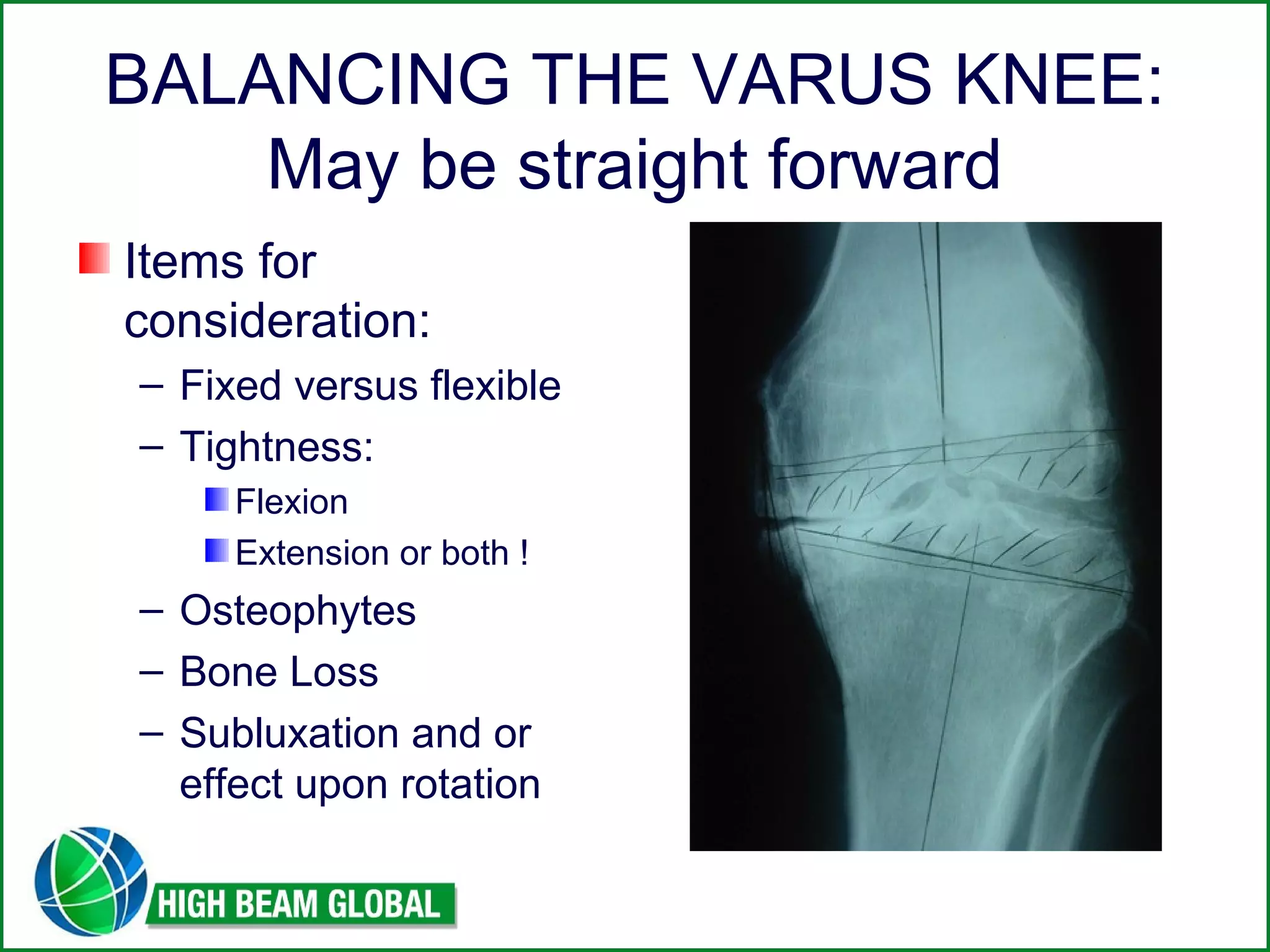
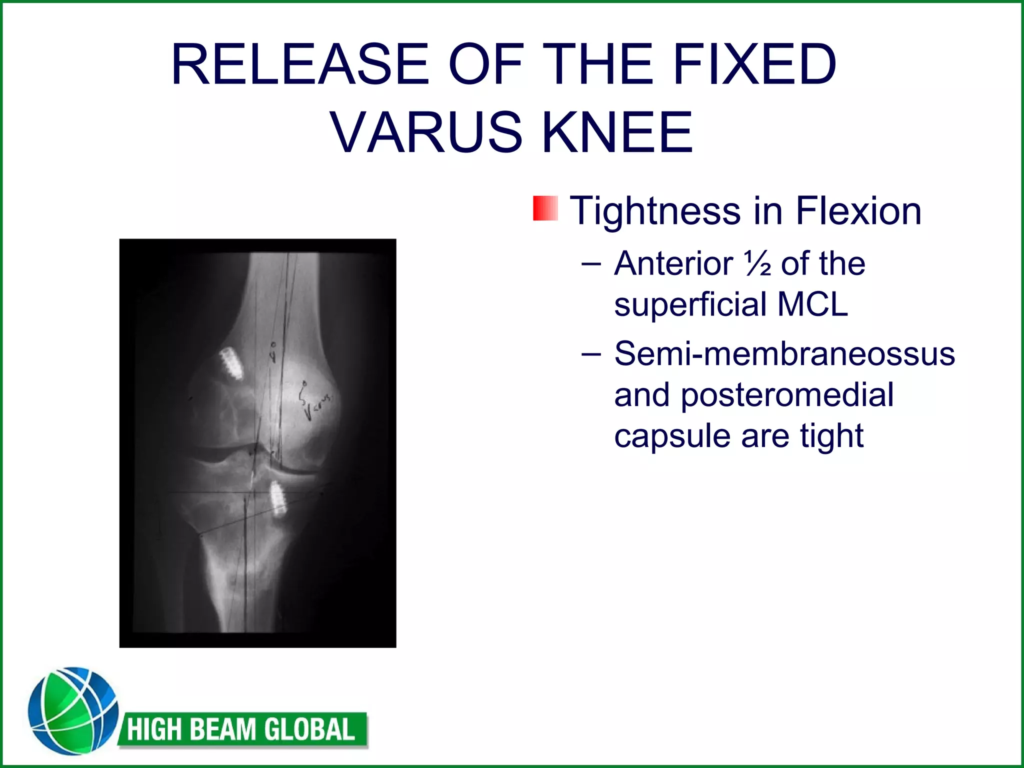
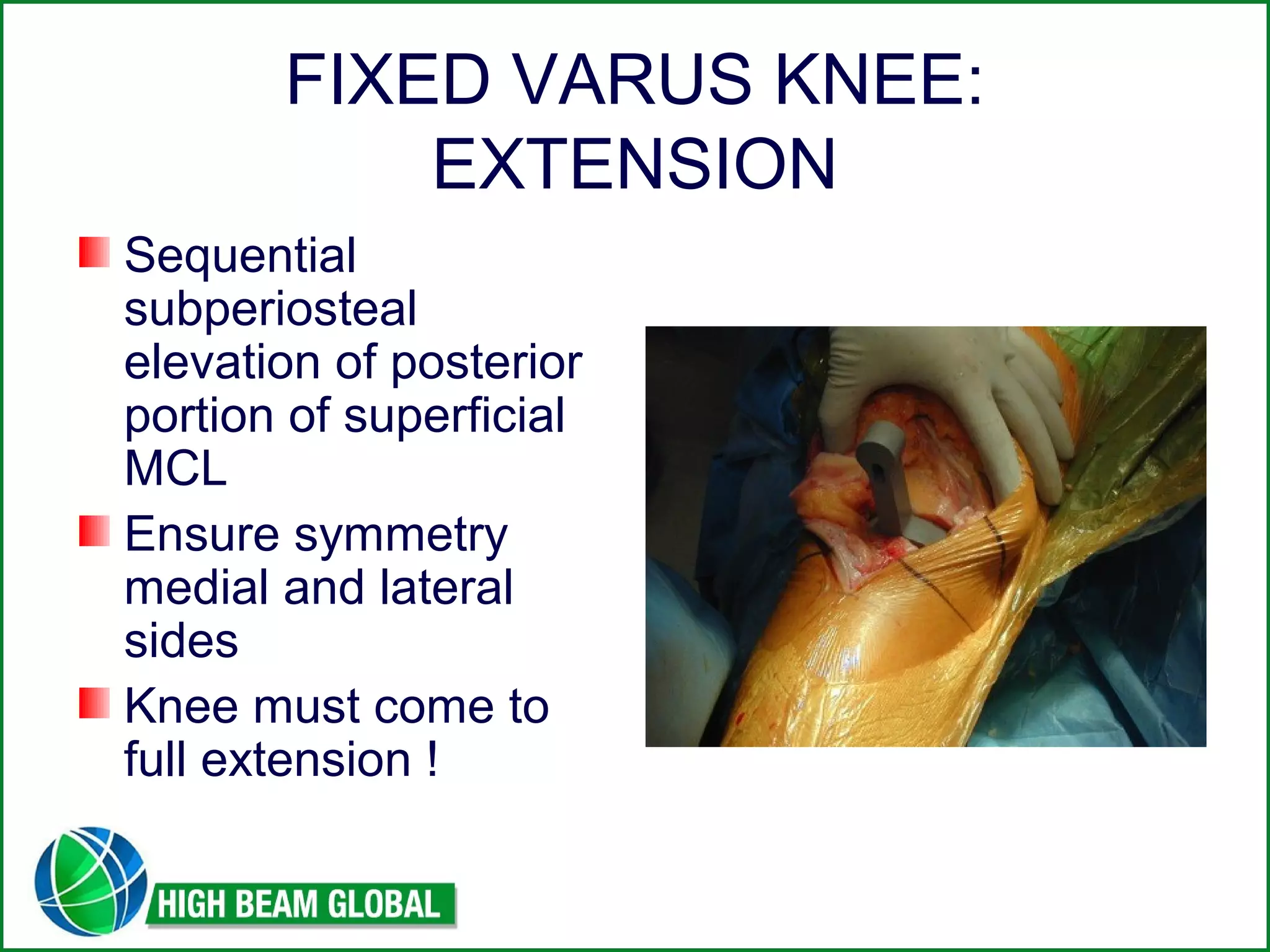
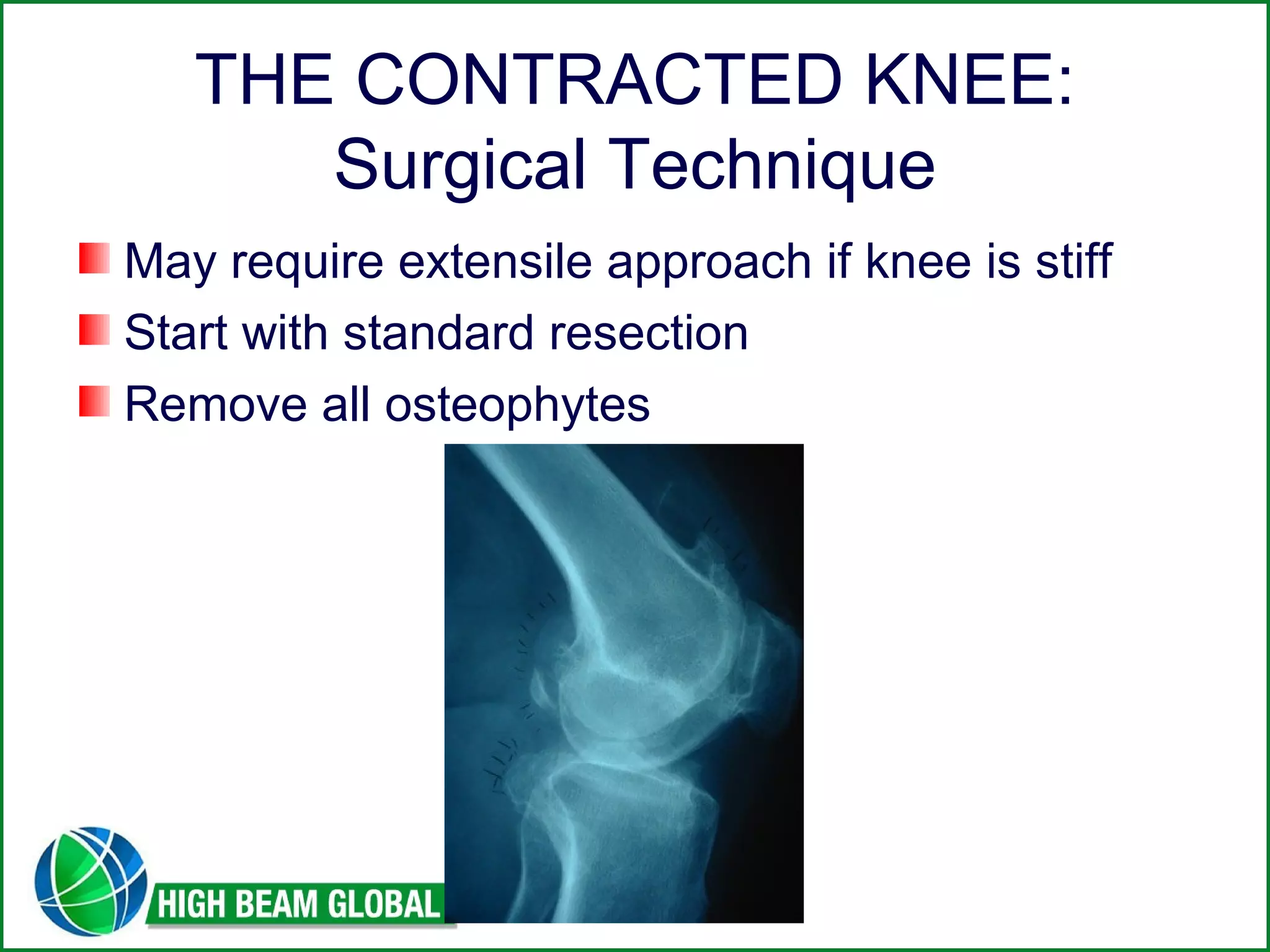
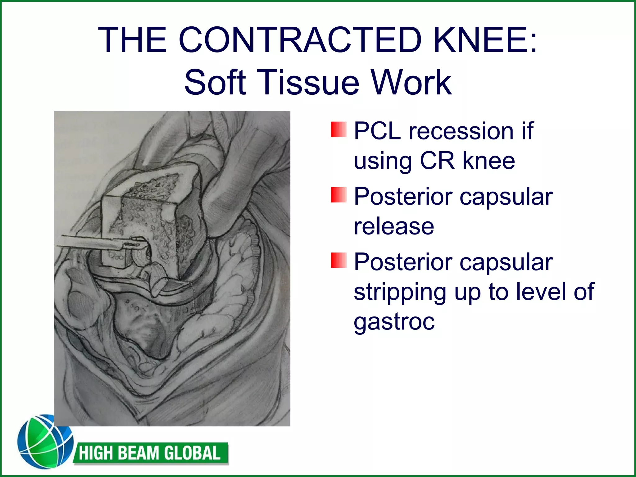
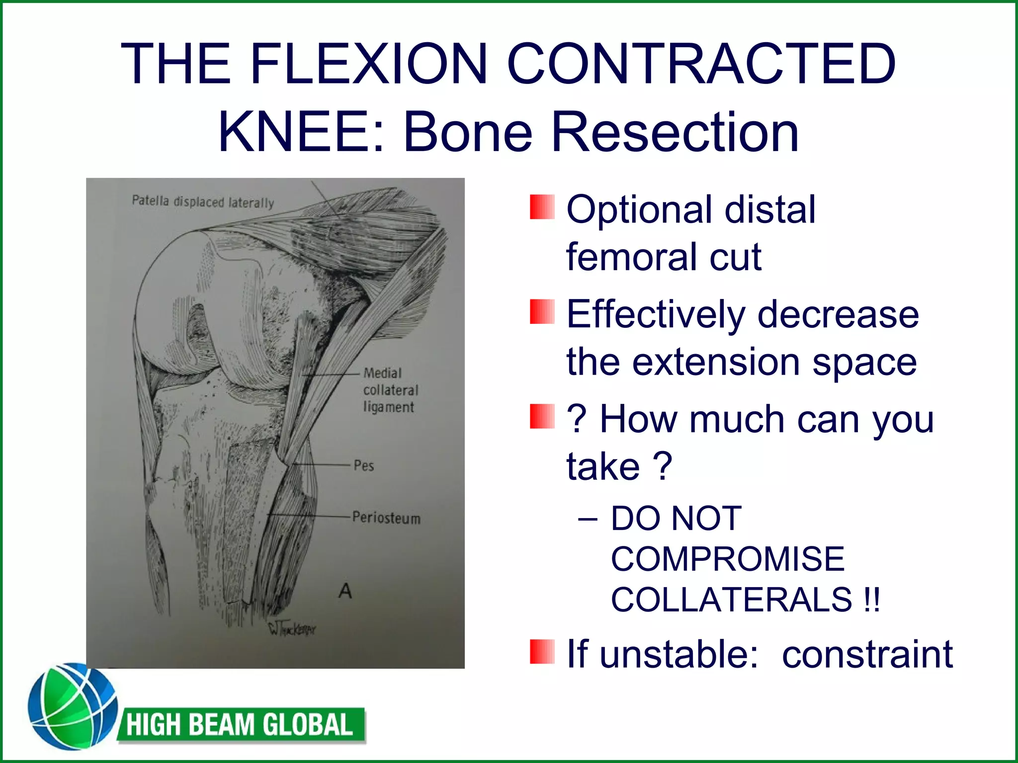
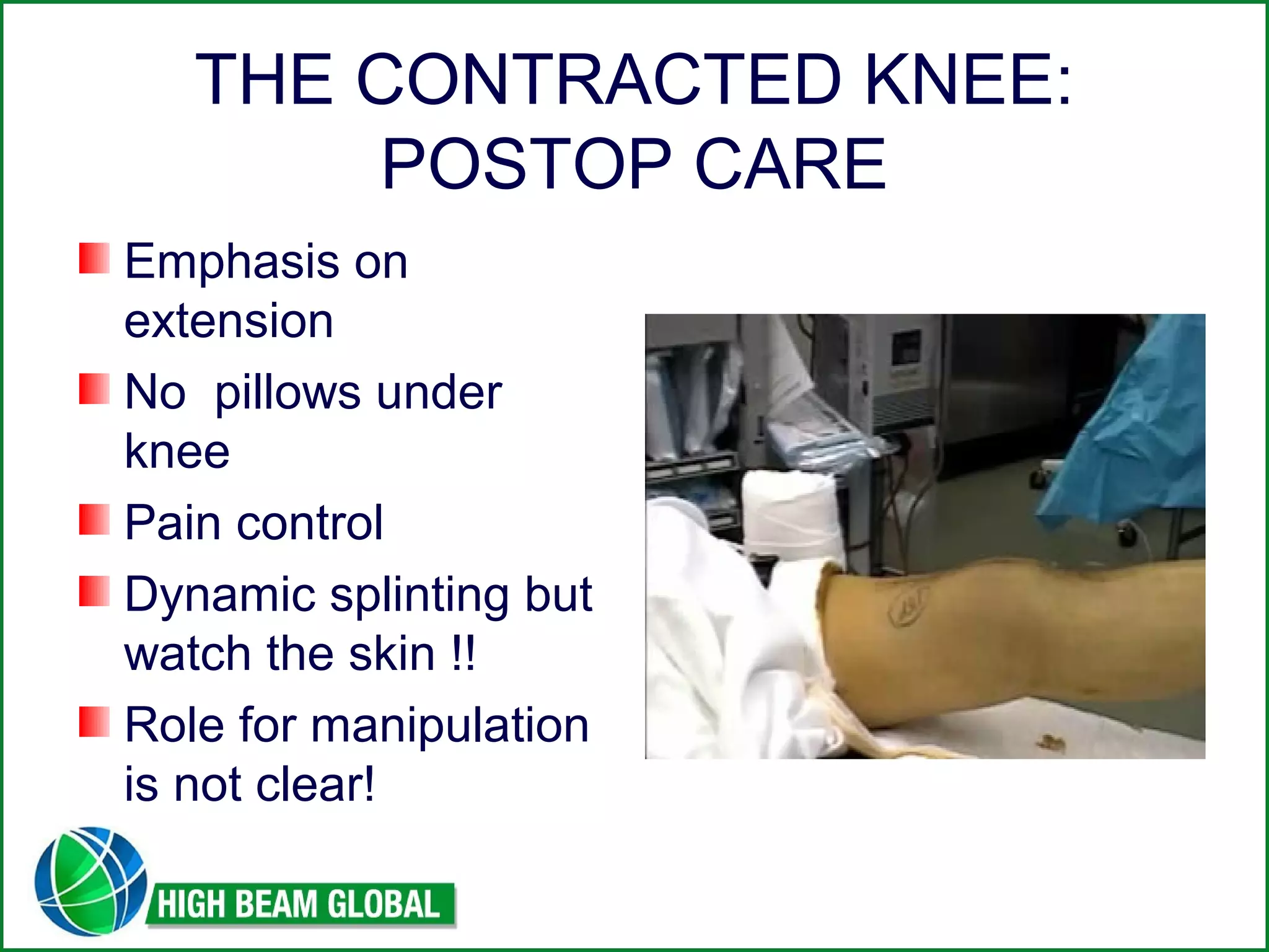
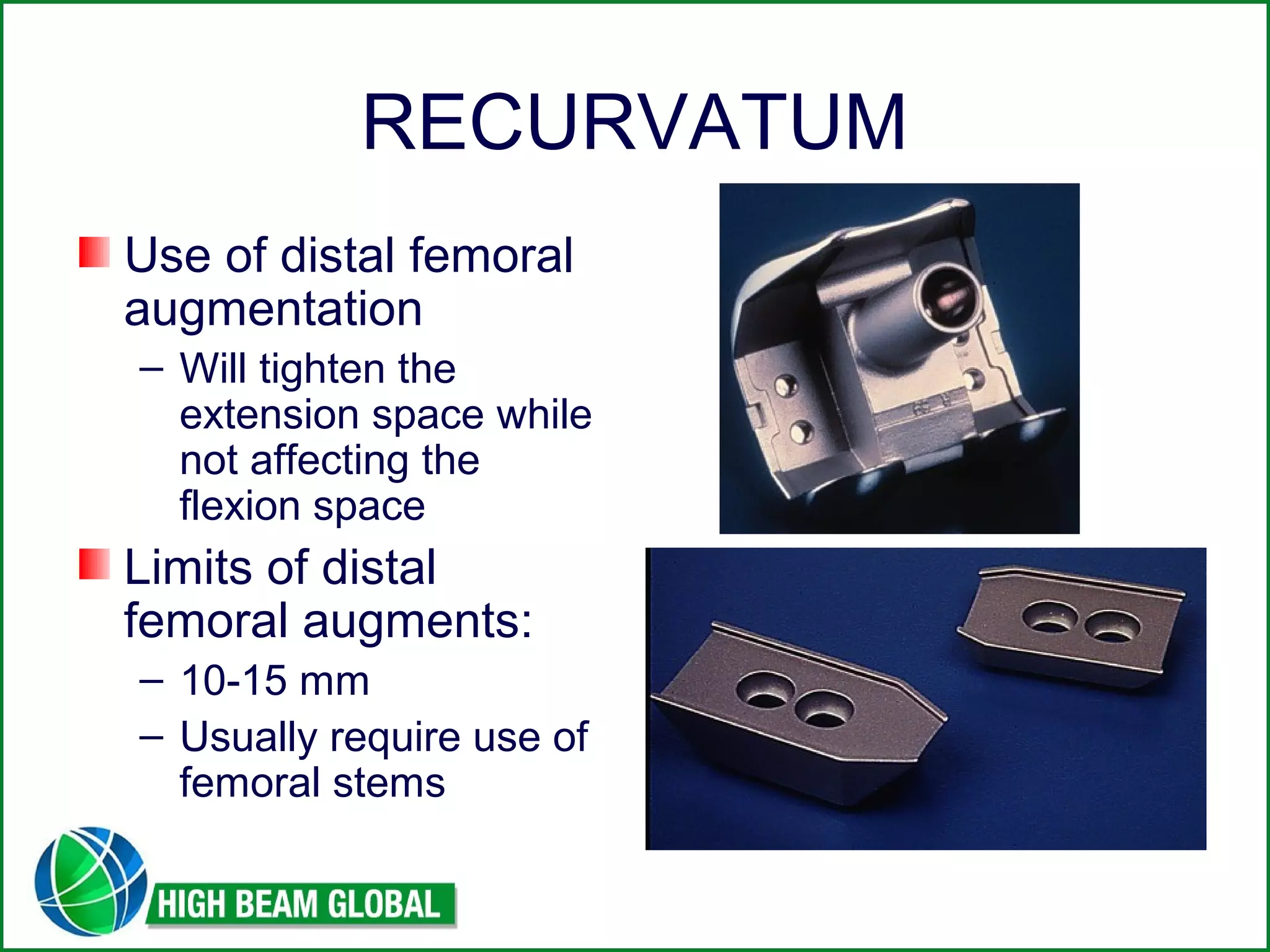
TECHNIQUES IN PRIMARY 
TOTAL KNEE 
ARTHROPLASTY: 
Balancing ! 
Douglas E. Padgett, M.D. 
Adult Reconstruction and Joint Replacement 
Hospital for Special Surgery 
New York, New York
DDIISSCCLLOOSSUURREESS 
None Related to the topic of this 
presentation 
Consultant: Mako, Stryker 
AAHKS: Board Member
Success in TKR 
Basic tenets of Dr. 
John Insall: 
– TKR should be 
thought of as a soft 
tissue operation 
– Failure to address the 
soft tissue envelope 
will result in: 
Pain 
Stiffness 
Laxity
“Thinking is the hardest work 
there is, which is the probable 
reason so few engage in it “ 
Henry Ford 
American entrepreneurr
FAILURE IN TKR 
Often can be traced back 
to original patho-anatomy 
Varus knee 
– Preop: Tight medial side 
– Trial Components: knee 
noted to be tight medially 
– Additional tibia resected 
– Result: knee “books open” 
medially
Failure in TKR: 
The Valgus Knee 
Complex patho-anatomy 
Rotation often difficult to 
discern 
Effect of deficiency of 
posterolateral condyle 
upon landmarks 
Insufficient lateral side 
release: results is 
unstable arthroplasty
The Happy TKR
THE VARUS KNEE
BALANCING THE VARUS KNEE: 
May be straight forward 
Items for 
consideration: 
– Fixed versus flexible 
– Tightness: 
Flexion 
Extension or both ! 
– Osteophytes 
– Bone Loss 
– Subluxation and or 
effect upon rotation
The Varus Knee: 
At times, a surgical challenge
APPROACH TO THE 
VARUS KNEE 
Standard medial 
parapatella approach 
Sharp dissection onto 
the proximo-medial 
tibia 
Dissection interval 
above the pes but 
below the joint line 
Subperiosteal 
elevation
The Varus Knee 
Remove all 
osteophytes 
STOP !!! 
– Assess ligamentous / 
soft tissue tightness 
before proceeding
RELEASE OF THE FIXED 
VARUS KNEE 
TIGHTNESS IN 
EXTENSION 
– Posterior ½ of the 
superficial MCL is 
primary factor 
– Anteromedial capsule 
can also contribute
RELEASE OF THE FIXED 
VARUS KNEE 
Tightness in Flexion 
– Anterior ½ of the 
superficial MCL 
– Semi-membraneossus 
and posteromedial 
capsule are tight
FIXED VARUS KNEE: 
FLEXION 
Check gap symmetry 
(medial and lateral 
sides) 
– Release anterior 
portion of superficial 
MCL 
Flexion space should 
not be excessively 
tight (I prefer some 
ability to translate 
forward)
FIXED VARUS KNEE: 
EXTENSION 
Sequential 
subperiosteal 
elevation of posterior 
portion of superficial 
MCL 
Ensure symmetry 
medial and lateral 
sides 
Knee must come to 
full extension !
ROLE OF BONE RESECTION 
EFFECT UPON BALANCING 
While majority of 
balancing is soft 
tissue in nature: 
– tibial resection in 
coronal plane (varus-valgus) 
will effect soft 
tissues 
– Femoral rotation 
clearly affects soft 
tissue tension 
especially in flexion
BALANCING THE 
VALGUS KNEE
THE VALGUS KNEE 
Fixed vs correctable 
Associated bone loss 
Tightness: 
– Flexion ? 
– Extension ? 
– Both ? 
Status of MCL
STEP #1: EXPOSURE / 
RESECTION 
Medial parapatella 
Minimal medial side 
release 
Tibial resection: 
– Minimal cut 
perpendicular to shaft 
Femoral resection: 
– I favor 2-3 degrees off 
of femoral line
FLEXION SPACE RELEASES 
Laminar spreader is 
the best device 
Pie-crusting of the 
posterolateral capsule 
and arcuate complex 
Leave the popliteus 
intact if possible. 
– Can result in flexion 
instability in varus
EXTENSION SPACE RELEASE 
Tight structures: 
– ITB 
– Posterolateral capsule 
Pie-crusting 
technique with SLOW 
gradual releases work 
best in my hands.
FINAL PRODUCT: 
Ligament balance: M=L 
Gap Balance: Flexion=Extension
THE VALGUS KNEE 
Adhering to the 
concept of sequential 
releases, majority of 
knees can be 
corrected with the use 
of additional 
constraint from the 
articulation.
THE FLEXION CONTRACTED 
KNEE
THE FLEXION CONTRACTED 
KNEE 
Considerations: 
– Definition: 15 degrees 
or greater loss of 
extension 
– ? Length of 
contracture 
– Status of skin, prior 
incisions 
– Neurologic exam
THE FLEXION CONTRACTED 
KNEE 
Deformity 
Bone loss 
Patella height
THE CONTRACTED KNEE: 
Surgical Technique 
May require extensile approach if knee is stiff 
Start with standard resection 
Remove all osteophytes
THE CONTRACTED KNEE: 
Soft Tissue Work 
PCL recession if 
using CR knee 
Posterior capsular 
release 
Posterior capsular 
stripping up to level of 
gastroc
THE FLEXION CONTRACTED 
KNEE: Bone Resection 
Optional distal 
femoral cut 
Effectively decrease 
the extension space 
? How much can you 
take ? 
– DO NOT 
COMPROMISE 
COLLATERALS !! 
If unstable: constraint
THE CONTRACTED KNEE: 
POSTOP CARE 
Emphasis on 
extension 
No pillows under 
knee 
Pain control 
Dynamic splinting but 
watch the skin !! 
Role for manipulation 
is not clear!
TOTAL KNEE 
ARTHROPLASTY 
RECURVATUM
RECURVATUM 
Seen predominantly 
in neuromuscular 
diseases: 
– Polio 
– Neuropathic joints 
– Spinal cord patients 
Can occasionally be 
seen in rheumatic 
conditions
RECURVATUM: 
Options 
Hyperextension up to 
20 degrees: 
– Attempt to “overstuff” 
the extension space 
– Must balance the 
temptation to leave the 
knee with a flexion 
contracture: 
If recurvatum is due to 
quad weakness, flexion 
contracture will lead to 
knee giving out !
RECURVATUM 
Use of distal femoral 
augmentation 
– Will tighten the 
extension space while 
not affecting the 
flexion space 
Limits of distal 
femoral augments: 
– 10-15 mm 
– Usually require use of 
femoral stems
RECURVATUM 
Posterior stabilized 
implants are preferred 
Less constraining 
implants may lead to 
instability
RECURVATUM 
Reliance upon 
standard implants will 
lead to excessive 
anterior polyethylene 
impingement 
Wear and or 
loosening is clearly a 
consequence
RECURVATUM: 
The larger deformities 
In instances where 
there is more than 20 
degrees of 
recurvatum: consider 
a more constrained 
implant with an 
extension stop 
DO NOT RELY 
UPON STANDARD 
CONDYLAR 
DESIGNS !!
Primary TKR: 
Summary 
Understanding of pathoanatomy crucial 
Correction of deforming forces is vital to 
successful outcome 
Know the limits of your prosthetic implant
THANK YOU FOR YOUR 
ATTENTION !

Techniques in primary total knee arthroplasty

  • 2.
    TECHNIQUES IN PRIMARY TOTAL KNEE ARTHROPLASTY: Balancing ! Douglas E. Padgett, M.D. Adult Reconstruction and Joint Replacement Hospital for Special Surgery New York, New York
  • 3.
    DDIISSCCLLOOSSUURREESS None Relatedto the topic of this presentation Consultant: Mako, Stryker AAHKS: Board Member
  • 4.
    Success in TKR Basic tenets of Dr. John Insall: – TKR should be thought of as a soft tissue operation – Failure to address the soft tissue envelope will result in: Pain Stiffness Laxity
  • 5.
    “Thinking is thehardest work there is, which is the probable reason so few engage in it “ Henry Ford American entrepreneurr
  • 6.
    FAILURE IN TKR Often can be traced back to original patho-anatomy Varus knee – Preop: Tight medial side – Trial Components: knee noted to be tight medially – Additional tibia resected – Result: knee “books open” medially
  • 7.
    Failure in TKR: The Valgus Knee Complex patho-anatomy Rotation often difficult to discern Effect of deficiency of posterolateral condyle upon landmarks Insufficient lateral side release: results is unstable arthroplasty
  • 8.
  • 9.
  • 10.
    BALANCING THE VARUSKNEE: May be straight forward Items for consideration: – Fixed versus flexible – Tightness: Flexion Extension or both ! – Osteophytes – Bone Loss – Subluxation and or effect upon rotation
  • 11.
    The Varus Knee: At times, a surgical challenge
  • 12.
    APPROACH TO THE VARUS KNEE Standard medial parapatella approach Sharp dissection onto the proximo-medial tibia Dissection interval above the pes but below the joint line Subperiosteal elevation
  • 13.
    The Varus Knee Remove all osteophytes STOP !!! – Assess ligamentous / soft tissue tightness before proceeding
  • 14.
    RELEASE OF THEFIXED VARUS KNEE TIGHTNESS IN EXTENSION – Posterior ½ of the superficial MCL is primary factor – Anteromedial capsule can also contribute
  • 15.
    RELEASE OF THEFIXED VARUS KNEE Tightness in Flexion – Anterior ½ of the superficial MCL – Semi-membraneossus and posteromedial capsule are tight
  • 16.
    FIXED VARUS KNEE: FLEXION Check gap symmetry (medial and lateral sides) – Release anterior portion of superficial MCL Flexion space should not be excessively tight (I prefer some ability to translate forward)
  • 17.
    FIXED VARUS KNEE: EXTENSION Sequential subperiosteal elevation of posterior portion of superficial MCL Ensure symmetry medial and lateral sides Knee must come to full extension !
  • 18.
    ROLE OF BONERESECTION EFFECT UPON BALANCING While majority of balancing is soft tissue in nature: – tibial resection in coronal plane (varus-valgus) will effect soft tissues – Femoral rotation clearly affects soft tissue tension especially in flexion
  • 19.
  • 20.
    THE VALGUS KNEE Fixed vs correctable Associated bone loss Tightness: – Flexion ? – Extension ? – Both ? Status of MCL
  • 21.
    STEP #1: EXPOSURE/ RESECTION Medial parapatella Minimal medial side release Tibial resection: – Minimal cut perpendicular to shaft Femoral resection: – I favor 2-3 degrees off of femoral line
  • 22.
    FLEXION SPACE RELEASES Laminar spreader is the best device Pie-crusting of the posterolateral capsule and arcuate complex Leave the popliteus intact if possible. – Can result in flexion instability in varus
  • 23.
    EXTENSION SPACE RELEASE Tight structures: – ITB – Posterolateral capsule Pie-crusting technique with SLOW gradual releases work best in my hands.
  • 24.
    FINAL PRODUCT: Ligamentbalance: M=L Gap Balance: Flexion=Extension
  • 25.
    THE VALGUS KNEE Adhering to the concept of sequential releases, majority of knees can be corrected with the use of additional constraint from the articulation.
  • 26.
  • 27.
    THE FLEXION CONTRACTED KNEE Considerations: – Definition: 15 degrees or greater loss of extension – ? Length of contracture – Status of skin, prior incisions – Neurologic exam
  • 28.
    THE FLEXION CONTRACTED KNEE Deformity Bone loss Patella height
  • 29.
    THE CONTRACTED KNEE: Surgical Technique May require extensile approach if knee is stiff Start with standard resection Remove all osteophytes
  • 30.
    THE CONTRACTED KNEE: Soft Tissue Work PCL recession if using CR knee Posterior capsular release Posterior capsular stripping up to level of gastroc
  • 31.
    THE FLEXION CONTRACTED KNEE: Bone Resection Optional distal femoral cut Effectively decrease the extension space ? How much can you take ? – DO NOT COMPROMISE COLLATERALS !! If unstable: constraint
  • 32.
    THE CONTRACTED KNEE: POSTOP CARE Emphasis on extension No pillows under knee Pain control Dynamic splinting but watch the skin !! Role for manipulation is not clear!
  • 33.
  • 34.
    RECURVATUM Seen predominantly in neuromuscular diseases: – Polio – Neuropathic joints – Spinal cord patients Can occasionally be seen in rheumatic conditions
  • 35.
    RECURVATUM: Options Hyperextensionup to 20 degrees: – Attempt to “overstuff” the extension space – Must balance the temptation to leave the knee with a flexion contracture: If recurvatum is due to quad weakness, flexion contracture will lead to knee giving out !
  • 36.
    RECURVATUM Use ofdistal femoral augmentation – Will tighten the extension space while not affecting the flexion space Limits of distal femoral augments: – 10-15 mm – Usually require use of femoral stems
  • 37.
    RECURVATUM Posterior stabilized implants are preferred Less constraining implants may lead to instability
  • 38.
    RECURVATUM Reliance upon standard implants will lead to excessive anterior polyethylene impingement Wear and or loosening is clearly a consequence
  • 39.
    RECURVATUM: The largerdeformities In instances where there is more than 20 degrees of recurvatum: consider a more constrained implant with an extension stop DO NOT RELY UPON STANDARD CONDYLAR DESIGNS !!
  • 40.
    Primary TKR: Summary Understanding of pathoanatomy crucial Correction of deforming forces is vital to successful outcome Know the limits of your prosthetic implant
  • 41.
    THANK YOU FORYOUR ATTENTION !