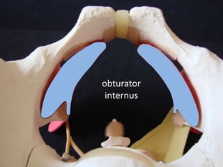
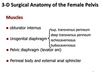
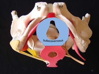
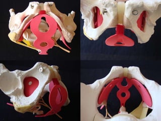
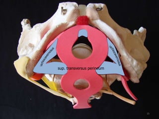
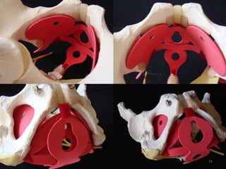
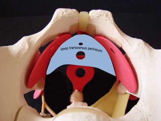
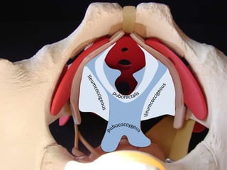
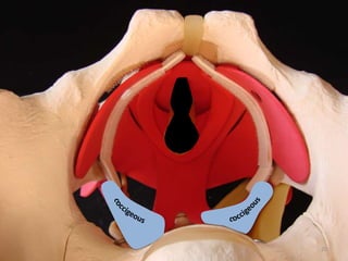
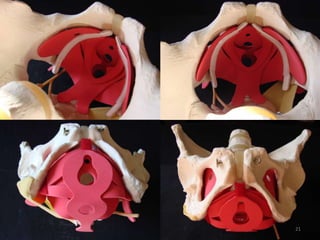
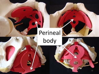
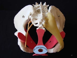
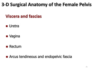
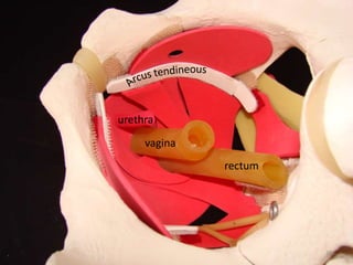
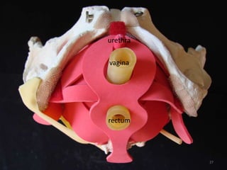
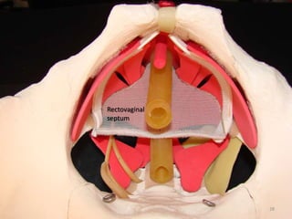
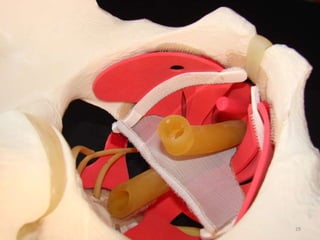
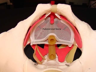
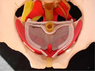
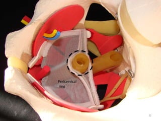
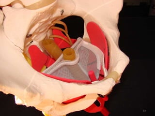
3-D Surgical
Anatomy of the
Female Pelvis
1
Coordinators
Cassio Riccetto - Associate Professor of Urology - University of
Campinas – Unicamp Sao Paulo – Brazil – cassioriccetto@gmail.com
Virginia Roncati – Head of Urogynecology Section – Hospital Heliopolis
Joana – SP – Brazil - virginia@gmail.com
Faculty
3
Carlos Del Roy – Head of Urogynecology Section – Hospital S. Joana –
SP – Brazil - carlosdelroy@gmail.com
José Tadeu Nunes Tamanini – Associate Professor of Urology – Federal
University of S. Carlos – SP – Brazil tadeutamanini@gmail.com
Simone Vidotti – Associate Physician of Urogynecology Section –
Hospital Heliopolis Joana – SP – Brazil simonevidotti@yahoo.com
Sophia Souto – Ft. PhD in Sciences of Surgery – University of Campinas
– SP – Brazil sophiasouto@hotmail.com
3-D Surgical Anatomy of the Female Pelvis
Bones benchmarks
 Symphysis pubis
 Obturator foramen
 Ischial spine
 Sacrum
4
5
Obturator
membrane
6
3-D Surgical Anatomy of the Female Pelvis
Ligaments
 Uterosacral
 Cardinal
 Pubourethral
7
 Sacrospinous
 Sacrotuberous
8
9
3-D Surgical Anatomy of the Female Pelvis
Muscles
 Urogenital diaphragm
 Pelvic diaphragm (levator ani)
 obturator internus
 Perineal body and external anal sphincter
10
obturator
internus
11
3-D Surgical Anatomy of the Female Pelvis
Muscles
 Urogenital diaphragm
 Pelvic diaphragm (levator ani)
 obturator internus
 Perineal body and external anal sphincter
sup. transversus perineum
deep transversus perineum
ischiocavernosus
bulbocavernosus
12
13
14
sup. transversus perineum
15
16
17
3-D Surgical Anatomy of the Female Pelvis
Muscles
 Urogenital diaphragm
 Pelvic diaphragm (levator ani)
 obturator internus
 Perineal body and external anal sphincter
18
19
20
21
3-D Surgical Anatomy of the Female Pelvis
Muscles
 Urogenital diaphragm
 Pelvic diaphragm (levator ani)
 obturator internus
 Perineal body and external anal sphincter
22
Perineal
body
23
24
3-D Surgical Anatomy of the Female Pelvis
Viscera and fascias
 Vagina
 Rectum
 Uretra
 Arcus tendineous and endopelvic fascia
25
urethra
vagina
rectum
26
urethra
vagina
rectum
27
Rectovaginal
septum
28
29
Pubocervical fascia
30
Cardinal
ligaments
31
Pericervical
ring
32
33
3-D Surgical Anatomy of the Female Pelvis
Thank you !
34

ALAPP 2017 3D Anatomy

Editor's Notes

  • #2 The objective of this presentation is to highlight the main concepts related to the surgical anatomy of the female pelvic floor. The main contributors for the vaginal support are: the bony pelvis, to which the pelvic tissues attach; the ligaments and fascia, including the cardinal and uterosacral ligament complex and endopelvic fascia, which attaches the vagina to the arcus tendineous and to the pericervical ring; and muscles, which are grouped in the urogenital and pelvic diaphragms and are the main components of the perineal body.
  • #3 The objective of this presentation is to highlight the main concepts related to the surgical anatomy of the female pelvic floor. The main contributors for the vaginal support are: the bony pelvis, to which the pelvic tissues attach; the ligaments and fascia, including the cardinal and uterosacral ligament complex and endopelvic fascia, which attaches the vagina to the arcus tendineous and to the pericervical ring; and muscles, which are grouped in the urogenital and pelvic diaphragms and are the main components of the perineal body.
  • #4 The objective of this presentation is to highlight the main concepts related to the surgical anatomy of the female pelvic floor. The main contributors for the vaginal support are: the bony pelvis, to which the pelvic tissues attach; the ligaments and fascia, including the cardinal and uterosacral ligament complex and endopelvic fascia, which attaches the vagina to the arcus tendineous and to the pericervical ring; and muscles, which are grouped in the urogenital and pelvic diaphragms and are the main components of the perineal body.
  • #5 Bones benchmarks The bones are the ultimate fixed attachment of the pelvic soft tissues. For surgical purposes, it have to be highlighted: pubic symphysis, in which pubourethral ligaments are inserted. obturator foramen, which are used as an approach for the transobturator slings and vaginal meshes. ischial spine, which represents the upper level of the vagina and in which the sacrospinous ligament are inserted. Sacrum, in which sacrospinous and sacrotuberous ligaments origin and where the levator ani bundles are inserted. The promontorium sacrum can be used for the fixation of the vaginal vault in abdominal / laparoscopic / robotic vaginal apical prolapse treatment.
  • #6 Detail of the obturator foramen and symphysis pubis
  • #7 Detail of the obturator foramen, which is closed by the following structures (deeper within the skin): Skin Subcutaneous tissue Adductor magnus muscle and fascia Gracilis / adductor brevis muscles and fascias Obturator externus muscle and fascia Obturator membrane Obturator internus muscle and fascia (arcus tendineous) Peraurethral fascia
  • #8 Urethra is suspended from pubic bones for the most of its length by arched, bilaterally, symmetrical anterior, posterior and intermediate pubourethral ligaments (Zacharin R. The suspensory mechanism of the females urethra. J Anat 1963; 97:423-427). Its posterior aspect blends with the arcus tendineous of the levator ani. They are attached to the lateral sides of the urethra an includes dense collagen, both smooth and striated (from pubococcigeous muscle) muscle and elastic fibers. The urogenital diaphragm form an envelope around the midurethra, composed of superior and inferior fascial layers separated by a layer of striated muscle (the deep transverse muscle of the perineum. Although there ins not a real sphincter, it acts a support for the midurethra by pressure from the nearby puboccocigeous muscle. The complex ins also called urethrovaginal “ligament”. Although sacrospinous ligament did not exert a direct function in the vaginal support, it is a common used landmark for vaginal prolapse repair, as it correspond to the DeLancey’s level one, in which an optimal cervix and/or vaginal apex repair has to reach. In this model, sacrotuberous ligament is also represented, because of its proximity and anatomic relationship to sacrospinous ligament.
  • #9 Detail of sacrospinous ligament and schial spine.
  • #10 Detail of sacrotuberous ligament and its relationship to sacrospinous ligament.
  • #12 The obturator internus muscle and its fascia are considered anatomical references for the modern female pelvic surgery because: It is an regular used access for mesh attachment The arcus tendineous, which helps the vaginal support, lies on its inner surface and divide pelvic and perineal compartments. The obturator canal, which is localized in its anterior and medial aspect, is a potential site of vascular and neural damage during vaginal surgery.
  • #14 The urogenital diaphragm is composed by the following muscles: sup. transversus perineum deep transversus perineum Ischiocavernosus Bulbocavernosus
  • #15 Detail of the bulbocavernosus muscle
  • #16 Detail of the ischiocavernosus and superficial transversus perineum muscles. The superficial transverse perineum arise from the pubic rami and attach to the perineal body and deep part of external anal sphincter. The superficial transversus perineum muscle, ischiocavernosus muscle and bulboespongiosus muscle are superficial to the urogenital diaphragm and appear to be considerably less important in urogenital support.
  • #17 Cranial and perineal view of the superficial perineal muscles.
  • #18 The deep transverse perineum muscle arises from the inferior ramus of the ischium, and is enclosed within the layers of the urogenital diaphragm in the each site of the vagina. Because of the presence of the vaginal hiatus, only few fibers can cross the midline, between rectum and vagina.
  • #20 The pelvic diaphragm is composed mainly by the levator ani and its fascias. The levator ani is composed of four portions, according to its origin and insertion: Puborectalis: arises from the lowest portion of the symphysis pubis and passes downward and backward on either side of the vagina and lateral aspect of the rectum. Posteriorly, it fuses in the midline and with the external anal sphincter. Puborectalis plays a role in rectal continence. Puboccocygeus: sweep downward and posteriorly along the sides of the urethra, vagina, perineal body and rectum. It is considered the most important portion for urinary continence. The right and left pubococcygei fuse in the midline posterior to the rectum and continue to the coccyx, forming the levator plate, in which the upper portion of the vagina and rectum lie horizontally. If the levator ani function is impared, the plate moves downward and the hiatus sags. Iliococcygeus: is thinner and flatter than pubococcygeus. It origins from the surface of obturator internus fascia (tendineous arcus) and inserts along the lateral margin of the coccyx and lower sacrum. Coccygeus (ischiococcygeus): is the most posterior portion of the levator ani. And originates in the ischial spine and inserts along the 4th and 5th lateral margins of the coccyx. It lies over the sacrospine ligament.
  • #21 Detail of the coccygeus muscles. Observe that they cover the sacrospine ligaments.
  • #22 Detail of levator ani
  • #24 The perineal body is a fibromuscular structure between the rectum and the vagina, at the level of the ischial tuberosities, and is composed by the fusion of the superficial and deep transverse muscles of the perineum, the bulbocavernous muscle, the external anal sphincter and distal aspect of the levator ani. The increase of perineal body tonus pulls it forward and upward in order to compensate abdominal pressure and prevent prolapses.
  • #25 Detail of the external anal sphincter. Its is divided in a subcutaneous portion, which continues with fibers ofbulbocavernosous and a deep portion, which cannot be separated posteriorly from fibers of the pubococcigeous. It originates from the coccyx and surrounds the anus, and then inserts into the perineal body.
  • #26 After including the urethra, vagina and rectum in the model, it is recommended the students verify its relationship to the nearby muscles. The arcus tendineous of the levator ani runs from the back of the pubis to the ischial spine. Somewhat medial to this is the arcus tendineous of the endopelvic connective tissue (pelvic fascia). The distance between these two arcus varies at their origin, and they may differ in their lateral extension; however, the come together at the ischial spine. The arcus tendineous of the levator ani provides a soft tissue attachment for the connective tissue bundle of fibers that is attached to the anteror vaginal sulcus (From Nichols DH & Randall CL. Pelvic anatomy of the living. In From Nichols DH & Randall CL. Vaginal Surgery, Willians & Wilkins, 4th edition, 1996).
  • #27 Detail of the arcus tendineous. Observed its direction from pubis to the schial spine. For vaginal reconstructive surgery, it is recommended that the vaginal axis is kept in the same direction of the arcus tendineous.
  • #28 Perineal view of the female pelvis.
  • #29 The rectovaginal septum is a fibromuscular tissue fused to posterior vaginal wall, which extends from the caudal margin of cul-de-sac of Douglas, at the posterior aspect of the pericervical ring, to the proximal edge of the perineal body. Laterally, its extends till the levator ani fascia, closing the ischial rectal space from vagina. It is composed of a dense collagen tissue as well as a smooth muscle and coarse elastic fibers.
  • #30 Rectovaginal fascia. Observe its relationship to the proximal aspect of the arcus tendineous, at the level of ischial spine.
  • #31 The pubocervical fascia correspond to the fibroelastic tissue between the vagina and bladder, which define the vesicovaginal space and thus, is not a real fascia. In fact its existence as an unique structure is on debate, and some ones consider it as the external vaginal layer. Pubocervical fascia supports the anterior vaginal wall along its length: The lower third is attached mainly to the pelvic diaphragm, arcus tendineous and urogenital diaphragm. The middle third support is contributed by lateral fusion with fibers in the pelvic diaphragm, but even stronger lateral support is obtained by attachments to the inferior portions of the cardinal ligaments. The upper third (anterior pericervical ring) and cervix are supported by their lateral attachments to the cardinal and uretrosacral ligaments.
  • #32 The ligaments in the cardinal and uterosacral complex, which include a fine meshwork of muscle fibers, collagen and vessels, are part of the suspensory system which hold the cervix and vaginal apex over the levator plate. Although near the cervix, these ligaments are definite bands of tissue covered by peritoneum, they thin out as they course posteriorly, forming the superior boundary of cul-de-sac of Douglas. Theses ligaments vary in thickness and strength, and they increase in prominence when tension or traction is applied to them. The posterior third of each uterosacral ligament is fan-shaped and consists of more delicate strands of tissue that attach to presacral fascia opposite to lower portion of the sacroiliac articulation. (From Nichols DH & Randall CL. Pelvic anatomy of the living. In From Nichols DH & Randall CL. Vaginal Surgery, Willians & Wilkins, 4th edition, 1996)
  • #33 The pericervical ring composed by fibromuscular tissues which surround the cervix and keep it in its proper position. It is composed by: Anterior aspect: pubocervical fascia Lateral aspect: cardinal ligaments Posterior aspect: rectovaginal fascia and uterosacral ligaments
  • #34 Detail of the pericervical ring