
Recommended
Recommended
More Related Content
What's hot
What's hot (20)
Similar to Acetabular fracture fixation surgical approaches
Similar to Acetabular fracture fixation surgical approaches (20)
Recently uploaded
Recently uploaded (20)
Acetabular fracture fixation surgical approaches
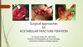
- 1. Surgical approaches for ACETABULAR FRACTURE FIXATION Dr Sharan Achar, PG- MS Ortho, Institute of Orthopaedics & Traumatology, Coimbatore Medical College Hospital.
- 2. Kocher Langenback approach Ilioinguinal approach Iliofemoral approach Modified Stoppa’s approach Modified Gibson’s approach SURGICAL EXPOSURES
- 3. ANATOMIC RELATIONSHIP OF ACETABULUM:
- 4. POSTERIOR STRUCTURES Muscular relations: The outer muscle layer consists of gluteus maximus which inserts into gluteal tuberosity and iliotibial tract. Gluteus medius is a fan shaped muscle originating from gluteal surface of ilium and inserts into greater trochanter. Short external rotators form the inner layer. They are pyriformis superior gemellus , tendon of obturator internus , inferior gemelli , and quadratus femoris. The critical structures of importance in deep surgical dissection are: • Superior gluteal nerve and vessels ( above pyriformis) • Inferior gluteal nerve and vessels • Sciatic nerve • Quadratus femoris muscle protecting the ascending branch of medial circumflex femoral artery
- 6. Vascular relations: • SUPERIOR GLUTEAL ARTERY: Commonly injured in greater sciatic notch, can be damaged by aggressive superior or lateral retraction of the abductor muscles during Kocher-Langenbeck exposure • ASCENDING BRANCH OF MEDIAL FEMORAL CIRCUMFLEX: It is the main blood supply to femoral head. It lies deep to quadratus, obturator internus, and piriformis, superficial to obturator externus
- 9. Nerve relations: SCIATIC NERVE: Most common traumatic & iatrogenic nerve injury. It exits from greater sciatic notch below the pyriformis. Sciatic nerve must be isolated and protected through out the procedure. Variations of its coarse must be kept in mind.
- 11. INDICATIONS Posterior wall fractures Posterior column fractures with or without an associated posterior wall fracture. Transverse and T-shaped fractures, treated within 15 days of injury. T-shaped fractures, the major displacement should be posterior, with only minor displacement occurring anteriorly at the pelvic brim
- 12. PATIENT POSITION Prone position- Most commonly used Lateral position
- 13. 1. J shaped skin incision was placed lateral to the posterior superior iliac spine, extended to the greater trochanter, and then continued along the axis of the femur to almost the midpoint of the thigh. 2. Gluteus maximus muscle is bluntly divided along the skin incision. Its insertion to femur is released. SURGICAL STEPS
- 14. 3. The sciatic nerve was identified on the posterior surface of the quadratus femoris and followed proximally until it disappears beneath the piriformis. The short external rotators and pyriformis tendon are tagged and divided. The knee joint is kept in adequate flexion through out the procedure to prevent iatrogenic sciatic nerve palsy
- 15. 4. The retractor is positioned is placed in lesser sciatic notch. 5. Subperiosteal elevation was done to exposes the inferior aspect of the iliac wing. Fracture reduced and fixed with contoured recon plate and lag screws.
- 17. ILIOINGUINAL APPROACH INDICATIONS: Anterior wall and Anterior column fractures. Anterior column/wall and posterior hemitransverse fractures Both-column fractures. Transverse fractures in which the major displacement is anterior with minimal posterior displacement Both-column fractures having a noncomminuted posterior column fragment .
- 18. SURGICAL STEPS 1. The skin incision extends from just posterior to the gluteus medius tubercle, paralleling the iliac crest to the anterior superior iliac spine, and then coursing medially to the midline ending two finger-breadths above the pubic symphysis
- 19. 2. Dissect through subcutaneous fat in the line of the skin incision to expose the aponeurosis of the external oblique muscle. 3. The external oblique aponeurosis has been incised from the anterior superior iliac spine to the midline, passing at least 1 cm superior to the superficial inguinal ring, and reflected distally.
- 20. 4. The spermatic cord in the male or the round ligament in the female is bluntly isolated along with the ilioinguinal nerve and retracted using a rubber sling. 5. Divide the rectus abdominis muscle 1 cm proximal to its insertion into the symphysis pubis. Detach the external, internal oblique and the transversus abdominis muscles from the inguinal ligament leaving 2 mm of the ligament attached to the muscles.
- 21. 6. Using a swab, push the peritoneum upward to reveal the femoral vessels. Mobilize the iliacus muscle from the inner aspect of the ilium. Note the iliopectineal fascia covering the muscle and separating it from the femoral sheath. 7. The iliopectineal fascia has been released and the exposure is complete. Pass the sling around the femoral sheath. Pass a sling around the iliopsoas deep to the iliopectineal fascia.
- 22. WINDOWS OF ILIOINGUINAL APPROACH 1. Lateral window: Exposes the internal iliac fossa to the sacroiliac joint and the pelvic brim. 2. Middle window: Exposes pelvic brim to pectineal eminence, quadrilateral surface & anterior wall. 3. Medial window: The space of Retzius, superior ramus, and symphysis pubis are visualized.
- 23. WINDOWS OF ILIOINGUINAL APPROACH
- 25. Indications (EXTENDED ILIOFEMORAL APPROACH) Transverse component with an extended posterior wall fracture (involving the posterior border of the bone), T-shaped and posterior wall fracture, and those associated with dislocation of the symphysis pubis or fracture of the contralateral pubis ramus. Select T-shaped fractures include those with a transtectal transverse component, those with a wide separation along the vertical stem of the T, and those associated with dislocation of the symphysis pubis or fracture of the contralateral pubic ramus. Select both-column fractures include those having a complex fracture of the posterior column, a displaced fracture line crossing the sacroiliac joint, or a wide separation of the anterior and posterior columns at the rim of the acetabulum.
- 26. SURGICAL STEPS 1. The incision extends from just posterior to the gluteus medius tubercle, paralleling the iliac crest, to the anterior superior iliac spine and then coursing distally for approximately 15 cm along the lateral aspect of the sartorius muscle.
- 27. 2. The iliopsoas muscle is elevated off the inner aspect of the iliac crest. 3. The sartorius origin and inguinal ligament are released from the anterior superior iliac spine. 4. The interval between the Sartorius and the tensor fascia lata is developed to allow exposure of the anterior hip joint capsule, the anteroinferior iliac spine, and the anterior column as far medial as the iliopectineal eminence
- 28. EXTENDED ILIOFEMORAL APPROACH 1. The skin incision runs from the posterior superior iliac spine to the anterior spine and then curves to lie on the anterior-lateral thigh. 2. The abductors have been reflected subperiosteally from the external ilium and reflected posteriorly with the tensor fascia lata muscle. The fascia separating the tensor from the rectus is split and the ascending branch of the lateral femoral circumflex vessels is ligated
- 29. 3. The abductor tendons are here transected from the greater trochanter. Alternatively, a trochanteric osteotomy can be performed. 4. The piriformis and obturator internus tendons have been transected and the exposure to the external ilium is completed.
- 31. INDICATIONS Anterior column Transverse T-shaped Anterior column/wall and posterior hemitransverse Both-column fractures. With this approach, it is often necessary to make a second approach through a skin incision along the iliac crest for fracture reduction or hardware insertion, essentially using the equivalent of the lateral and medial “windows” of the ilioinguinal approach.
- 32. SURGICAL STEPS 1. A transverse skin incision is made 2 cm above the pubic symphysis extending approximately from one external inguinal ring to the other. 2. The linea alba is incised at the midline and split vertically from inferior to superior with care taken to remain extraperitoneal in the proximal portion.
- 33. 3. Protecting the bladder, the rectus abdominis muscle is then retracted upward. 4. Sharp dissection is used to elevate the rectus to expose the symphysis body and pubic ramus
- 34. 6. Coronal mortis, anastomotic vessel between major arteries, such as the inferior epigastric and obturator vessels, is identified and ligated (don’t cauterize). 5. Rectus and neurovascular structures are subsequently retracted laterally and anteriorly so that they are protected.
- 35. 7. It is imperative to pay strict attention at all times to the location of the obturator neurovascular bundle and lumbosacral trunk which traverse the operative field. 8. Full access is then developed from anterior to posterior along the pelvic brim, sharply dividing and elevating the iliopectineal fascia superiorly and the obturator fascia inferiorly andexposing the medial wall of the acetabulum, the fracture, and the pelvic brim.
- 37. INDICATIONS Differs from the Kocher–Langenbeck approach in its proximal dissection.
- 38. 1. The interval between the gluteus maximus and tensor fasciae lata muscles is developed,rather than splitting the gluteus maximus muscle. 2. In this way, the neurovascular supply to the anterior portion of the gluteus maximus muscle is not at risk. In addition, anterosuperior visualization and access are extended
- 39. OUR EXPERIENCE
- 41. MODIFIED STOPPA AND ILIOFEMORAL LATERLAL WINDOW
- 42. MODIFIED STOPPA AND ILIOINGUINAL LATERAL WINDOW
- 43. THANKYOU
Editor's Notes
- Iliinguinal lateral window had a similar approach with incision overinguinal ligament without osteomitising ASIS