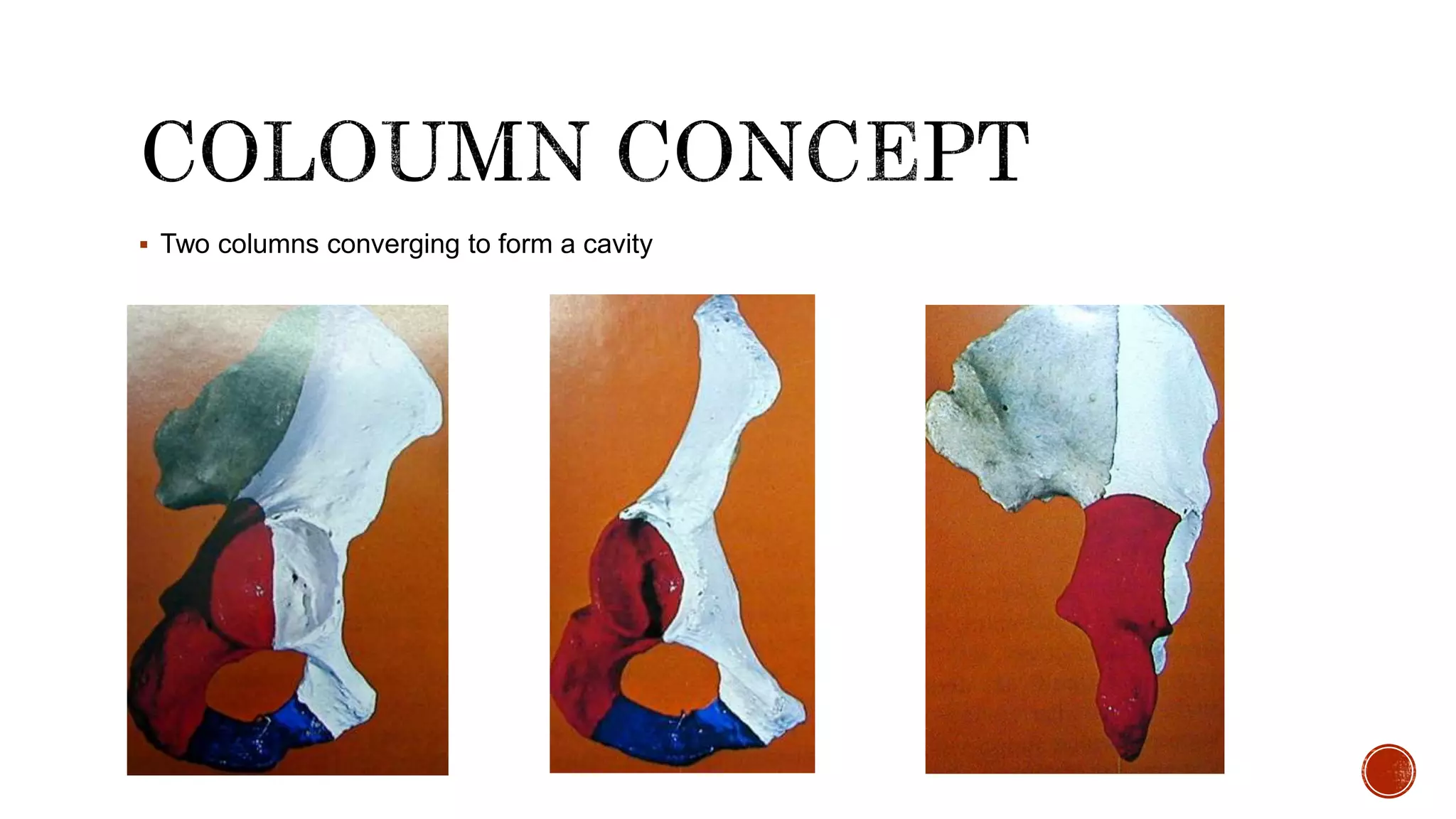
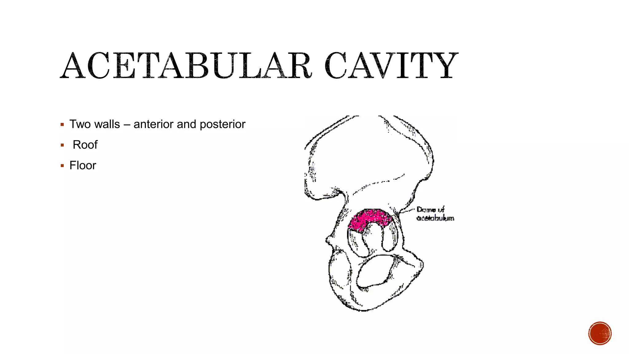
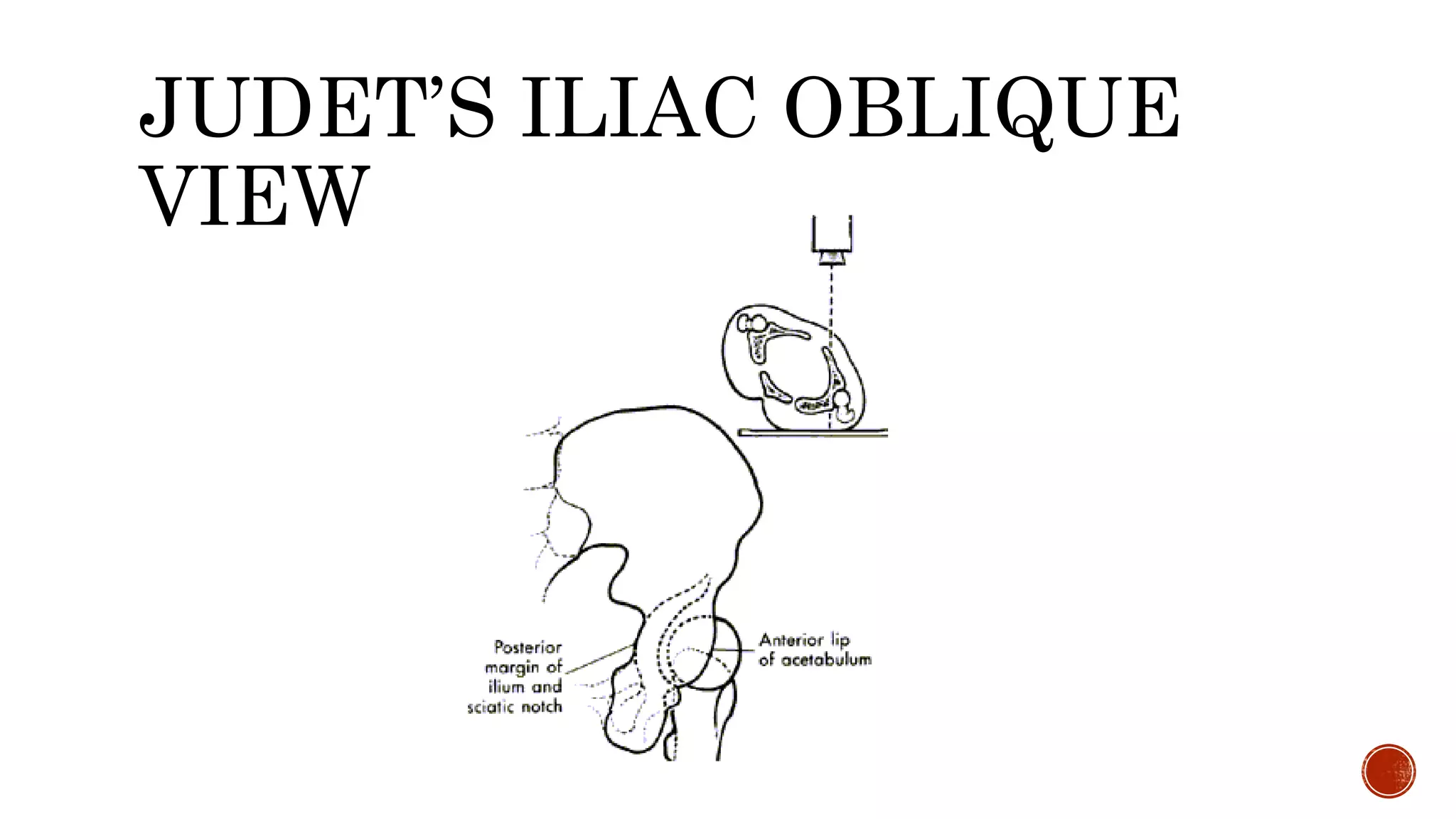
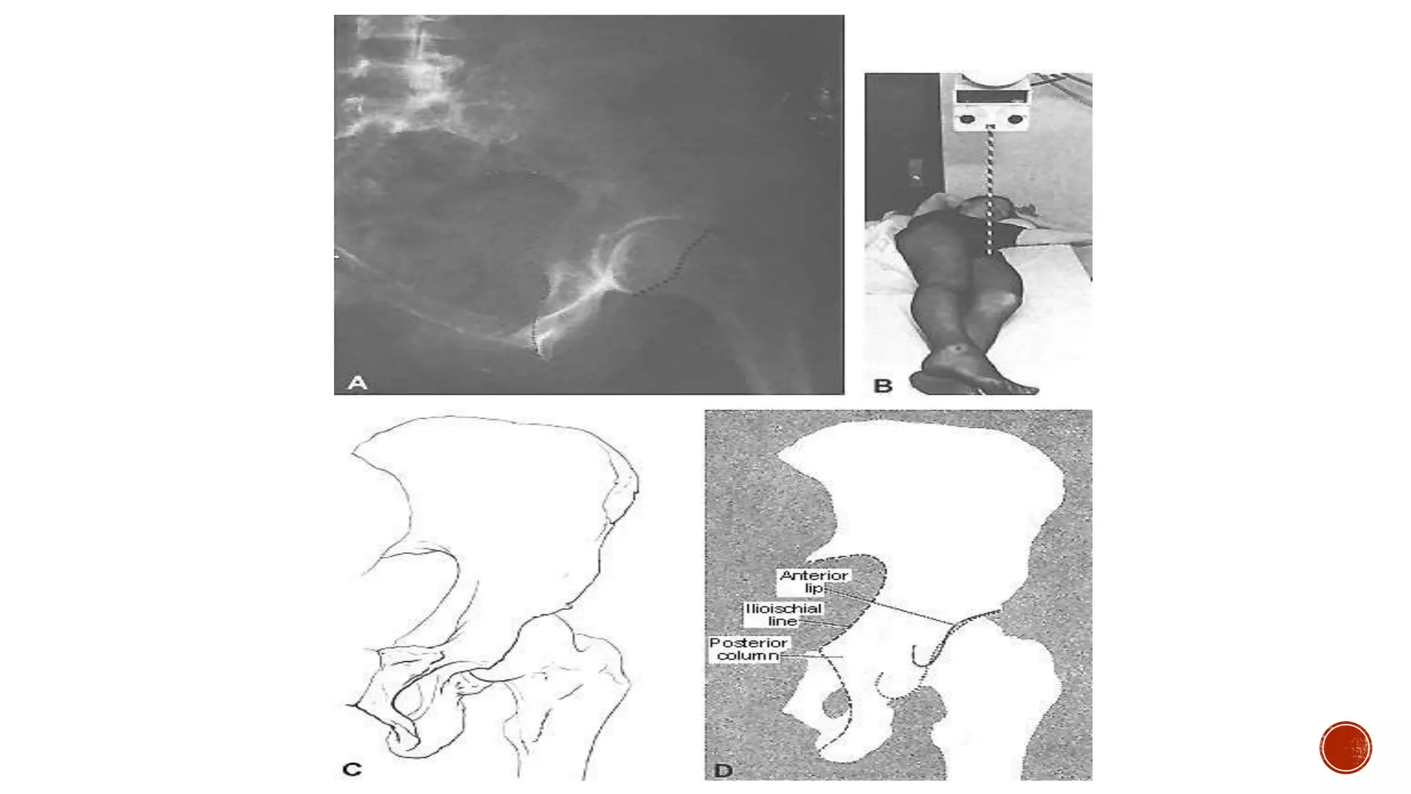
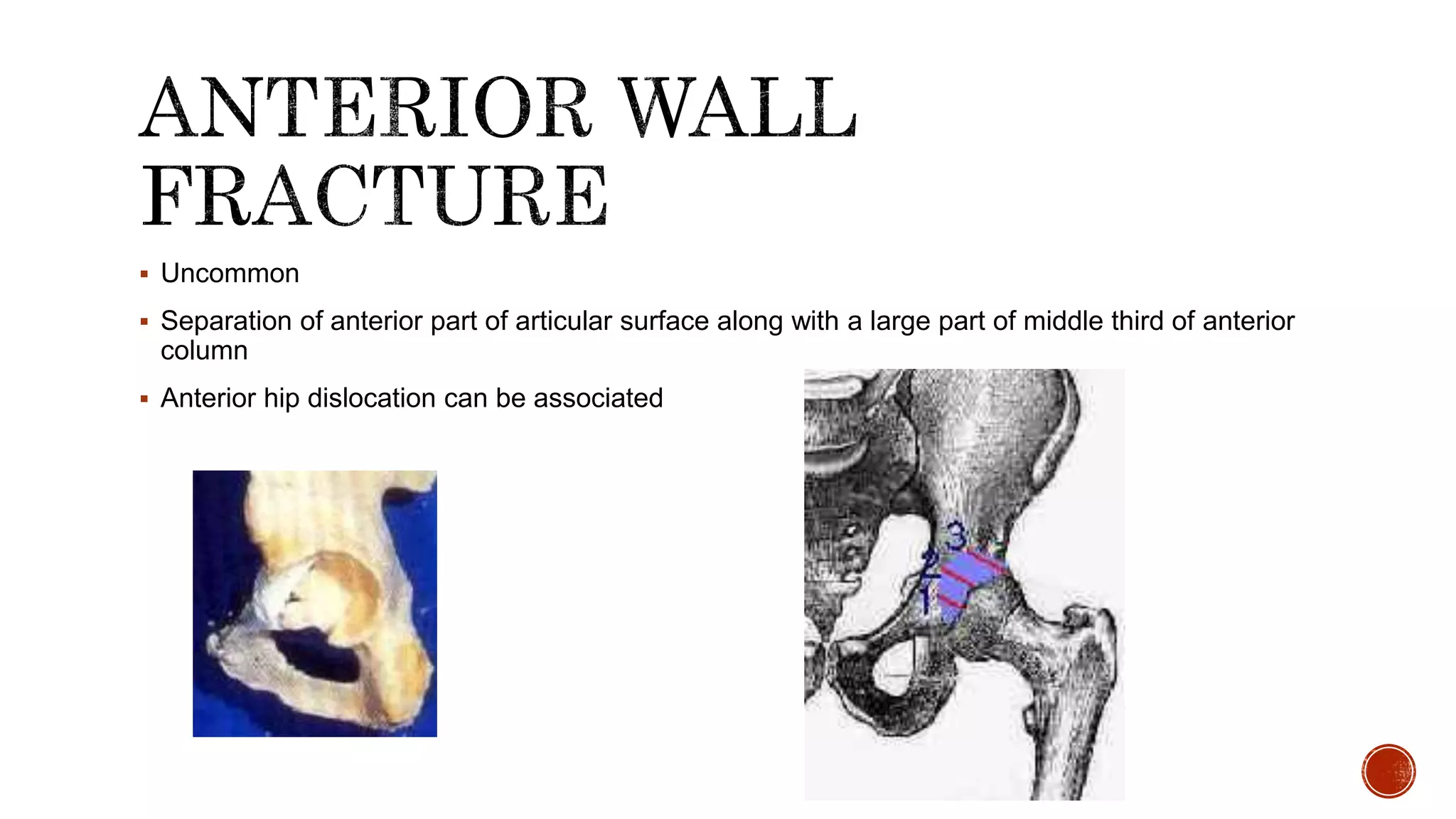
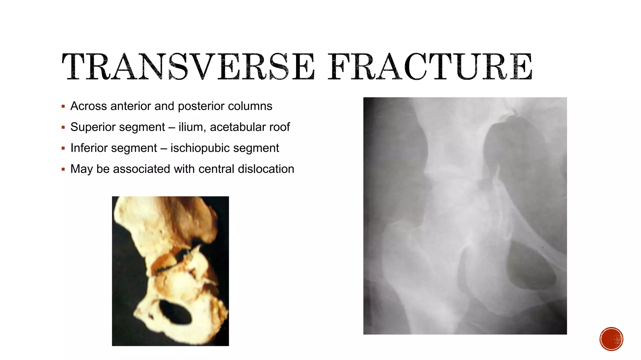
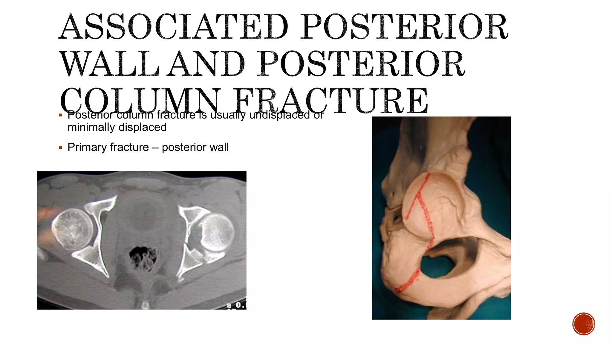
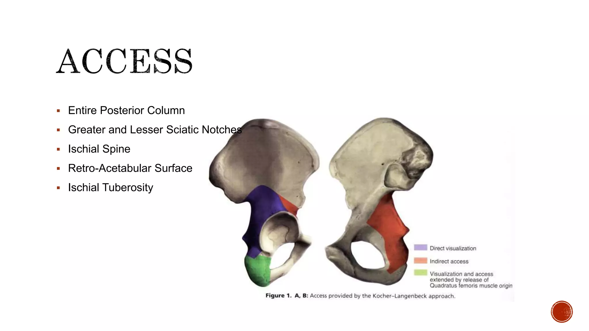
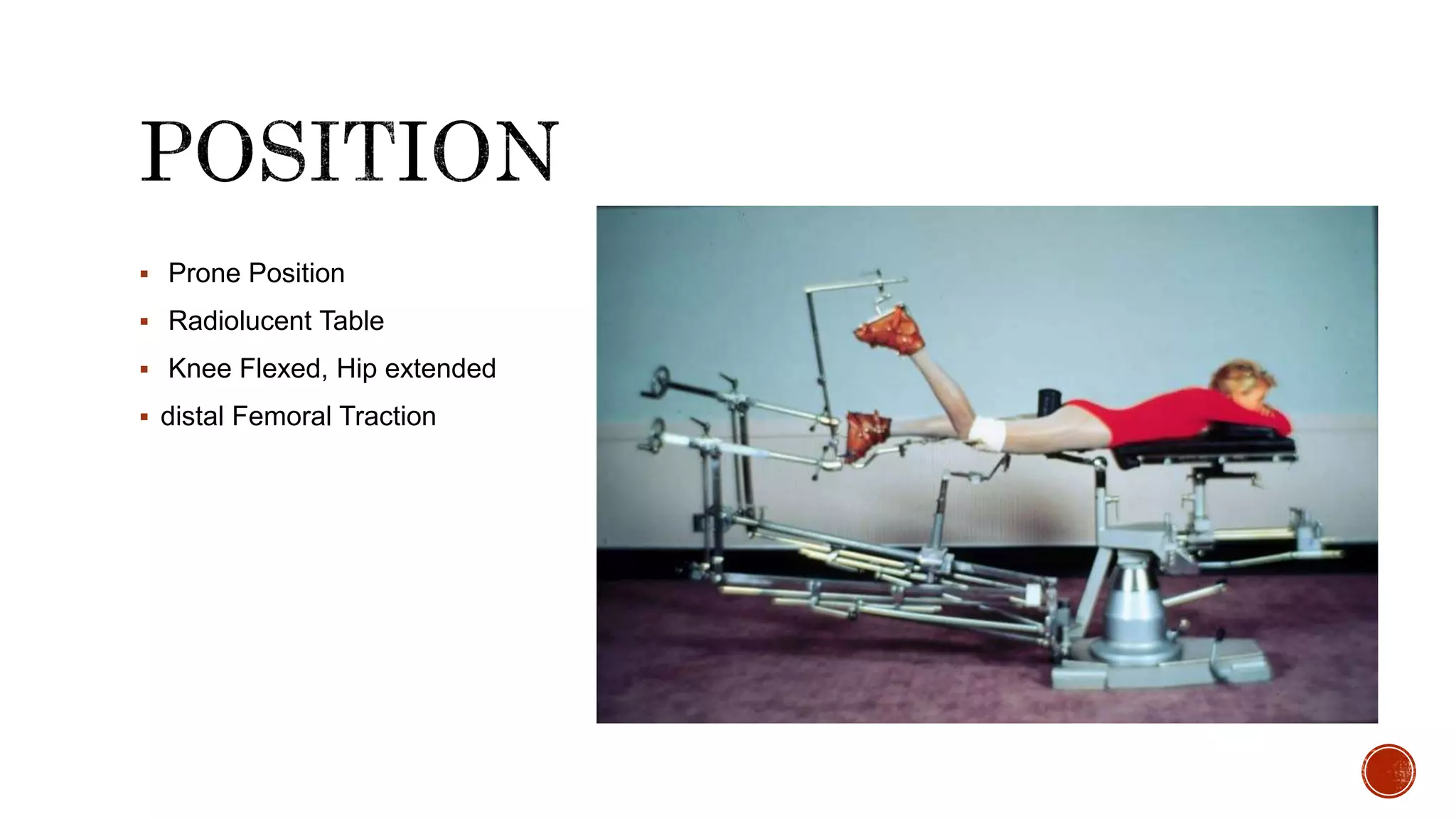
16. Extends from PSIS to ischio pubic ramus
Involves posterior articular surface and ilio ischial line
17. Uncommon
Separation of anterior part of articular surface along with a large part of middle third of anterior
column
Anterior hip dislocation can be associated
18. Extends from symphysis pubis to iliac crest
Most commonly fracture line exits below AIIS
Often comminution into the quadrilateral plate
19. Across anterior and posterior columns
Superior segment – ilium, acetabular roof
Inferior segment – ischiopubic segment
May be associated with central dislocation
20. Posterior column fracture is usually undisplaced or
minimally displaced
Primary fracture – posterior wall
22. Commonly posterior dislocation
Sometimes central dislocation
Highest incidence of pre op sciatic palsy and AVN of femoral head
23. Transverse and vertical components
Acetabular cavity is split into at least 3 fragments
24. All segments of fractured acetabulum are detached from the ilium
25. Fracture Displaced >2mm
Irreducible fracture dislocation
Intra articular fragment interfering with joint movement
Instability of the joint
To prepare the joint for hip replacement
26. Severe osteoporosis
Very old patients
Severe associated injuries
Poor local skin condition
Limited experience of the surgeon
27. 0-7 DAYS 74%
8-14 DAYS 71%
15-21 DAYS 57%
POOR RESULTS AFTER 3 WEEKS
28. Open acetabular fracture
New onset sciatic nerve palsy after closed reduction of hip dislocation
29. Irreducible posterior hip dislocation
Medial dislocation of femoral head against cancellous bone surface of intact ilium
30. Type of fracture pattern
Posterior dislocation
Initial displacement
Presence if intraarticular fragments
Presence of acetabular impaction
31. Injury to cartilage or bone of femoral head
Anatomical reduction
36. Prone Position
Radiolucent Table
Knee Flexed, Hip extended
distal Femoral Traction
37. Prone Position
Aids in Reduction of Transverse Fractures
Improves Quadrilateral Surface Access
Allows Clamp Placement through Greater
Sciatic Notch
Controls Position of Hip, Minimizes Sciatic
Nerve Stretch
38. 6 to 8 cm from PSIS
Tip of Greater Trochanter
Parallel Shaft of Femur 15-20 cm
39.
40. Divide Iliotibial Band
Separate Fibers of Gluteus Maximus
– Superior Gluteal Artery
– Inferior Gluteal Artery
Split to Inferior Gluteal Nerve Branch
41. Release Gluteus Maximus Insertion 1 cm from its insertion
Identify Sciatic Nerve on Border of Quadratus Femoris Muscle
42.
43. Release Piriformis Tendon >1cm from trochanter
Release Conjoint Tendon
Open Obturator Internus Bursa for Sciatic Nerve Retractor
44. FEMORAL HEAD BLOOD SUPPLY
Deep Branch of Medial Femoral Circumflex
May be injured by:
– Detaching quadratus
– Reflecting obturator internus or piriformis too close to trochanter
45. 84%: Anterior to Piriformis
12%: Peroneal Division through Piriformis
3%: Peroneal Division Posterior to
Piriformis / Tibial Division anterior to
Piriformis
1%: Entire Nerve through Piriformis
46.
47. Subperiosteal Elevation of:
– Greater Sciatic Notch
– Quadrilateral Surface
– Gluteus Minimus
Debridement of Fracture Edges
Avoid Devascularization of Fx Fragments
49. Developed by Letournel (1960) after extensive
cadaveric anatomical study
Approach to the
anterior column and anterior articular surface
Hip abductors are not disturbed (rapid post op
rehabilitation)
51. SI Joint
Internal Iliac Fossa
Pelvic Brim
Quadrilateral Surface
Superior Pubic Ramus
Limited Access to External Iliac Wing
52. The surgical exposure requires development of three wound intervals
Lateral
Middle
medial
Mobilization of the femoral vessels and nerve, as well as the spermatic cord (male) or round
ligament (female), is key to the development of these intervals.
53. Supine
Distal Femoral Traction
Traction avoided in contralateral pubic ramus fracture
Access to Greater Trochanter (Lateral Traction)
Hip flexed 20°
54. 3-4 cm cranial to Symphysis pubis
Curve to ASIS
Parallel Iliac Crest -Past Most Convex Portion of Ilium
anterior 2/3
55.
56. Subperiosteal Dissect Internal Iliac Fossa
release the external oblique insertion
mobilize the iliacus muscle
subperiosteally
• Expose Sacroiliac Joint
Dissect over Pelvic Brim
57. Incise External Oblique Aponeurosis
– From ASIS to midline
– 1 cm proximal to External Inguinal Ring
Expose Floor of Inguinal Canal
Retract Spermatic Cord/Round Ligament
Protect Ilioinguinal Nerve
Lies proximal to the inguinal ligament after
penetrating the abdominal wall
58.
59. Incise Inguinal Ligament
Leave 1-2 mm with Internal Oblique and Transversus
Abdominis origin
Protect External Iliac Vessels
Protect Lateral Femoral Cutaneous
usually encountered just deep to the conjoint tendon
(of the internal oblique and the transversus abdominis)
approximately 1-2 cm medial to the anterior superior iliac
spine.
60. Divide conjoint tendon& rectus abdominis at their insertion on
pubis to open retropubic space
Structures beneath inguinal ligament lie in 2 compartment
Lacuna muscularom
Lateral
Contains iliopsoas muscle,femoral nerve & lateral
cutaneous nerve
Lacuna vasorum
Medial
Contans external iliac vessels & lymphatics
Incise Iliopectineal Fascia to Superior Ramus and from Pelvic
Brim
62. Ecompasses the entire internal iliac fossa from the
sacroiliac joint posteriorly to the iliopectineal eminence
anteriorly.
This window is optimized with hip flexion to relax the
iliopsoas
Medial retraction usually requires placement of retractors
on the quadrilateral surface
Internal Iliac Fossa
Sacroiliac Joint
Pelvic Brim - Upper 1/3
63. Pelvic Brim - SI joint to pectineal eminence
Quadrilateral Surface
Anterior Rim
Medial retraction of the femoral vessels should be gentle
and must be carefully monitored.
64. The most limited leaves the ipsilateral rectus insertion
attached and visualization is provided between the
rectus and the spermatic cord (or round ligament)
Superior Pubic Ramus
Symphysis Pubis
65. Medial window may also be created utilizing Stoppa approach
– Midline rectus split
– Subperiosteal dissection of quadrilateral surface
– Retractor in lesser sciatic notch
– Protect obturator nerve/artery
68. Intrapelvic approach
Hirvensalo et al 1993 – first described
Substitute-ilioinguinal approach
69. INDICATIONS
Anterior wall
Anterior column
Transverse
T type
Post hemitransverse
Both column
70. ADVANTAGE
Improved quadrilateral surface exposure & post column
Minimise dissection( avoid middle window)
Iliac vessels dissection not required
DISADVANTAGE
Lack of acess to middle window
71. Position : supine
Skin incision
Pfannensteil or
Midline incision starting 1cm inf to pubic symphysis ending 2-3 cm inf to umbilicus
72. Subcutaneous tissue dissected in line with skin incision
Fascia over both rectus muscle exposed
Fascia incise along linea alba
73. Rectus muscle both bellies retracted laterally
In proximal aspect do not enter peritoneum
Entire approach periperitoneal space
74. Loosely packed wet sponge in retropubic space to protect bladder
75. The thick periosteum from the superior pubic bone is dissected sharply, allowing for deeper blunt
dissection.
At the beginning, dissection should be enlarged also on the anterior part of the symphysis.
The upper border of the superior pubic ramus is identified (pecten pubis) and the dissection is carried
laterally along the pelvic brim. The iliopectineal fascia is detached from the pelvic brim
76. Dissecting carefully along the medial surface of the superior ramus, the corona mortis vessels
are identified and ligated (or clipped) as necessary
77. Dissection of the periosteum is continued further laterally following the upper border of the
superior pubic bone to the direction of the pelvic brim exposing the beginning of the iliopectineal
eminence
78. At this point the beginning of the iliopectineal arch should be dissected from the bone. This enables the
elevation of the femoral vessels and nerve.
The dissection is continued subperiosteally more laterally following the upper border of the pelvic brim.
At this point the entire internal surface of the superior pubic ramus has been exposed adequately for plate
fixation
79. At this level, the obturator neurovascular bundle is crossing the quadrilateral surface. In some cases it
should be mobilized. A spatula or malleable retractor is used to protect the obturator neurovascular
bundle and pelvic floor.
With a Cobb elevator, the periosteum and obturator internus is elevated and the quadrilateral surface can
be sufficiently exposed.
One Hohmann retractor should be put in the middle part of the superior pubic ramus and another curved
Hohmann retractor is placed on the posterior top of the acetabulum on the iliac part of the pelvic brim.
Great care should be taken not to injure the external iliac vein which may be in close proximity to the
elevators
80. In some rare cases, the internal iliac artery bifurcates very distally and makes the dissection of
the posterior part of the quadrilateral surface risky and limits the further dissection
81. Developed by Letournel (1975)
Based on Smith- Peterson Approach
Maximal Simultaneous access to both
columns of the acetabulum
82. INDICATIONS:
Transtectal Transverse +PW or T-shaped fractures
Transverse fractures with extended posterior wall
T-shaped fractures with wide separations of the vertical stem of the "T" or those with associated
pubic symphysis dislocations.
Certain Associated Both Column Fractures.
Associated fracture patterns or transverse fractures which are operated greater than 21 days
following injury.
83. INDICATIONS IN BOTH COLOUMN FRACTURES
Inability to reduce Posterior Column through Ilioinguinal
Wide displacement at the rim
Complex posterior column involvement
Associated SI joint disruption
Small posterior wall component
84. External Aspect of Ilium
Anterior Column as far medial as
Iliopectineal eminence
Posterior Column to the Upper Ischial
Tuberosity
87. INCISION:
Inverted J incision
Parallel Iliac Crest from PSIS to ASIS
Incise along anteriorlateral thigh
88. Release Origins of Gluteals and Tensor
Fascia Lata from Iliac Crest
Dissect Subperiosteal Iliac Wing
Elevate Periosteum from Greater Sciatic
Notch
Incise Fascia Lata to end of muscle belly
89. Retract Tensor Fascia Lata Muscle
Posteriorly
Incise Sheath of Rectus Femoris
Ligate Lateral Femoral Circumflex Artery
and Vein
90.
91. Release Gluteus Medius and Minimus
Tendons from Greater Trochanter
Alternatively, Greater Trochanteric
Osteotomy
Reflect Gluteals and Tensor Fascia Lata
Posteriorly pedicled on Superior Gluteal
92.
93. Incise and Retract:
– Piriformis Tendon
– Obturator Internus Tendon with Gemelli muscles
Place Sciatic Nerve Retractor in Lesser Sciatic Notch
Capsulotomy if Required
94.
95. If Internal Iliac Fossa Exposure Required:
– Elevate Abdominal Muscles from Iliac Crest
– Elevate Iliacus Subperiosteally
– Release Sartorius and Inguinal Ligament from ASIS
– Preserve Anterior Capsule and Direct Head of Rectus for Blood Supply to Anterior Column
97. Kocher-Langenbeck + Ilioinguinal
• May be simultaneous or sequential
– Simultaneous may compromise both approaches but can aid in assessment of transverse
fracture reduction
– Care with sequential not to block anterior reduction during posterior fixation
98. Rarely necessary
– T-shaped fractures if unable to reduce anterior column from KL
– AW+PHT if hemitransverse is segmental or widely displaced
99.
100. The bony pelvis lies in close proximity to various vascular , neural and soft tissue structures making these
structures vulnerable in the event of pelvic ring disruption
101.
102.
103.
104.
105. Anterior ring fixation may provide structural protection of posterior fixation
If combined open and percutaneus techniques are used, the open portion is often done first to aid in
reduction of the percutaneusly treated injury
LETOURNEL’s Golden rule: Posterior stabilization to be done before anterior as posterior is the main
weight bearing part.
106. Indications for ORIF
Symphyseal dislocation >2.5cm(static or dynamic)
To augment posterior fixation in vertically dislaced fractures.
Locked symphysis.
109. The cut edges of the rectus abdominal
muscles superiorly to reveal the symphysis
and pubic crest.
If access to the back of the symphysis is
required, use the fingers to push the bladder
gently off the back of the bone
110. Anterior External Fixation for controlling rotation but Internal fixation >>> for resisting vertical
displacements
Ex fix particularly useful in open injuries or pts requiring GI/GU procedures.
111. Apply circumferential wrap at the level of the GT.
Internally rotate the legs and tape them.
Anterior approach to pubic symphysis.
Place reduction forceps anteriorly so that plate can be put on the superior surface.
112.
113. Inlet view: judge the alignment of the plate;
Outlet view judge the length of screws; screws should have a bicortical purchase.
114. Fractures medial to insertion of inguinal ligament should be treated like symphyseal dislocations.
• Comminuted fractures: ORIF
• Minimal comminution: Ramus screw
115. Indications for ORIF:-
1. Displaced iliac wing fractures that enter and exit both the crest and GSN/SIJ.
2. Multiplanar instability(disruption of ligaments)
3. Non impacted comminuted displaced sacral fractures.
4. Vertical or cephalad displacement.
5. U shaped fractures with spino-pelvic dissociation
116. Pt is placed prone with longitudinal traction.
In severely displaced fractures we can rigidly fix the contralateral pelvis
117.
118.
119. Make a curved incision over the iliac
crest, beginning 7 cm posterior to the
anterior superior iliac spine. Curve the
incision anteriorly and medially along
the line of the inguinal ligament for 5
cm.
120. Subperiosteally dissect the iliacus muscle and retract medially to reach the anterior part of the SIJ.
Care should be taken not to injure L5 nerve root.
121.
122.
123. Inlet projection - screw towards anterior aspect of
promontory
Outlet - screw is above the S1 foramen
Screw to be directed anteriorly; superiorly and
medially.
124. Iliac wing fractures exiting through the SIJ are crescent #.
Crescent fragment is the variable sized that contains the PSIS and PIIS and remains attached to the
sacrum.
Smaller the “CRESCENT” fragment > damage to posterior structures
ALWAYS POSTERIOR APPROACH
125. Can be regarded as a pelvic injury, spinal injury or both.
Indications for fixation:-
Ant and post ring disruption with vertical sheer sacrum fracture.
Comminuted # with rotation
Spinal-pelvic dissociation
Rarely in impacted # with Internal rotation deformity
127. 1. Spinal point of fixation- L5(usually)
2. Iliac screw just inf to PSIS
3. Iliac screw is connected to pedicle screw with appropriate rods and screw-rod clamps
128. This bypasses the lines of force transmission from
spine to illium through the construct instead of the
sacrum