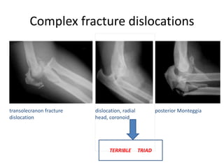
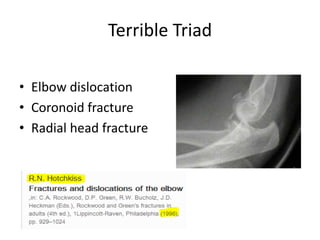
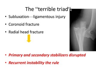
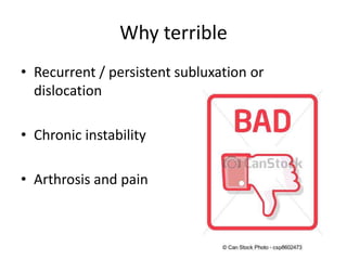
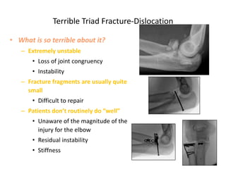
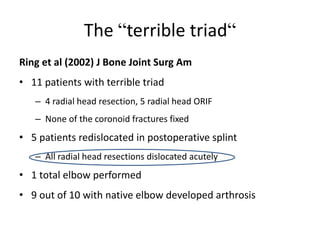
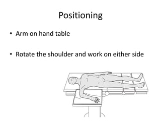
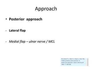
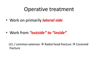
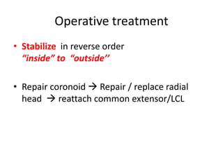
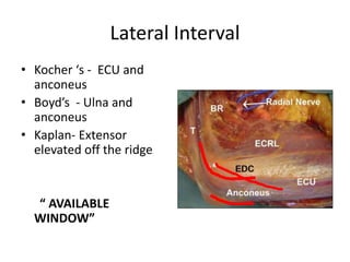
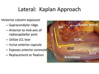
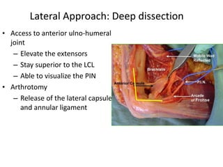
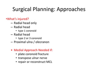
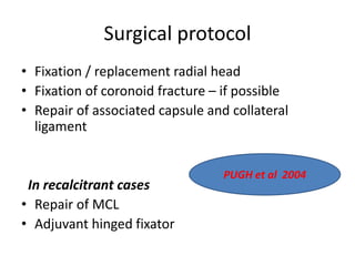
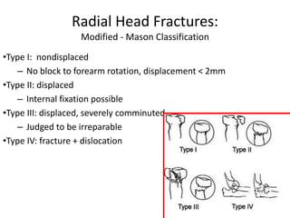
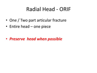
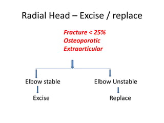
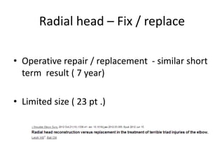
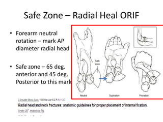
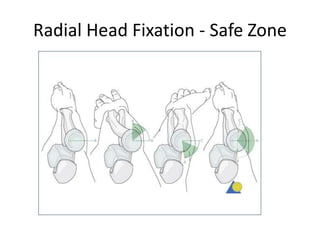
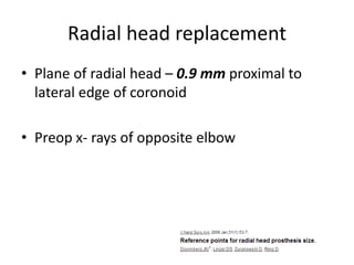
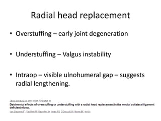
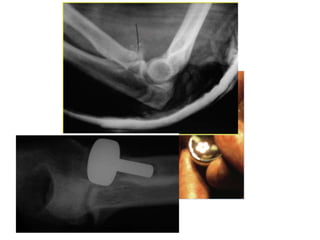
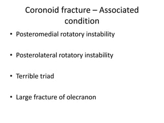
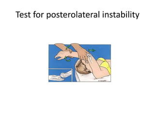
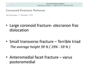
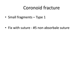
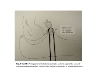
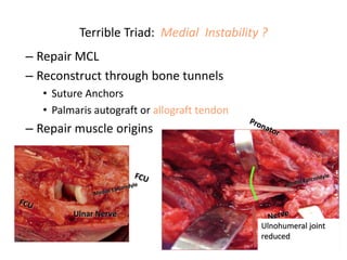
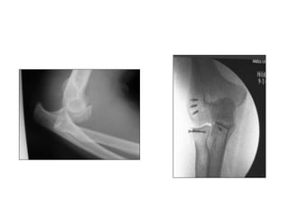
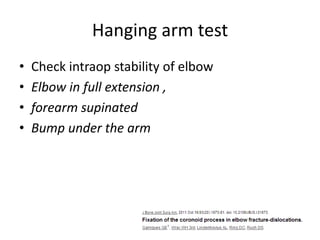
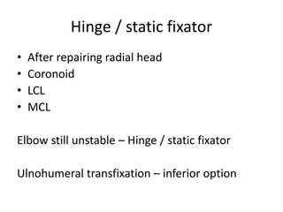
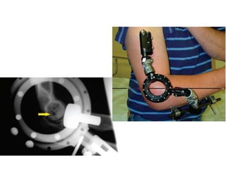
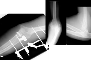
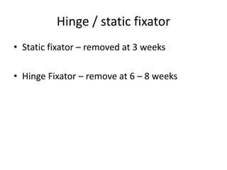
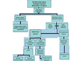
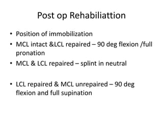
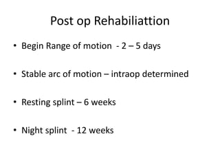
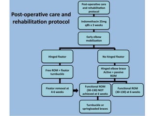
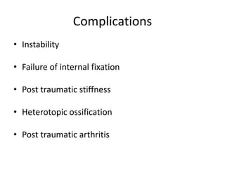
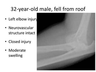
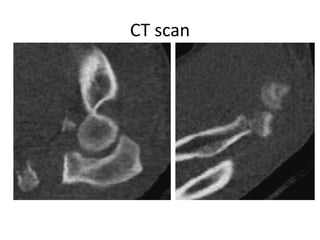
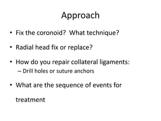
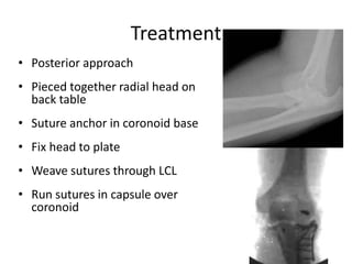
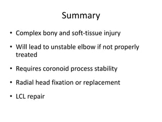
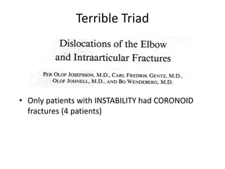
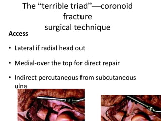
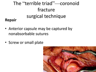
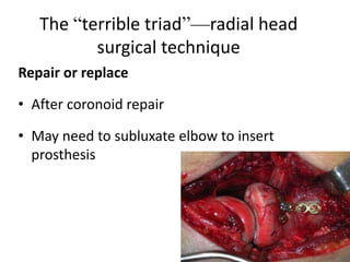
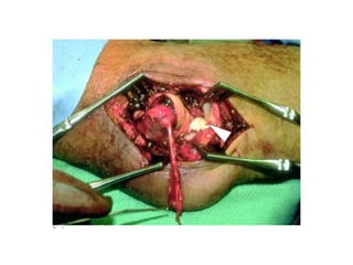
Terrible Triad - Elbow
Elbow anatomy—coronoid process
• Anterior aspect of the
greater sigmoid notch
– Articulates with trochlear
– Brachialis insertion
• Laterally
– Lesser semilunar notch
articulates with radial
head
• Medially
– Attachment of anterior
fibers of MCL
Medial Collateral Ligament
Lateral Collateral Ligament
Stabilizers of elbow
• Primary stabilizers
Ulnohumeral joint
MCL -( Ant. Bundle)
LCL -
• Secondary stabilizers
Radiohumeral joint
Capsule
Origin of flexor & extensor
tendons
Dynamic stabilizers - Muscle crossing elbow
Anconeus
Brachialis
Triceps
Simple dislocations
• Universal disruption of the LCL
• MCL partially or completely torn
Bony congruence
• Secondary stabilizers intact
• Recurrent instability rare
Complex fracture dislocations
transolecranon fracture
dislocation
posterior Monteggiadislocation, radial
head, coronoid
TERRIBLE TRIAD
Terrible Triad
• Elbow dislocation
• Coronoid fracture
• Radial head fracture
The “terrible triad“
• Subluxation—ligamentous injury
• Coronoid fracture
• Radial head fracture
• Primary and secondary stabilizers disrupted
• Recurrent instability the rule
Why terrible
• Recurrent / persistent subluxation or
dislocation
• Chronic instability
• Arthrosis and pain
Terrible Triad Fracture-Dislocation
• What is so terrible about it?
– Extremely unstable
• Loss of joint congruency
• Instability
– Fracture fragments are usually quite
small
• Difficult to repair
– Patients don’t routinely do “well”
• Unaware of the magnitude of the
injury for the elbow
• Residual instability
• Stiffness
The “terrible triad“
Ring et al (2002) J Bone Joint Surg Am
• 11 patients with terrible triad
– 4 radial head resection, 5 radial head ORIF
– None of the coronoid fractures fixed
• 5 patients redislocated in postoperative splint
– All radial head resections dislocated acutely
• 1 total elbow performed
• 9 out of 10 with native elbow developed arthrosis
Mechanism of injury
• Fall on outstretched hand
• Axial load, supination & Valgus stress
Stages
I Ulnar lateral collateral
ligament disruption
II Anterior and posterior
soft issue disruption with
coronoid under trochlea
III a Intact MCL anterior
band
III b Ruptured MCL anterior
band
III c All soft tissue stripped
Terrible triad - Presentation
• Pain
• Clicking
• Locking of elbow in extension
• Varus instability
• Valgus instability – ( If MCL injured )
What are the Dilemna
• Surgical techniques challenging
• Debate in surgical steps
• Choices in management
Critical components to achieve treatment goals
• Obtaining and
maintaining
a concentrically
reduced
articulation
• Management of
coronoid & radial head
fracture if present
• Early range of motion
Examination
• Unstable elbow with wrist injury - High risk of
compartment syndrome
• Combined distal radius and elbow fracture –
9/59 ( 15%)
• Isolated distal radius # - 3/869 ( .3%)
• Baseline neural examination
• 20% patient – Terrible ulnar nerve palsy
• High risk of developing heterotopic
ossification
Management
• Dislocated elbow – reduce in emergency dept
• Unstable – Do not perform rpt rereduction
• Plan under anaesthesia
Imaging
• X- rays – Ap and lateral
• Ct scan – Include 3D reconstruction
Pathoanatomy
• Capsuloligamentous injury
• Avulsion of flexor & extensor muscle from
epicondyle
• Coronoid fracture – transverse fragment with
anterior capsule attached, involves 30% of height
• Radial head – anterolateral or entire radial head
Standard treatment protocols
Pugh DMW, et al (2004) J Bone Joint Surg Am
• Fixation or replacement of radial head
• Fixation of coronoid fracture
• Repair of associated capsular and lateral soft-tissue
injuries
• Evaluation of stability and repair of MCL as necessary
• Adjuvant hinged external fixation if residual instability
Aim of management
• Ulnohumeral joint reduced – 4 - 6 weeks
• Prevent injury and treatment related
complication
Non operative treatment
• Small coronoid and radial head fracture
• Concentrically reduced ulnohumeral and
radiocapitellar joint
• Ct scan – insignificant fracture
• Elbow unstable in only < 30 deg flexion
IMMOBILIZE IN 90 Deg
FLEXION
•Planning operative
treatment of terrible triad
Positioning
• Arm on hand table
• Rotate the shoulder and work on either side
Approach
• Posterior approach
- Lateral flap
- Medial flap – ulnar nerve / MCL
Operative treatment
• Work on primarily lateral side
• Work from “outside” to “inside”
LCL / common extensor  Radial head fracture  Coronoid
fracture
Operative treatment
• Stabilize in reverse order
“inside” to “outside’’
• Repair coronoid  Repair / replace radial
head  reattach common extensor/LCL
Lateral Interval
• Kocher ‘s - ECU and
anconeus
• Boyd’s - Ulna and
anconeus
• Kaplan- Extensor
elevated off the ridge
“ AVAILABLE
WINDOW”
Lateral: Kaplan Approach
•Anterior column exposure
– Supracondylar ridge
– Anterior to mid-axis of
radiocapitellar joint
– Utilize LCL tear
– Incise anterior capsule
– Exposes anterior coronoid
– Replacement or fixation
Lateral Approach: Deep dissection
• Access to anterior ulno-humeral
joint
– Elevate the extensors
– Stay superior to the LCL
– Able to visualize the PIN
• Arthrotomy
– Release of the lateral capsule
and annular ligament
Medial Interval
• Medial
- Between the two head
FCU
- Over the top - Hotchkis
Approach
• Medial and lateral approach
- Large repairable radial head in the way
Surgical Planning: Approaches
•What’s injured?
– Radial head only
– Radial head
• type 1 coronoid
– Radial head
• type 2 or 3 coronoid
– Proximal ulna / olecranon
• Medial Approach Needed if:
• plate coronoid fracture
• transpose ulnar nerve
• repair or reconstruct MCL
Surgical protocol
• Fixation / replacement radial head
• Fixation of coronoid fracture – if possible
• Repair of associated capsule and collateral
ligament
In recalcitrant cases
• Repair of MCL
• Adjuvant hinged fixator
PUGH et al 2004
Radial Head Fractures:
Modified - Mason Classification
•Type I: nondisplaced
– No block to forearm rotation, displacement < 2mm
•Type II: displaced
– Internal fixation possible
•Type III: displaced, severely comminuted
– Judged to be irreparable
•Type IV: fracture + dislocation
Radial Head - ORIF
• One / Two part articular fracture
• Entire head – one piece
• Preserve head when possible
Radial Head – Excise / replace
Fracture < 25%
Osteoporotic
Extraarticular
Elbow stable Elbow Unstable
Excise Replace
Radial head – Fix / replace
• Operative repair / replacement - similar short
term result ( 7 year)
• Limited size ( 23 pt .)
Do not excise without replacement
• Restore radial head
• If not possible replace
• Repair lateral collateral lig
• Orif of coronoid
Safe Zone – Radial Heal ORIF
• Forearm neutral
rotation – mark AP
diameter radial head
• Safe zone – 65 deg.
anterior and 45 deg.
Posterior to this mark
Radial Head Fixation - Safe Zone
Radial head replacement
• Plane of radial head – 0.9 mm proximal to
lateral edge of coronoid
• Preop x- rays of opposite elbow
Radial head replacement
• Overstuffing – early joint degeneration
• Understuffing – Valgus instability
• Intraop – visible ulnohumeral gap – suggests
radial lengthening.
Coronoid fracture
• Classification
- Regan and Moorey
- O’ Driscoll
Coronoid Fracture – Regan & Moorey
Classification
• Type 1 - # tip
• Type 2 - < 50 %
• Type 3 - >50%
Classification: Coronoid fractures
•O’Driscoll Classification
•Type I: tip
•Type II: anteromedial facet
•Type III: base
Coronoid fractures—nonoperative
treatment
Type I
• Usually early motion
Type II
• Early motion, unless unstable
• Internal fixation if associated injuries
Coronoid fractures—surgical
treatment
Type III
• Internal fixation
• Screw or anterior plate
• Reconstruction with
bone bone graft (tip of
olecranon)
Coronoid fracture – Associated
condition
• Posteromedial rotatory instability
• Posterolateral rotatory instability
• Terrible triad
• Large fracture of olecranon
Test for posterolateral instability
• Large coronoid fracture- olecranon frac
dislocation
• Small transverse fracture – Terrible triad
The average height 39 % ( 19% - 59 % )
• Anteromedial facet fracture – varus
posteromedial
Coronoid fracture
• Small fragments – Type 1
• Fix with suture - #5 non absorbale suture
Type 1 & 2 – No fixation
Repair / replacement of radial
head and LUCL complex –
stable elbow
Coronoid fracture
• Type 2 ( < 50%)
• Type 3 ( >50%)
Fix with screw passed
from ulnar cortex
Large fragement – plate
fixation – medial
approach
Coronoid fracture
• Approach – lateral – Thru the fracture radial
head
• Large fragment – separate medial approach
Lateral Collateral Ligament Complex
• Avulsed from lateral condyle along with
common extensor
• Unstable elbow to varus test
• Local bruising
Lateral Collateral ligament
• Repair done elbow – 90 deg
• MCL intact forearm – pronated
• MCL injured – forearm supinated
Lateral Collateral ligament
• Repair with suture
anchors
• Transosseous tunnels
Medial Collateral ligament
• After repairing radial head
• Coronoid
• LCL
• Test elbow stability – Fluoroscopically
• Elbow unstable from 30 to 130 – repair MCL
Terrible Triad: Medial Instability ?
– Repair MCL
– Reconstruct through bone tunnels
• Suture Anchors
• Palmaris autograft or allograft tendon
– Repair muscle origins
Ulnohumeral joint
reduced
Hanging arm test
• Check intraop stability of elbow
• Elbow in full extension ,
• forearm supinated
• Bump under the arm
Hinge / static fixator
• After repairing radial head
• Coronoid
• LCL
• MCL
Elbow still unstable – Hinge / static fixator
Ulnohumeral transfixation – inferior option
Hinge / static fixator
• Static fixator – removed at 3 weeks
• Hinge Fixator – remove at 6 – 8 weeks
Post op Rehabiliattion
• Position of immobilization
• MCL intact &LCL repaired – 90 deg flexion /full
pronation
• MCL & LCL repaired – splint in neutral
• LCL repaired & MCL unrepaired – 90 deg
flexion and full supination
Post op Rehabiliattion
• Begin Range of motion - 2 – 5 days
• Stable arc of motion – intraop determined
• Resting splint – 6 weeks
• Night splint - 12 weeks
Complications
• Instability
• Failure of internal fixation
• Post traumatic stiffness
• Heterotopic ossification
• Post traumatic arthritis
32-year-old male, fell from roof
• Left elbow injury
• Neurovascular
structure intact
• Closed injury
• Moderate
swelling
CT scan
Approach
• Fix the coronoid? What technique?
• Radial head fix or replace?
• How do you repair collateral ligaments:
– Drill holes or suture anchors
• What are the sequence of events for
treatment
Treatment
• Posterior approach
• Pieced together radial head on
back table
• Suture anchor in coronoid base
• Fix head to plate
• Weave sutures through LCL
• Run sutures in capsule over
coronoid
Terrible Triad Injuries: Summary
• Not so Terrible
– Isolated injury & cooperative patient
– Stable repairs & motion
• Coronoid fixation
• Radial head arthroplasty vs. ORIF
• LCL repair
• Terrible
– Poor stability after repairs complete
– Multi-trauma
• ICU stay
• Head injuries
• Non-weight bearing on lower extremities
– Uncooperative patient
Summary
• Complex bony and soft-tissue injury
• Will lead to unstable elbow if not properly
treated
• Requires coronoid process stability
• Radial head fixation or replacement
• LCL repair
Terrible Triad
• Only patients with INSTABILITY had CORONOID
fractures (4 patients)
The “terrible triad”—coronoid
fracture
surgical technique
Access
• Lateral if radial head out
• Medial-over the top for direct repair
• Indirect percutaneous from subcutaneous
ulna
The “terrible triad”—coronoid
fracture
surgical technique
Repair
• Anterior capsule may be captured by
nonabsorbable sutures
• Screw or small plate
The “terrible triad”—radial head
surgical technique
Repair or replace
• After coronoid repair
• May need to subluxate elbow to insert
prosthesis
Final check for stability
• Excessive valgus instability  repair MCL
• If unstable in progressive extension or the
fixation is tenuous
– Hinged external fixation
– Splint in flexion and plan staged capsular release
Radial Head fracture
• Mason Classification
• Hotchkiss modification
Terrible triad - elbow

Terrible triad - elbow

  • 1.
  • 3.
    Elbow anatomy—coronoid process •Anterior aspect of the greater sigmoid notch – Articulates with trochlear – Brachialis insertion • Laterally – Lesser semilunar notch articulates with radial head • Medially – Attachment of anterior fibers of MCL
  • 4.
  • 5.
  • 6.
    Stabilizers of elbow •Primary stabilizers Ulnohumeral joint MCL -( Ant. Bundle) LCL - • Secondary stabilizers Radiohumeral joint Capsule Origin of flexor & extensor tendons Dynamic stabilizers - Muscle crossing elbow Anconeus Brachialis Triceps
  • 7.
    Simple dislocations • Universaldisruption of the LCL • MCL partially or completely torn Bony congruence • Secondary stabilizers intact • Recurrent instability rare
  • 8.
    Complex fracture dislocations transolecranonfracture dislocation posterior Monteggiadislocation, radial head, coronoid TERRIBLE TRIAD
  • 9.
    Terrible Triad • Elbowdislocation • Coronoid fracture • Radial head fracture
  • 11.
    The “terrible triad“ •Subluxation—ligamentous injury • Coronoid fracture • Radial head fracture • Primary and secondary stabilizers disrupted • Recurrent instability the rule
  • 12.
    Why terrible • Recurrent/ persistent subluxation or dislocation • Chronic instability • Arthrosis and pain
  • 13.
    Terrible Triad Fracture-Dislocation •What is so terrible about it? – Extremely unstable • Loss of joint congruency • Instability – Fracture fragments are usually quite small • Difficult to repair – Patients don’t routinely do “well” • Unaware of the magnitude of the injury for the elbow • Residual instability • Stiffness
  • 14.
    The “terrible triad“ Ringet al (2002) J Bone Joint Surg Am • 11 patients with terrible triad – 4 radial head resection, 5 radial head ORIF – None of the coronoid fractures fixed • 5 patients redislocated in postoperative splint – All radial head resections dislocated acutely • 1 total elbow performed • 9 out of 10 with native elbow developed arthrosis
  • 15.
    Mechanism of injury •Fall on outstretched hand • Axial load, supination & Valgus stress
  • 16.
    Stages I Ulnar lateralcollateral ligament disruption II Anterior and posterior soft issue disruption with coronoid under trochlea III a Intact MCL anterior band III b Ruptured MCL anterior band III c All soft tissue stripped
  • 17.
    Terrible triad -Presentation • Pain • Clicking • Locking of elbow in extension • Varus instability • Valgus instability – ( If MCL injured )
  • 18.
    What are theDilemna • Surgical techniques challenging • Debate in surgical steps • Choices in management
  • 19.
    Critical components toachieve treatment goals • Obtaining and maintaining a concentrically reduced articulation • Management of coronoid & radial head fracture if present • Early range of motion
  • 20.
    Examination • Unstable elbowwith wrist injury - High risk of compartment syndrome • Combined distal radius and elbow fracture – 9/59 ( 15%) • Isolated distal radius # - 3/869 ( .3%)
  • 21.
    • Baseline neuralexamination • 20% patient – Terrible ulnar nerve palsy
  • 22.
    • High riskof developing heterotopic ossification
  • 23.
    Management • Dislocated elbow– reduce in emergency dept • Unstable – Do not perform rpt rereduction • Plan under anaesthesia
  • 24.
    Imaging • X- rays– Ap and lateral • Ct scan – Include 3D reconstruction
  • 25.
    Pathoanatomy • Capsuloligamentous injury •Avulsion of flexor & extensor muscle from epicondyle • Coronoid fracture – transverse fragment with anterior capsule attached, involves 30% of height • Radial head – anterolateral or entire radial head
  • 26.
    Standard treatment protocols PughDMW, et al (2004) J Bone Joint Surg Am • Fixation or replacement of radial head • Fixation of coronoid fracture • Repair of associated capsular and lateral soft-tissue injuries • Evaluation of stability and repair of MCL as necessary • Adjuvant hinged external fixation if residual instability
  • 27.
    Aim of management •Ulnohumeral joint reduced – 4 - 6 weeks • Prevent injury and treatment related complication
  • 28.
    Non operative treatment •Small coronoid and radial head fracture • Concentrically reduced ulnohumeral and radiocapitellar joint • Ct scan – insignificant fracture • Elbow unstable in only < 30 deg flexion IMMOBILIZE IN 90 Deg FLEXION
  • 29.
  • 30.
    Positioning • Arm onhand table • Rotate the shoulder and work on either side
  • 31.
    Approach • Posterior approach -Lateral flap - Medial flap – ulnar nerve / MCL
  • 32.
    Operative treatment • Workon primarily lateral side • Work from “outside” to “inside” LCL / common extensor  Radial head fracture  Coronoid fracture
  • 33.
    Operative treatment • Stabilizein reverse order “inside” to “outside’’ • Repair coronoid  Repair / replace radial head  reattach common extensor/LCL
  • 34.
    Lateral Interval • Kocher‘s - ECU and anconeus • Boyd’s - Ulna and anconeus • Kaplan- Extensor elevated off the ridge “ AVAILABLE WINDOW”
  • 35.
    Lateral: Kaplan Approach •Anteriorcolumn exposure – Supracondylar ridge – Anterior to mid-axis of radiocapitellar joint – Utilize LCL tear – Incise anterior capsule – Exposes anterior coronoid – Replacement or fixation
  • 36.
    Lateral Approach: Deepdissection • Access to anterior ulno-humeral joint – Elevate the extensors – Stay superior to the LCL – Able to visualize the PIN • Arthrotomy – Release of the lateral capsule and annular ligament
  • 37.
    Medial Interval • Medial -Between the two head FCU - Over the top - Hotchkis
  • 38.
    Approach • Medial andlateral approach - Large repairable radial head in the way
  • 39.
    Surgical Planning: Approaches •What’sinjured? – Radial head only – Radial head • type 1 coronoid – Radial head • type 2 or 3 coronoid – Proximal ulna / olecranon • Medial Approach Needed if: • plate coronoid fracture • transpose ulnar nerve • repair or reconstruct MCL
  • 40.
    Surgical protocol • Fixation/ replacement radial head • Fixation of coronoid fracture – if possible • Repair of associated capsule and collateral ligament In recalcitrant cases • Repair of MCL • Adjuvant hinged fixator PUGH et al 2004
  • 41.
    Radial Head Fractures: Modified- Mason Classification •Type I: nondisplaced – No block to forearm rotation, displacement < 2mm •Type II: displaced – Internal fixation possible •Type III: displaced, severely comminuted – Judged to be irreparable •Type IV: fracture + dislocation
  • 42.
    Radial Head -ORIF • One / Two part articular fracture • Entire head – one piece • Preserve head when possible
  • 43.
    Radial Head –Excise / replace Fracture < 25% Osteoporotic Extraarticular Elbow stable Elbow Unstable Excise Replace
  • 44.
    Radial head –Fix / replace • Operative repair / replacement - similar short term result ( 7 year) • Limited size ( 23 pt .)
  • 45.
    Do not excisewithout replacement • Restore radial head • If not possible replace • Repair lateral collateral lig • Orif of coronoid
  • 46.
    Safe Zone –Radial Heal ORIF • Forearm neutral rotation – mark AP diameter radial head • Safe zone – 65 deg. anterior and 45 deg. Posterior to this mark
  • 47.
  • 48.
    Radial head replacement •Plane of radial head – 0.9 mm proximal to lateral edge of coronoid • Preop x- rays of opposite elbow
  • 49.
    Radial head replacement •Overstuffing – early joint degeneration • Understuffing – Valgus instability • Intraop – visible ulnohumeral gap – suggests radial lengthening.
  • 51.
    Coronoid fracture • Classification -Regan and Moorey - O’ Driscoll
  • 52.
    Coronoid Fracture –Regan & Moorey Classification • Type 1 - # tip • Type 2 - < 50 % • Type 3 - >50%
  • 53.
    Classification: Coronoid fractures •O’DriscollClassification •Type I: tip •Type II: anteromedial facet •Type III: base
  • 54.
    Coronoid fractures—nonoperative treatment Type I •Usually early motion Type II • Early motion, unless unstable • Internal fixation if associated injuries
  • 55.
    Coronoid fractures—surgical treatment Type III •Internal fixation • Screw or anterior plate • Reconstruction with bone bone graft (tip of olecranon)
  • 56.
    Coronoid fracture –Associated condition • Posteromedial rotatory instability • Posterolateral rotatory instability • Terrible triad • Large fracture of olecranon
  • 57.
  • 58.
    • Large coronoidfracture- olecranon frac dislocation • Small transverse fracture – Terrible triad The average height 39 % ( 19% - 59 % ) • Anteromedial facet fracture – varus posteromedial
  • 59.
    Coronoid fracture • Smallfragments – Type 1 • Fix with suture - #5 non absorbale suture
  • 62.
    Type 1 &2 – No fixation Repair / replacement of radial head and LUCL complex – stable elbow
  • 63.
    Coronoid fracture • Type2 ( < 50%) • Type 3 ( >50%) Fix with screw passed from ulnar cortex Large fragement – plate fixation – medial approach
  • 64.
    Coronoid fracture • Approach– lateral – Thru the fracture radial head • Large fragment – separate medial approach
  • 65.
    Lateral Collateral LigamentComplex • Avulsed from lateral condyle along with common extensor • Unstable elbow to varus test • Local bruising
  • 66.
    Lateral Collateral ligament •Repair done elbow – 90 deg • MCL intact forearm – pronated • MCL injured – forearm supinated
  • 67.
    Lateral Collateral ligament •Repair with suture anchors • Transosseous tunnels
  • 68.
    Medial Collateral ligament •After repairing radial head • Coronoid • LCL • Test elbow stability – Fluoroscopically • Elbow unstable from 30 to 130 – repair MCL
  • 69.
    Terrible Triad: MedialInstability ? – Repair MCL – Reconstruct through bone tunnels • Suture Anchors • Palmaris autograft or allograft tendon – Repair muscle origins Ulnohumeral joint reduced
  • 71.
    Hanging arm test •Check intraop stability of elbow • Elbow in full extension , • forearm supinated • Bump under the arm
  • 72.
    Hinge / staticfixator • After repairing radial head • Coronoid • LCL • MCL Elbow still unstable – Hinge / static fixator Ulnohumeral transfixation – inferior option
  • 75.
    Hinge / staticfixator • Static fixator – removed at 3 weeks • Hinge Fixator – remove at 6 – 8 weeks
  • 77.
    Post op Rehabiliattion •Position of immobilization • MCL intact &LCL repaired – 90 deg flexion /full pronation • MCL & LCL repaired – splint in neutral • LCL repaired & MCL unrepaired – 90 deg flexion and full supination
  • 78.
    Post op Rehabiliattion •Begin Range of motion - 2 – 5 days • Stable arc of motion – intraop determined • Resting splint – 6 weeks • Night splint - 12 weeks
  • 81.
    Complications • Instability • Failureof internal fixation • Post traumatic stiffness • Heterotopic ossification • Post traumatic arthritis
  • 82.
    32-year-old male, fellfrom roof • Left elbow injury • Neurovascular structure intact • Closed injury • Moderate swelling
  • 83.
  • 84.
    Approach • Fix thecoronoid? What technique? • Radial head fix or replace? • How do you repair collateral ligaments: – Drill holes or suture anchors • What are the sequence of events for treatment
  • 85.
    Treatment • Posterior approach •Pieced together radial head on back table • Suture anchor in coronoid base • Fix head to plate • Weave sutures through LCL • Run sutures in capsule over coronoid
  • 88.
    Terrible Triad Injuries:Summary • Not so Terrible – Isolated injury & cooperative patient – Stable repairs & motion • Coronoid fixation • Radial head arthroplasty vs. ORIF • LCL repair • Terrible – Poor stability after repairs complete – Multi-trauma • ICU stay • Head injuries • Non-weight bearing on lower extremities – Uncooperative patient
  • 89.
    Summary • Complex bonyand soft-tissue injury • Will lead to unstable elbow if not properly treated • Requires coronoid process stability • Radial head fixation or replacement • LCL repair
  • 90.
    Terrible Triad • Onlypatients with INSTABILITY had CORONOID fractures (4 patients)
  • 91.
    The “terrible triad”—coronoid fracture surgicaltechnique Access • Lateral if radial head out • Medial-over the top for direct repair • Indirect percutaneous from subcutaneous ulna
  • 92.
    The “terrible triad”—coronoid fracture surgicaltechnique Repair • Anterior capsule may be captured by nonabsorbable sutures • Screw or small plate
  • 93.
    The “terrible triad”—radialhead surgical technique Repair or replace • After coronoid repair • May need to subluxate elbow to insert prosthesis
  • 94.
    Final check forstability • Excessive valgus instability  repair MCL • If unstable in progressive extension or the fixation is tenuous – Hinged external fixation – Splint in flexion and plan staged capsular release
  • 95.
    Radial Head fracture •Mason Classification • Hotchkiss modification