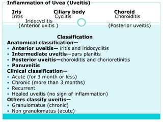
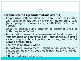
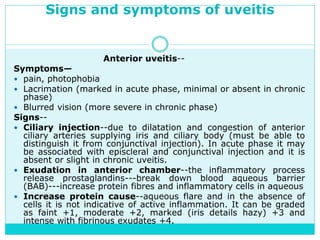
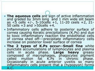
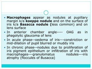
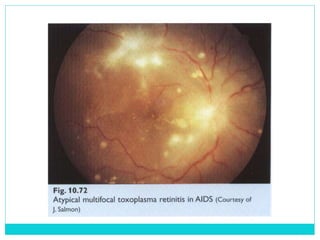
The document provides information on the uveal tract, which consists of the iris, ciliary body, and choroid. It describes the anatomy, histology, and functions of each part of the uvea. Signs and symptoms of uveal inflammation/uveitis are discussed, including anterior uveitis characterized by pain, photophobia, cells and flare in the anterior chamber, and keratic precipitates. Chronic/granulomatous uveitis can cause nodules on the iris and chronic inflammation. Posterior uveitis may involve the choroid and retina without anterior chamber signs.
you will get information about the staphyloma, its types, its etiology, its pathogenesis and its treatment.
you will know about the clinical types of staphyloma in detailed such as anterior staphyloma, posterior staphyloma, equatorial staphyloma, ciliary staphyloma, and Intercalary staphyloma.
you will get information about the staphyloma, its types, its etiology, its pathogenesis and its treatment.
you will know about the clinical types of staphyloma in detailed such as anterior staphyloma, posterior staphyloma, equatorial staphyloma, ciliary staphyloma, and Intercalary staphyloma.
This presentation describes the background of the cornea and the corneal diseases in general, also it describes in detailed manner how to manage the corneal ulcer with its different causes
fungal / mycotic corneal ulcer power point presentation for O.A 2nd year stud...Vinitkumar MJ
Belong suborder Acanthopodina and the genus Acanthamoeba
• Family of free-living cyst-forming protozoans that are ubiquitous in air, soil, dust and water.
• 11 species of which A. Castellanii and A. polyphaga are the most common in keratitis
• Life cycle consist of motile trophozoite and cyst dormant stage
Uveitis
• Inflammation of uveal tissue.
• Associated inflammation of adjacent structures, such as Retina, Vitreous, Sclera and Cornea.
Figure 1 uveitis
Anatomical classification
Clinical classification
Pathological classification
Etiological classification
(Duke Elder’s)
1. Anterior uveitis
Can be divided as follow;
1) Iritis_ inflammation mainly the iris
2) Iridocyclitis _iris and pars plicata involved
3) Cyclitis_ pars plicata is affected
Acute uveitis
Onset is sudden,
Last for less than 3 weeks Granulomatous uveitis
Infective nature
Inflammation is insidious in onset
Chronic in nature with minimum clinical features Infective uveitis
2. Intermediate uveitis
Inflammation of pars plana, peripheral retina and choroid.
Also called as “pars planitis”. Chronic uveitis
Onset is insidious
Duration is more than 3 weeks
Non-granulomatous uveitis
due to allergic or immune related reaction
acute onset
short duration
Allergic uveitis or immune related uveitis
3. Posterior uveitis
Inflammation of choroid(choroiditis)
Associated inflammation of retina (chorioretinitis) Recurrent uveitis
uveitis keeps reoccurring periodically
Toxic uveitis
4. Panuveitis
Inflammation of whole uveal tract Traumatic uveitis
5. Uveitis associated with non-infective systemic diseases
6. Idiopathic uveitis
7. Neoplastic
Figure 2 anatomical classification of uveitis
Panuveitis
Endophthalmitis
Panophthalmitis
Inflammation of all layers of uvea of eye
Can also affect lens, retina, optic nerve and vitreous causing reduced vision or blindness. Inflammation of internal structures of the eye, I;e choroid, retina and vitreous Purulent inflammation of all structures of eye
Including all the three coats and Tenon’s capsule as well.
Etiology
1. Idiopathic
After ruling out other causes
2. Infectious
Tuberculosis
Syphilis
Lyme disease
Leptospirosis
Infectious endophthalmitis
3. Immune related
Sarcoidosis
Vogt-koyanagi-Harada syndrome
Sympathetic ophthalmitis
Behcet syndrome
Etiology
Acute process 1-7 days following intraocular surgery such as Cataract surgery and filtering operation
Commonly caused by Bacteria-staphylococcus, pseudomonas, pneumococcus, streptococcus, E. coli,
Fungus -aspergillus fumigatus, candida albicans, fusarium,
Etiology
1.Exogenous
Due to infected wounds
Common pathogens are pneumococcus, staphylococcus, pseudomonas, pneumococcus, streptococcus, E. coli.
2.Endogenous
Due to metastasis of infected embolus in retinal artery and choroidal vessels.
Clinical Features
• Sudden onset of unilateral pain, redness, photophobia
• Maybe associated with lacrimation
• Visual acuity is usually good at presentation except in eyes with severe hypopyon.
• Low IOP
• Fibrinous exudate
• Posterior synechiae
• Miosis
• Aqueous flare and cells
• Endothelial dusting
Clinical Features
Bacterial endophthalmitis
• Sudden onset with severe pain
• Redness
• Visual loss
• Lid oedema, chemosis, corneal haze
• Low
Uveitis is an interesting disease of the with such a varied and diverse pathogenesis, various systemic causes and Dangerous complications in relation to the eye which makes it difficult and challenging to treat in a proper way. I hope this share will help.
The prostate is an exocrine gland of the male mammalian reproductive system
It is a walnut-sized gland that forms part of the male reproductive system and is located in front of the rectum and just below the urinary bladder
Function is to store and secrete a clear, slightly alkaline fluid that constitutes 10-30% of the volume of the seminal fluid that along with the spermatozoa, constitutes semen
A healthy human prostate measures (4cm-vertical, by 3cm-horizontal, 2cm ant-post ).
It surrounds the urethra just below the urinary bladder. It has anterior, median, posterior and two lateral lobes
It’s work is regulated by androgens which are responsible for male sex characteristics
Generalised disease of the prostate due to hormonal derangement which leads to non malignant enlargement of the gland (increase in the number of epithelial cells and stromal tissue)to cause compression of the urethra leading to symptoms (LUTS
Title: Sense of Smell
Presenter: Dr. Faiza, Assistant Professor of Physiology
Qualifications:
MBBS (Best Graduate, AIMC Lahore)
FCPS Physiology
ICMT, CHPE, DHPE (STMU)
MPH (GC University, Faisalabad)
MBA (Virtual University of Pakistan)
Learning Objectives:
Describe the primary categories of smells and the concept of odor blindness.
Explain the structure and location of the olfactory membrane and mucosa, including the types and roles of cells involved in olfaction.
Describe the pathway and mechanisms of olfactory signal transmission from the olfactory receptors to the brain.
Illustrate the biochemical cascade triggered by odorant binding to olfactory receptors, including the role of G-proteins and second messengers in generating an action potential.
Identify different types of olfactory disorders such as anosmia, hyposmia, hyperosmia, and dysosmia, including their potential causes.
Key Topics:
Olfactory Genes:
3% of the human genome accounts for olfactory genes.
400 genes for odorant receptors.
Olfactory Membrane:
Located in the superior part of the nasal cavity.
Medially: Folds downward along the superior septum.
Laterally: Folds over the superior turbinate and upper surface of the middle turbinate.
Total surface area: 5-10 square centimeters.
Olfactory Mucosa:
Olfactory Cells: Bipolar nerve cells derived from the CNS (100 million), with 4-25 olfactory cilia per cell.
Sustentacular Cells: Produce mucus and maintain ionic and molecular environment.
Basal Cells: Replace worn-out olfactory cells with an average lifespan of 1-2 months.
Bowman’s Gland: Secretes mucus.
Stimulation of Olfactory Cells:
Odorant dissolves in mucus and attaches to receptors on olfactory cilia.
Involves a cascade effect through G-proteins and second messengers, leading to depolarization and action potential generation in the olfactory nerve.
Quality of a Good Odorant:
Small (3-20 Carbon atoms), volatile, water-soluble, and lipid-soluble.
Facilitated by odorant-binding proteins in mucus.
Membrane Potential and Action Potential:
Resting membrane potential: -55mV.
Action potential frequency in the olfactory nerve increases with odorant strength.
Adaptation Towards the Sense of Smell:
Rapid adaptation within the first second, with further slow adaptation.
Psychological adaptation greater than receptor adaptation, involving feedback inhibition from the central nervous system.
Primary Sensations of Smell:
Camphoraceous, Musky, Floral, Pepperminty, Ethereal, Pungent, Putrid.
Odor Detection Threshold:
Examples: Hydrogen sulfide (0.0005 ppm), Methyl-mercaptan (0.002 ppm).
Some toxic substances are odorless at lethal concentrations.
Characteristics of Smell:
Odor blindness for single substances due to lack of appropriate receptor protein.
Behavioral and emotional influences of smell.
Transmission of Olfactory Signals:
From olfactory cells to glomeruli in the olfactory bulb, involving lateral inhibition.
Primitive, less old, and new olfactory systems with different path
New Directions in Targeted Therapeutic Approaches for Older Adults With Mantl...i3 Health
i3 Health is pleased to make the speaker slides from this activity available for use as a non-accredited self-study or teaching resource.
This slide deck presented by Dr. Kami Maddocks, Professor-Clinical in the Division of Hematology and
Associate Division Director for Ambulatory Operations
The Ohio State University Comprehensive Cancer Center, will provide insight into new directions in targeted therapeutic approaches for older adults with mantle cell lymphoma.
STATEMENT OF NEED
Mantle cell lymphoma (MCL) is a rare, aggressive B-cell non-Hodgkin lymphoma (NHL) accounting for 5% to 7% of all lymphomas. Its prognosis ranges from indolent disease that does not require treatment for years to very aggressive disease, which is associated with poor survival (Silkenstedt et al, 2021). Typically, MCL is diagnosed at advanced stage and in older patients who cannot tolerate intensive therapy (NCCN, 2022). Although recent advances have slightly increased remission rates, recurrence and relapse remain very common, leading to a median overall survival between 3 and 6 years (LLS, 2021). Though there are several effective options, progress is still needed towards establishing an accepted frontline approach for MCL (Castellino et al, 2022). Treatment selection and management of MCL are complicated by the heterogeneity of prognosis, advanced age and comorbidities of patients, and lack of an established standard approach for treatment, making it vital that clinicians be familiar with the latest research and advances in this area. In this activity chaired by Michael Wang, MD, Professor in the Department of Lymphoma & Myeloma at MD Anderson Cancer Center, expert faculty will discuss prognostic factors informing treatment, the promising results of recent trials in new therapeutic approaches, and the implications of treatment resistance in therapeutic selection for MCL.
Target Audience
Hematology/oncology fellows, attending faculty, and other health care professionals involved in the treatment of patients with mantle cell lymphoma (MCL).
Learning Objectives
1.) Identify clinical and biological prognostic factors that can guide treatment decision making for older adults with MCL
2.) Evaluate emerging data on targeted therapeutic approaches for treatment-naive and relapsed/refractory MCL and their applicability to older adults
3.) Assess mechanisms of resistance to targeted therapies for MCL and their implications for treatment selection
Report Back from SGO 2024: What’s the Latest in Cervical Cancer?bkling
Are you curious about what’s new in cervical cancer research or unsure what the findings mean? Join Dr. Emily Ko, a gynecologic oncologist at Penn Medicine, to learn about the latest updates from the Society of Gynecologic Oncology (SGO) 2024 Annual Meeting on Women’s Cancer. Dr. Ko will discuss what the research presented at the conference means for you and answer your questions about the new developments.
- Video recording of this lecture in English language: https://youtu.be/lK81BzxMqdo
- Video recording of this lecture in Arabic language: https://youtu.be/Ve4P0COk9OI
- Link to download the book free: https://nephrotube.blogspot.com/p/nephrotube-nephrology-books.html
- Link to NephroTube website: www.NephroTube.com
- Link to NephroTube social media accounts: https://nephrotube.blogspot.com/p/join-nephrotube-on-social-media.html
Explore natural remedies for syphilis treatment in Singapore. Discover alternative therapies, herbal remedies, and lifestyle changes that may complement conventional treatments. Learn about holistic approaches to managing syphilis symptoms and supporting overall health.
Prix Galien International 2024 Forum ProgramLevi Shapiro
June 20, 2024, Prix Galien International and Jerusalem Ethics Forum in ROME. Detailed agenda including panels:
- ADVANCES IN CARDIOLOGY: A NEW PARADIGM IS COMING
- WOMEN’S HEALTH: FERTILITY PRESERVATION
- WHAT’S NEW IN THE TREATMENT OF INFECTIOUS,
ONCOLOGICAL AND INFLAMMATORY SKIN DISEASES?
- ARTIFICIAL INTELLIGENCE AND ETHICS
- GENE THERAPY
- BEYOND BORDERS: GLOBAL INITIATIVES FOR DEMOCRATIZING LIFE SCIENCE TECHNOLOGIES AND PROMOTING ACCESS TO HEALTHCARE
- ETHICAL CHALLENGES IN LIFE SCIENCES
- Prix Galien International Awards Ceremony
HOT NEW PRODUCT! BIG SALES FAST SHIPPING NOW FROM CHINA!! EU KU DB BK substit...GL Anaacs
Contact us if you are interested:
Email / Skype : kefaya1771@gmail.com
Threema: PXHY5PDH
New BATCH Ku !!! MUCH IN DEMAND FAST SALE EVERY BATCH HAPPY GOOD EFFECT BIG BATCH !
Contact me on Threema or skype to start big business!!
Hot-sale products:
NEW HOT EUTYLONE WHITE CRYSTAL!!
5cl-adba precursor (semi finished )
5cl-adba raw materials
ADBB precursor (semi finished )
ADBB raw materials
APVP powder
5fadb/4f-adb
Jwh018 / Jwh210
Eutylone crystal
Protonitazene (hydrochloride) CAS: 119276-01-6
Flubrotizolam CAS: 57801-95-3
Metonitazene CAS: 14680-51-4
Payment terms: Western Union,MoneyGram,Bitcoin or USDT.
Deliver Time: Usually 7-15days
Shipping method: FedEx, TNT, DHL,UPS etc.Our deliveries are 100% safe, fast, reliable and discreet.
Samples will be sent for your evaluation!If you are interested in, please contact me, let's talk details.
We specializes in exporting high quality Research chemical, medical intermediate, Pharmaceutical chemicals and so on. Products are exported to USA, Canada, France, Korea, Japan,Russia, Southeast Asia and other countries.
ARTIFICIAL INTELLIGENCE IN HEALTHCARE.pdfAnujkumaranit
Artificial intelligence (AI) refers to the simulation of human intelligence processes by machines, especially computer systems. It encompasses tasks such as learning, reasoning, problem-solving, perception, and language understanding. AI technologies are revolutionizing various fields, from healthcare to finance, by enabling machines to perform tasks that typically require human intelligence.
Pulmonary Thromboembolism - etilogy, types, medical- Surgical and nursing man...VarunMahajani
Disruption of blood supply to lung alveoli due to blockage of one or more pulmonary blood vessels is called as Pulmonary thromboembolism. In this presentation we will discuss its causes, types and its management in depth.
1. D R . M O H D N A J M U S S A D I Q K H A N
M S ( O P H T H ) D I S S O ( E S A S O )
Uveal Tract
2. UVEA
Is the middle coat of the eye consists of iris, ciliary
body and choroid.
Iris--It is the anterior extension of the ciliary body. Its
anterior surface is divided by an irregular circular line
called collarette into a large ciliary and small pupillary
zones, it contains a round aperture (pupil) in the
middle. Histologically it consist of--
Endothelium (continuous with corneal endothelium
and anterior limiting layer)
Stroma (contains sphincter and dilator pupillae
muscles, vessels, nerve, pigment cells)
Posterior pigment epithelium (continues with pigment
epithelium)
Function-- controls amount of light entering the eye
3. Ciliary body--It extends from anterior end of choroid to the
root of iris about 6mm length. Grossly it consists of 2
zones, pars plicata (cornea ciliaris) which contains ciliary
processes (2 mm anterior) & pars plana smoother and
flatter ( 4 mm posteriorly). Histologically--
Ciliary muscles (longitudinal, circular, radial fibres)
Stroma (vessels, nerve, pigment cells)
Basal lamina
Epithelium--external pigment and internal non pigment,
both of which continues as pigment layer over posterior
surface of iris
Functions--
Aqueous secretion into posterior chamber
Contraction and relaxation of zonule for near and far vision
(by ciliary muscles contraction (accommodation)
4. Choroid--The posterior portion of uvea and it lies
between sclera and retina. It extends from the
edge of optic nerve posteriorly to ciliary body
anteriorly at ora serrata. Highly vascular and
consists mainly of blood vessels. The largest
vessels are nearer to sclera while smallest vessels
(choriocapillaries) are towards retina being
separated from retinal pigment epithelium by
Bruch s membrane.
Suprachoroid is on either sides of stroma--
avascular layer of collagen and elastic fibres.
Function--nutrition of outer part of retina
5. Signs and symptoms of uveal diseases--Depends on site
of uvea affected
Pain, redness, visual loss, floaters
Iritis---pain, photophobia
Iridocyclitis---ciliary injection, pain
Most diseases of ciliary body---disturb accommodation
Inflammation of choroid---no ciliary injection, no pain if
affect retina--- interfere with vision
Pupil distortion in anterior uveitis
Release of cells and protein in the anterior chamber (iritis)
and vitreous cavity (choroiditis)
Keratic precipitates (Kps) in iridocyclitis
Ophthalmoscopically chorioretinal involvement in
choroiditis
6. Congenital and development anomalies--
Coloboma iridis--failure of complete iris development
(in 6 clocks and bilateral)
Aniridia--failure of anterior growth and differentiation
of optic disc ---- rudimentary iris, iris not visible, black
cornea, 60% is autosomal dominant inheritance.
Albinism--patient with photophobia and myopia
Heterochromia iridis--the 2 irises different in colour
Iridocorneal endothelial syndrome---glaucoma, iris
atrophy, abnormal corneal endothelium. P.A.S, multiple
iris nodules, transparent membrane covering anterior
surface of iris.
Rubeosis iridis--neovascularization of the iris occur as
irregularly, distributed networks of new vessels on
surface and stroma of iris and they may cover
trabecular meshwork, peripheral anterior synechae
formation, angle closer glaucoma, more common in
diabetics, intraocular tumours.
7. Inflammation of Uvea (Uveitis)
Iris Ciliary body Choroid
Iritis Cyclitis Choroiditis
Iridocyclitis
(Anterior uvitis ) (Posterior uveitis)
Classification
Anatomical classification—
Anterior uveitis— iritis and iridocyclitis
Intermediate uveitis—pars planitis
Posterior uveitis—choroiditis and chorioretinitis
Panuveitis
Clinical classification—
Acute (for 3 month or less)
Chronic (more than 3 months)
Recurrent
Healed uveitis (no sign of inflammation)
Others classify uveitis—
Granulomatus (chronic)
Non granulomatus (acute)
8. Aetiological classification---
Idiopathic–25-30% of cases
Infections---either exo or endogenous
Exogenous--introduction of microorganisms after
perforated wound or ulcer
Endogenous--spread of infection from other site in the
body
Bacterial (T.B, Syphilis)
Viral (mumps, herpes, influenza)
Parasites (Toxoplasma, malaria)
Fungal (candidiasis)
Allergy--delayed bacterial allergy to T.B
Autoimmune (sympathetic ophthalmia)
Trauma--after contusion or surgery
9. Secondary to ocular pathology--
Suppurative keratitis, scleritis, retinitis
Hyper-mature cataract
Orbital cellulitis
After extra-capsular cataract extraction
Dislocated lens
Intraocular tumours
Intraocular haemorrhage
Secondary to systemic diseases—sarcoidosis, T.B, Crohn s
diseases, behcet s disease, ulcerative colitis, Vogt–
Koyanagi syndrome, Still disease, rheumatoid arthritis,
Reiter syndrome
Malignant tumours (masquerade syndrome)
Sinusitis, otitis media, tonsillitis
Specific HLA type (B-27 in acute anterior uveitis, B-5 in
behcets disease, DW-22j in VKH syndrome)
10. Acute uveitis (non granulomatous)--mostly anterior
uveitis, non suppurative, recurrent inflammation of
uveal tract characterized by oedema, capillary
dilatation, exudation of inflammatory cells (PMN,
Lymphocytes, plasma cells)
Onset is acute, marked pain, photophobia, blurred
vision (symptoms are severe)
Ciliary injection, fine Keratic precipitates, exudation in
to A.C, posterior synechiae
No nodules on iris
In most cases it affects anterior uvea but posterior
uvea (choroid) is rarely affected
In acute uveitis---retinal oedema without exudation,
fine opacities in vitreous
Generally idiopathic in nature
11. Chronic uveitis (granulomatous uveitis)--
Progressive inflammation of uveal tract associated
with cellular infiltration by chronic inflammatory cells
(macrophages, mononuclear, epithelioid cells) with
fibrosis
Onset is insidious and anterior or posterior uvea or
both may be affected
In anterior uvea involvement---minimal signs of
inflammation and minimal pain, photophobia, blurred
vision, slight ciliary injection or none, mutton fat
Keratic precipitates.
The eye is relatively white
In post uvea involvement---slight retinal oedema with
exudation, heavy vitreous opacities, heavy choroidal
exudation
T.B, sarcoidosis and leprosy
12. Signs and symptoms of uveitis
Anterior uveitis--
Symptoms—
pain, photophobia
Lacrimation (marked in acute phase, minimal or absent in chronic
phase)
Blurred vision (more severe in chronic phase)
Signs--
Ciliary injection--due to dilatation and congestion of anterior
ciliary arteries supplying iris and ciliary body (must be able to
distinguish it from conjunctival injection). In acute phase it may
be associated with episcleral and conjunctival injection and it is
absent or slight in chronic uveitis.
Exudation in anterior chamber--the inflammatory process
release prostaglandins---break down blood aqueous barrier
(BAB)---increase protein fibres and inflammatory cells in aqueous
Increase protein cause--aqueous flare and in the absence of
cells it is not indicative of active inflammation. It can be graded
as faint +1, moderate +2, marked (iris details hazy) +3 and
intense with fibrinous exudates +4.
13.
14. The aqueous cells are sign of active inflammation
and graded by 3mm long and 1 mm wide slit beam
as <5 cells +/-, 5-10cells +1, 11-20 ceels +2, 21-
50 cells +3 and >50cells +4.
Inflammatory cells adhere to posterior surface of
cornea causing Keratic precipitations (K.Ps) and due
to toxic inflammatory reaction the endothelial cells
of cornea shed off---precipitate inflammatory cells
increase on posterior lower surface of cornea
The 2 types of K.Ps occur--Small fine white
punctate accumulations of lymphocytes and plasma
cells in acute phase. Large heavy greasy
composed of macrophages, phagocytes pigment
called mutton fat K.Ps in chronic phase.
Occasionally in acute anterior uveitis so many
inflammatory cells present that a (hypopyon) forms
A.C.
15.
16.
17.
18.
19. Macrophages appear as nodules at pupillary
margin k/a koeppe nodule and on the surface of
iris k/a Basacca nodule (less common) and on
lens surface
In anterior chamber angle---- OAG as in
phagolyitc glaucoma of lens
In acute phase--oedema of iris---constriction or
mid-dilation of pupil blurred or muddy iris
In chronic phase--nodules due to proliferation of
iris pigment epithelium or infiltration of iris with
macrophages---granulomatus nodules---iris
atrophy (floccules of Busacca)
20.
21.
22. Posterior synechiae--Adhesion between iris and lens---cause pupil to
be small irregular constricted non reactive to light (complete or partial)
Ring or annular synechiae—causing seclusion pupillae and iris
bombe
Total posterior synechiae
Peripheral anterior synechiae
Constricted pupil (miosis)--due to toxin and irritation of iris
sphincter muscles contraction leads to constriction of pupil and
sluggish or non reacting pupil or irregular festooned pupil after
dilatation
If posterior synechiae involves whole pupillary margin then aqueous
humour cannot flow from posterior to anterior chambers leading to
pupillary block (occlusion papillae)---iris bombe---2ry glaucoma
Lens changes—
Anterior surface pigmentation
Complicated cataract with polychromatic lusture and bread crum
appearance
Cyclitic m embrane behind the lens
23.
24.
25. Intermediate uveitis/pars Planitis/chronic cyclitis — it is
due to some immunological reaction at the pars plana and
outer layers of the peripheral retina. It is an idiopathic,
insidious, generally bilateral (80%) inflammatory disease of
pars plana, peripheral retina and underlying choroid. 10-15%
patients develop multiple sclerosis due to common HLA-DR
15. The symptoms are floaters and impairment of visual
acuity in young adults of either sex in 2-4th decade of life and
the signs are
Some signs of anterior uveitis
Vitritis showing snowballs, cotton balls or sheet like
opacities with a few cells in the anterior chamber
Peripheral retinal periphlebitis
Greyish white plaque like snowbanking involving inferior
pars plana
Absence of focal lesions in the posterior fundus
Complications are cystoids macular oedema, cyclitic
membrane formation, secondary cataract and tractional
retinal detachment
26.
27.
28. Posterior Uveitis (usually chronic form)—the symptoms
are floaters and impaired vision. The important signs are
Vitreous flare, cells, opacities---occur because of
exudation of inflammatory cells and protein rich fluid with
RBC and this appears as black dots ( more intense in
chronic phase)
Posterior hyaloids phase may be covered by
inflammatory precipitates
Choroiditis—active lesions appear as greyish white or
greyish yellow area with ill defined borders surrounded by
normal fundus (with ophthalmoscope) while inactive lesions
appear as white well defined atrophic areas with pigmented
borders.
Retina is nearly always involved (retinitis)---retina
oedematous, opaque, obscuration of retinal vessels
Vasculitis—involving retinal veins (periphlebitis) more in
comparisons to retinal arteries (periarteritis)
Spill over anterior uveitis is common
29.
30.
31. Classification of posterior uveitis—
Focal—toxoplasmosis
Multifocal—birdshot retinochoroidopathy
Geographical—cytomegalovirus retinitis
Diagnosis of uveitis--
History--symptoms, any systemic disease
Clinical exam--ophthalmic exam for red eye, KPs,
vitreous clouding aqueous flare
General exam for systemic disease
Lab exam--important in non responsive uveitis to
treatment chronic uveitis trying to find the etiology
Skin tests for T.B, histoplasmosis
Test for syphilis, toxoplasmosis, rheumatoid arthritis
X-rays
32. Treatment of uveitis--
Anterior Uveitis—
Treat the cause of known (T.B, Sarcoidosis)
Mydriatics and cycloplegics like long acting atropine 1% (up to 2
weeks) drops or ointment or short acting like cyclopentolate 24
hours, tropicamide 6 hours and phenylephrine (no cycloplegia) 3
hours which will produce dilation of pupil to prevent posterior
synechiae and relaxation of ciliary muscle will relief pain caused by
its spasm.
Systemic steroids like oral prednisolone or I/M injections of ACTH
Topical corticosteroids like betamethesone, dexamethasone,
prednisolone e. d. and eye ointment
Subconjunctival /anterior or posterior subtenon injections of
dexamethasone/prednisolone/triamcinolone
Nonsteroiddal anti-inflammatory drugs if steroids are
contraindicated
Local hot compresses 4 times/day and dark goggles.
Analgesics if needed
Immunosuppressive agents antimetabolites like azathioprine 1-3
mg/kg/day in Behcets disease, methotrexate 7.5-25 mg single
dose once/week, mycophenolate mofetil 1 gm b d and T-cell
inhibitors like cyclosporine 2-5 mg/kg/day, tacrolimus (FK-506)
0.05-0.15 mg/kg/day
33. Intermediate uveitis—Mild cases are self limiting, in
severe cases--
Topical corticosteroid
Sub tenon injection of depot steroids every 2-3 weeks
Systemic steroids for 2 weeks then tapering
Rarely immune suppressive agents
Cryotherapy if above treatment fails
Pars plana vitrectomy
Posterior uveitis--
Treat the cause
Atropine 1%
Posterior sub tenon injection of long acting steroids
Intravitreal injection of triamcinolone is under
evaluation
Systemic corticosteroids
34.
35. Treatment of complications--
Iris bombe--LASER iridotomy
2ry glaucoma--decrease corticosteroids
Systemic carbonic anhydrase inhibitor
Local timolol, epinephrine
Surgery for synechiae ,trabecular meshwork damage
Cataract--surgery if vision is poor
Phthisis bulbi--enucleation of eye if painful
and still infected
36. Complications of uveitis--
Formation of synechiae--
Anterior (decrease aqueous out flow at angle of A.C---
Glaucoma)
Posterior (occlusion of pupil by organized exudates causing
adhesion of pupillary borders to lens (occlusion pupillae)---
increase pressure behind iris---iris bombe---2ry angle
closure angle
Change in IOP (2ry Glaucoma)--In the early stage uveitis
cause hypo-secretion of aqueous humour and decrease IOP.
But later on IOP becomes high due to--
Inflammation or occlusion of trabecular meshwork by
inflammatory cells and protein (OAG)
Topical corticosteroids in genetically susceptible person
causes resistance of aqueous out flow from AC (OAG)
Posterior synechiae causes pupillary block---prevent passage
of aqueous from posterior to A.C---Iris bombe---2ry angle
closure glaucoma (ACG )
Peripheral anterior synechiae (between iris and cornea)---
2ry angle closure glaucoma (ACG )
37. Pseudoglioma--due to vitreous exudation
Band shaped keratopathy—more in children with juvenile
rheumatoid arthritis and chronic iridocyclitis
Keratitis and keratopathy--Cornea can be affected in uveitis due
to histological contraction. Keratitis-–damage to corneal
endothelium, vascularization of cornea, deposition of KPs, band
keratopathy (calcium deposition in Bowman s zone)
Phthisis bulbi—in severe uveitis---atrophy of ciliary body---no
aqueous humour secretion--–shrinking of eyeball
Cataract—it is due to interference with lens metabolism---
posterior sub-capsular cataract. In severe cases anterior and
posterior sub-capsular cataract
Retinal detachment (traction detachment)—it is due to fibro-
vascular proliferation after exudation in vitreous causes traction of
sensory retina. Severe effusion from uveal tract causes elevation of
retina
Cystoid macular oedema--Capillaries of central retina become
abnormally permeable---oedema of posterior pole
Macular ischaemia, epiretinal membrane formation
Choroidal neovascularisation
Consecutive optic neuropathy
39. Suppurative uveitis
It is the inflammation of uvea with pus formation. The
causes are staphylococci, proteus, pseudomonas,
bacillus subtitis, and fungi. Organisms introduced
into eye after--
Accidental injury or surgery penetration wound
Rupture of corneal ulcer
Through filtration bleb in glaucoma surgery
Endogenous cause--Septic emboli in bacterial
endocarditis, meningococcemia, bacteraemia,
Viraemia, fungaemia
After urologic surgery
Spread from adjacent structure--Orbital abscess,
Pharyngeal phycomycosis
40. Types--
A)Endophthalmitis--Suppurative inflammation of
intraocular contents, starts in uveal tract then some other
tissues are 2ry affected.
Clinically--
Severe pain, photophobia, diminution of vision,
Lacrimation, red tender oedematous lids, chemotic
conjunctiva with mixed (conjunctiva and ciliary) injection
Hazy cornea with KPs, muddy iris (pus in vitreous, yellow
reflex)
Cloudy vitreous, fundus not seen clearly
Cloudy aqueous
Decreased IOP
No Proptosis
No restriction of ocular movement
Ultimately no perception of light (PL –ve)
41. Complications--
Panophthalmitis
Retinal detachment
Shrinking eyeball
Treatment of endophthalmitis--
Aqueous and vitreous tap with gram stain, KOH
preparation, culture for bacteria and fundus
Intra-vitreal injections of gentamicin / vancomycin /
ceftazidime / amikacin / amphotericin-B, dexamethasone
Antibiotics (local, subconjunctival injection, systemic)
Systemic antifungal drugs like ketoconazole/fluconazole
Systemic corticosteriods
Pars plana vitrectomy in some case if medical treatment
fails to control within 48-72 hours
If eye still infected, blind, painful then enucleation
42. B)Panophthalmitis--Suppurative inflammation of all parts
of the eye.
Clinically--
Same symptoms as endophthalmitis but more severe
Same signs as endophthalmitis with--
Restriction of ocular movements
Corneal necrosis (hypopyon, corneal abscess)
Proptosis
Finally shrinking of eyeball (phthisis bulbi)
Treatment—
Systemic and local antibiotic
Systemic corticosteroids
If no cure then evisceration--removal of contents of eye
ball leaving the sclera around optic nerve to avoid opening
of meninges which can cause fatal meningitis
So Enucleation is contraindication in panophthalmitis, fear
of spread of infection along sheaths of optic nerve to brain
and causing meningitis
43. C)Lens–induced uveitis--An autoimmune disease
2ry to lens antigens liberated in hyper-mature
cataract or traumatic rupture of lens capsule.
Lens material passes into A.C causing an
inflammatory reaction.
D)Endophthalmitis phacoanaphylactica--More
severe form of lens induced uveitis and occurs
after extra-capsular lens extraction where the eye
become sensitive to its own lens material left
behind in the operation, causing Ag–Ab reaction.
Phacolytic glaucoma is a common complication of
lens induced uveitis
Treatment--Removal of all lens material and
local corticosteriods
44. Sympathetic ophthalmia (sympathetic uveitis)
Very rare diffuse granulomatus bilateral panuveitis which
may occur 10 days to years after perforating eye injury
or intraocular surgery (very rare).
Cause is unknown may be autoimmune response to
uveal pigment. The injured (exciting) eye becomes
inflamed first then the fellow (sympathizing) eye
becomes inflamed within 3 months of injury in most
cases.
65% cases between 2 weeks and 3 months after initial
injury and 90% cases within 1 year after trauma.
Pathology--
It is an auto immune reaction to antigens in the uveal
tissue, uveal pigments and retinal antigens.
Nodular aggregation of lymphocytes and plasma cells
in the uveal tract
Pigment cell of uveal tissue proliferates with epitheloid
cells and macrophages to form Dalen-Fuchs nonules
45. Symptoms and signs--
In sympathising eye pain, photophpbia (1st symptom),
difficult accommodation
In exciting eye pain, irritation, redness
Signs in exciting eye ciliary congestion and traumatic
injury
Signs in sympathising eye retrolental flare(1st sign),
signs of granulomatous uveitis, posterior fundus shows
yellow white Dalen Fuchs nodules, perivasculitis, retina
oedema, multifocal choroiditis and optic disc swelling.
Treatment--
In case of severe injury blind eye---immediate eye
excision to prevent sympathetic uveitis within 2 weeks.
If inflammation appear in fellow eye---local steroid,
atropine, systemic corticosteroids (80-100 mg/day) or
cyclosporine
46. Toxoplasmic uveitis--Toxoplasmosis is caused by Toxoplasma
gondi a protozoon parasite (cat is definitive host). There are
two clinical types--
Congenital--due to intrauterine infection (more severe)
Acquired--infection in later life (ingestion of oocyst)
Clinically there is chorioretinitis which appear in early stage as
white elevated masses covered by hazy vitreous. In congenital
toxoplasmosis chorioretinitis, convulsions and
calcification (3 Cs)
Symptoms and signs—
Visual loss due to media opacities, cystoids macular oedema,
maculopapillary bundle or fovea or optic disc involvement
In most patient bilateral healed choriretinal scars detected in
later life
Occasionally anterior uveitis
Severe vitritis and focal necrotising retinitis
papillitis
In late stage--punched out pigmented chorioretinal lesion
47.
48.
49.
50. Complications--
Cystoids macular oedema
Sub retinal neovascularisation
Tractional retinal detachment
Retinal and choroidal atrophy and loss of vision
Diagnosis--
Dye test (Sabin–Feldman)
ELISA for toxoplasmic antibody
Indirect fluorescein test
Treatment--
Sulphonamides (2 gm loading dose then 1 gm q i d)
Clindamycine orally 300 mg q i d for 3 weeks
Pyrimethamine 100 mg loading dose then 25 mg qid
(Daraprim). It is given with 10 mg/day folinic acid
51. Heterochromatic uveitis (Fuchs s iridocyclitis)--
Inflammation of anterior uvea with pigmentary changes in
iris. There is non granulomatus anterior uveitis with insidious
onset and generally unilateral in middle aged adults.
Unknown cause
Iris and ciliary body show atrophy but posterior synechiae
typically absent
Eyes are of different colour
Enlarged pupil (mydrasis) due to atrophy of sphincter
pupillae
Insidious onset
No synechiae--so no mydriatic is needed
Fine Kps scattered throughout the entire corneal
endothelium
flare and cells in A.C
fine neovascularisation of iris (rubeosis iridis) and angle
(Amsler sign)
Vitreous opacities
No symptoms until complicated cataract develops--blurred
vision
Glaucoma may also occur
Treatment—topcal corticosteroids and cataract surgery
52.
53.
54.
55.
56.
57. Uveitis associated with
spondylarthropathies
Anklyosing spondylitis (AS)— it is a chronic
inflammatory arthritis of unknown aetiology, affecting
mainly sacroiliac joint and axial skeleton. Males only
affected (20-40 yr) where in females it has benign
course. More than 90% of patients carry the histo-
compatibility antigen HLA – B27
Ocular manifestations--
Acute unilateral non granulomatus iridocyclitis
Dusting on corneal endothelium then small white KPs
Usually acute course < 6 weeks
Diagnosis :_
X-rays (Sacroiliac joint)
Tissue typing for HLA – B27
58. Reiter s syndrome—It is defined as an episode of
peripheral arthritis of more than one month duration. There
is triad of
Urethritis/cervicitis
Conjunctivitis bilateral mucoprulent
Arthritis (seronegative )
70% of patients show HLA-B27 and males more affected
60% of patients have associated sacroiliitis
The aetiology is unknown but some patients are presented
with previous bacillary dysentery or previous venereal
diseases gonococci and joint problem.
Other less common features are keratoderma of palm and
sole, nail dystrophy.
Ocular manifestation--
Conjunctivitis (mucopurulent)
Keratitis (punctate, sub-epithelial)
Iridocyclitis (unilateral acute non granulomatous anterior
uveitis in 20% of patient at 1st or recurrent attacks)
59.
60. Psoriatic arthritis—affects 7% patients with
psoriasis and associated with HLA-B27 & B17
Anterior uveitis—acute/chronic
Conjunctivitis
Keratitis
Secondary Sjogren syndrome
61. Uveitis in Juvenile (rheumatoid
arthritis) still disease
It is a inflammatory arthritis of at least 6 weeks duration in
children before 16 years of age. Patients are seronegative for
IgM rheumatoid factor. 20% of patients develop a chronic
bilateral (in 70%) non granulomatus iridocyclitis. The female
are more affected.
In most cases the onset is insidious; the disease discovered
only when child noted to have difference of colour of two
eyes, difference in size, shape of pupil or strabismus.
Clinically—
Eye is typically white (white iritis)
Band keratopathy, white medium sized KPs
Posterior synechiae very common
Hypopyon does not develop even in acute phase
Complications--
Complicated cataract
2ry glaucoma
Band shaped keratopathy is very common
62.
63.
64. Sarcoidosis-- Chronic granulomatus disease of unknown cause
characterized by multiple cutaneous and subcutaneous nodules with
similar invasion in viscera
Onset > 30 yr
Tissue reaction < T. B
No caseation
If parotid gland affected---uveo-parotid fever (Heerfordt s disease)
If Lacrimal gland affected---Mikulicz s syndrome
Clinically—in 30% of cases chronic bilateral anterior uveitis (less
commonly posterior uveitis)
Anterior uveitis nodules may lead to cataract and 2ry glaucoma,
papilloedema, optic atrophy
In posterior uveitis--multiple yellow retinal exudates--snow ball
floaters, occasionally severe periphlebitis (candlewax drippings),
BRVO, choroiditis with granuloma, optic disc granulomas, peripheral
retinal neovascularisation & palsy of extraocular muscles
Diagnosis--
Chest x rays showing bilateral hilar lymphadenopathy
Skin biopsy of nodules
Kveim test
Treatment--corticosteroids
In case of sarcoidosis in acute stage unilateral eye and in chronic
stage bilateral eye involvement
65.
66.
67.
68.
69.
70. Behcet s disease--Idiopathic multi-systemic disease, common
in young (3-4th decade) men of Middle East, mediterranean
region and Japan. + ve for HLA–B5. Present theory is
obliterative vasculitis caused by abnormal circulating immune
complex
Clinically--
Recurrent oral ulcer
Genital ulcers 90%
Skin lesions (erythema nodosum, pustules, ulcers)
70% bilateral non granulomatus iridocyclitis or sometime
posterior uveitis
Conjunctivitis, keratitis, episcleritis, transient hypopyon,
posterior uveitis, retinitis, retnal vasculitis, massive retinal
exudation, vitritis
Less common--- thrombophlebitis, cerebral ivolvement
(meningioencephalitis)
Poor visual prognosis due to phthisis bulbi
Treatment--
Topical and systemic steroids
Oral chlorambucil
71.
72.
73.
74.
75. Vogt-koyanagi-harada (VKH) syndrome—it
is a idiopathic, bilateral, granulomatous severe
panuveitis associated with cutaneous (alopecia,
vitiligo, poliosis), neurological (headache,
convulsion, cranial nerve palsy) and auditory
(tinnitus, vertigo, deafness) involvement.
More in japaneese with HLA-B 22j
Bilateral serofibrinous granulomatous
iridocyclitis
Multifocal exudative choroiditis
Exudative retinal detachment
Optic neuritis in some cases
76.
77.
78.
79. AIDS and uveitis—ocular complications occur in
75% patients of AIDS patients.
The lesions are retinal micro-vasculoangiopathy
with cotton wool spots and superficial and deep
haemorrhages
Kaposi sarcoma of eye lids and conjunctiva. It
appears as bright red mass
Opportunistic infectios--like CMV retinitis
(commonest), herpes zoster ophthalmicus,
candida endophthalmitis, toxoplasma
retinochoroiditis, herpes simplex infection,
molluscum contagiosum of lid
80.
81.
82.
83.
84. Toxocariasis—it is caused by toxocara canis and cati
which are common intestinal worms of dog and cat.
Human infected by accidental ingestion of
contaminated soil or food (usually children between 2-
9 years)
Clinically--
Choroidal granuloma
Macular oedema
Mild anterior uveitis
Chronic endophthalmitis with vitreous clouding
Retinal detachment
Cataract severe cyclitic membrane
Granuloma in fundus
Diagnosed by ELISA
Treatment--Majority of cases come late so no effective
treatment (thiabendozole, pars plana vitrectomy)
85.
86.
87.
88.
89. Tuberculous uveitis--T.B cause a granulomatus
uveitis (rare 1%). It is in the form of
chorioretinitis
Clinically--
Hazy vitreous
Yellow exudates on retina
If anterior segment involved--iris nodules, mutton
fat KPs
Treatment--
Systemic anti–Tuberculous drugs & Atropine 1%
90.
91. Tumours--
a) Melanoma benign (naevus)—flat or slightly elevated
lesions less than 3 disc diameter and generally asymptomatic
b) Malignant melanoma-- most common primary intra ocular
tumour in adults affecting choroid (vision may 6/6). Average
age being 50 years, more in males and its always unilateral
Clinically divided into 4 stages—
Quiscent stage
Glaucomatous stage
Stage of extra ocular extension
Stage of metastasis
Decrease vision defects in visual field
C A Glaucoma
Ophthalmoscopically appears as solitary unilateral brown
pigmented mass with choroidal folds, sub retinal and vitreous
haemorrhages
Cataract and posterior uveitis
Spread to orbit
Metastasis to liver
92. Investigations—
General examination, liver function test, X-ray chest
Indirect ophthalmoscopy
Fundus photography
Fluorescein angioscopy
A & B-scan(ultrasound)
CT & MRI
Differential diagnosis—
Rhegmatogenous retinal detachment
Metastatic tumour of choroid
Choroidal haemangioma
Large chorpidal naevus
Choroidal detachment
93. Management—
Observation for small and slow growing tumours, age
more than 65 years, with liver metastasis
Enucleation for very large melanoma when vision loss
is irreversible
Radioactive sclera plaques of cobalt 60 for medium
sized melanoma
Heavy charged particle irradiation (proton or helium)
Photocoagulation with xenon arc or argon laser for
small tumours away from fovea
Choroidectomy (local resection) but very difficult
Palliative therapy with chemotherapy or
immunotherapy for cases with distant metastasis