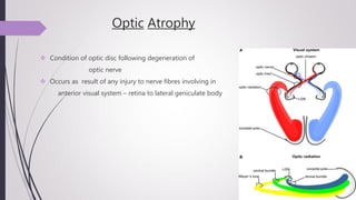
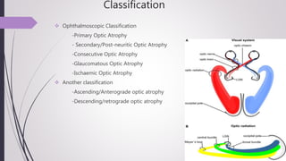
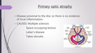
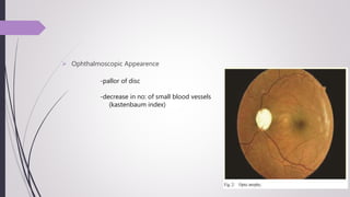
Optic atrophy
Angel Das
Optic Atrophy
 Condition of optic disc following degeneration of
optic nerve
 Occurs as result of any injury to nerve fibres involving in
anterior visual system – retina to lateral geniculate body
Classification
 Ophthalmoscopic Classification
-Primary Optic Atrophy
- Secondary/Post-neuritic Optic Atrophy
-Consecutive Optic Atrophy
-Glaucomatous Optic Atrophy
-Ischaemic Optic Atrophy
 Another classification
-Ascending/Anterograde optic atrophy
-Descending/retrograde optic atrophy
Primary optic atrophy
 Disease proximal to the disc so there is no evidence
of local inflammation.
 CAUSES: Multiple sclerosis
Space occupying lesions
Leber’s disease
Tabes dorsalis
o Disc is chalky white
o Margins well defined
o Lamina cribrosa seen
o Cupping is shallow
o Retina looks normal
 Ophthalmoscopic appearance
Secondary/Post neuritic Optic Atrophy
 Break in continuity of fibres in optic disc
 Occurs in longstanding papilloedema, papillitis or neuroretinitis
Ophthalmoscopic Appearance
-Disc- dirty white
-Edges blurred due to gliosis(proliferation
of astrocytes & glial tissue)
-Cup obliterated
Consecutive Optic Atrophy
 Occurs following destruction of ganglion cell-
secondary to lesions in choroid /retina
 Causes
-Diffuse chorioretinitis
-Retinitis pigmentosa
-Pathological myopia
-Occlusion of central retinal artery
 Ophthalmoscopic Appearence
-Disc – yellow waxy
-Edges – not well defined
-Retinal vessels attenuated
Glaucomatous Atrophy
 Results from long-standing raised IOP
Ophthalmoscopic Appearence
-Deep & wide cupping of optic disc
-Nasal shift of blood vessels
-Lamina cribrosa pores seen
(lamellar dot sign)
 Conditions producing disc ischaemia
 Causes
-Giant cell arteritis
-Severe haemorrhage
-Severe anaemia
-Quinine poisoning
Vascular / Ischaemic Optic Atrophy
 Ophthalmoscopic Appearence
-pallor of disc
-marked attenuation of vessels
Clinical Features
 Loss of vision
-partial /total
 Pupil
-semi-dilated
-direct light reflex –sluggish /absent
-Marcus Gunn pupil/RAPD
 Visual field loss
-Peripheral, Central /Eccentric
 Ophthalmoscopic Appearence
-pallor of disc
-decrease in no: of small blood vessels
(kastenbaum index)
Treatment
 Partial optic atrophy – treat underlying cause
 If complete atrophy – vision cannot be recovered
Optic atrophy

Optic atrophy

  • 1.
  • 2.
    Optic Atrophy  Conditionof optic disc following degeneration of optic nerve  Occurs as result of any injury to nerve fibres involving in anterior visual system – retina to lateral geniculate body
  • 3.
    Classification  Ophthalmoscopic Classification -PrimaryOptic Atrophy - Secondary/Post-neuritic Optic Atrophy -Consecutive Optic Atrophy -Glaucomatous Optic Atrophy -Ischaemic Optic Atrophy  Another classification -Ascending/Anterograde optic atrophy -Descending/retrograde optic atrophy
  • 4.
    Primary optic atrophy Disease proximal to the disc so there is no evidence of local inflammation.  CAUSES: Multiple sclerosis Space occupying lesions Leber’s disease Tabes dorsalis
  • 5.
    o Disc ischalky white o Margins well defined o Lamina cribrosa seen o Cupping is shallow o Retina looks normal  Ophthalmoscopic appearance
  • 6.
    Secondary/Post neuritic OpticAtrophy  Break in continuity of fibres in optic disc  Occurs in longstanding papilloedema, papillitis or neuroretinitis Ophthalmoscopic Appearance -Disc- dirty white -Edges blurred due to gliosis(proliferation of astrocytes & glial tissue) -Cup obliterated
  • 7.
    Consecutive Optic Atrophy Occurs following destruction of ganglion cell- secondary to lesions in choroid /retina  Causes -Diffuse chorioretinitis -Retinitis pigmentosa -Pathological myopia -Occlusion of central retinal artery
  • 8.
     Ophthalmoscopic Appearence -Disc– yellow waxy -Edges – not well defined -Retinal vessels attenuated
  • 9.
    Glaucomatous Atrophy  Resultsfrom long-standing raised IOP Ophthalmoscopic Appearence -Deep & wide cupping of optic disc -Nasal shift of blood vessels -Lamina cribrosa pores seen (lamellar dot sign)
  • 10.
     Conditions producingdisc ischaemia  Causes -Giant cell arteritis -Severe haemorrhage -Severe anaemia -Quinine poisoning Vascular / Ischaemic Optic Atrophy  Ophthalmoscopic Appearence -pallor of disc -marked attenuation of vessels
  • 11.
    Clinical Features  Lossof vision -partial /total  Pupil -semi-dilated -direct light reflex –sluggish /absent -Marcus Gunn pupil/RAPD  Visual field loss -Peripheral, Central /Eccentric
  • 12.
     Ophthalmoscopic Appearence -pallorof disc -decrease in no: of small blood vessels (kastenbaum index)
  • 13.
    Treatment  Partial opticatrophy – treat underlying cause  If complete atrophy – vision cannot be recovered