This document provides an overview of uveitis, including:
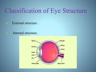
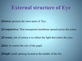
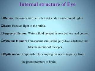
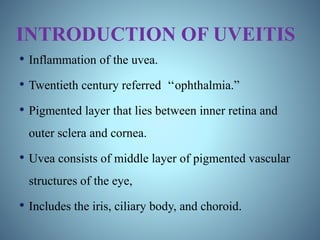
- Definitions of uveitis and the structures of the eye involved
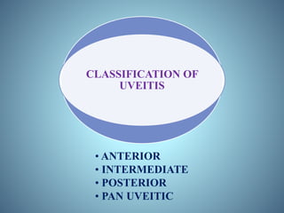
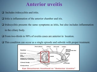
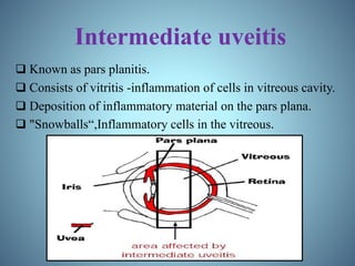
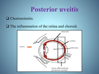
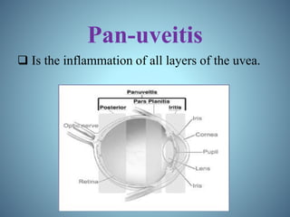
- Classification of uveitis into anterior, intermediate, posterior, and pan-uveitic types
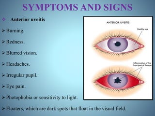
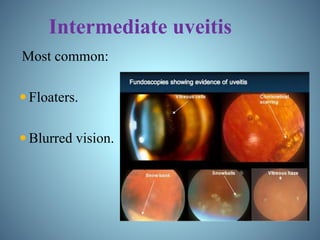
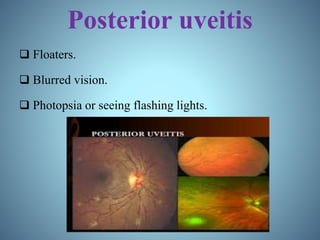
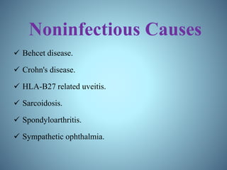
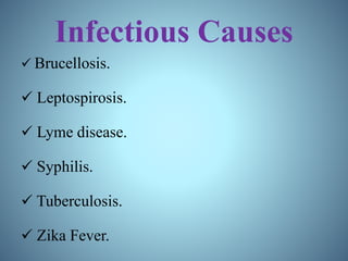
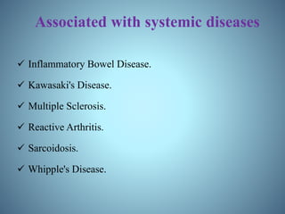
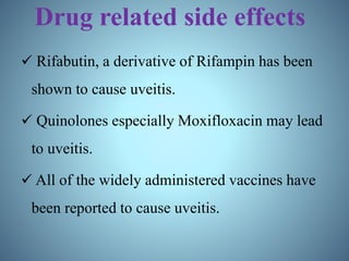
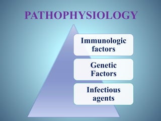
- Signs and symptoms, causes like infections and autoimmune diseases, and pathophysiology involving immune and genetic factors
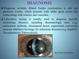
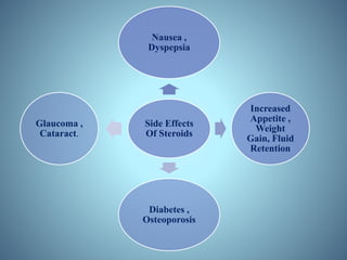
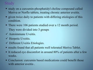
- Diagnosis through examination and testing, as well as treatment using steroids, immunosuppressants, mydriatics, and natural products like turmeric
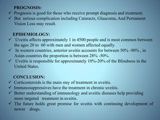
- Prognosis being generally good with treatment but potential for vision loss, and epidemiology with uveitis affecting approximately 1 in 5,000 people.
Simple eye education for EHW, Ophthalmic eye student, school eye education & first - second year optometry students only .
common eye lid inflammatory conditions .
stye or hordeolum ( external / internal hordeolum ), lid abscess , chalazion or mebomian retention cyst, accessory lacrimal glands , lacrimal gland etc...
Uveitis is an interesting disease of the with such a varied and diverse pathogenesis, various systemic causes and Dangerous complications in relation to the eye which makes it difficult and challenging to treat in a proper way. I hope this share will help.
www.ophthalclass.blogspot.com has the complete post.
In Part1 the topics discussed are the causes of anterior, intermediate, posterior and panuveitis. There is also a section on the associated features like history, demographics and examination findings that help to narrow down the differential diagnosis.
www.ophthalclass.blogspot.com has the complete class on uveitis for undergraduate medical students. This presentation is the first in the series and deals with the classification of uveitis.
www.ophthalclass.blogspot.com has the complete class on uveitis for undergraduate medical students. This presentation is the fourth in the series and deals with the management of uveitis.
This is appt presentation done by me and my colleagues zakaria Abul-Nasser and Sara Hassan ( agroup of medical undergarduates , school of Medicine, Ain-shams university , Cairo , Egypt ) ...
This work was presented at the end of our Ophthalmolgy clinical round ..
I Hope every one to get the best out of the presentaion ..Any commentaries are even more appreciated :)
The corneal diseases are one of the leading causes of blindness in the world. in most cases, these infections are preventable or treatable.
This seminar provides an overview of the anatomy and physiology of the cornea, as well as an overview of common conditions.
The important diseases recognized as responsible for visual impairment and blindness in India are cataract, refraction errors, glaucoma, conjunctivitis.
Mal nutrition and systemic disease are also important contributing factors.
Other causes include are eye injury, congenital disorders, retinal detachment, tumors, leprosy etc.
The visual disorder can be found as the following problems.
Amblyopia or subnormal vision in one or both eyes in spite of correction of significant refractive error.
Night blindness – it means inability to well at night or in faint light. It may occur in retinitis, choroidoretinistis, Vit A deficiency, retina toxic drugs.
Double Vision – It is found in squint, ptosis. It may be warning sign of increase IOP, brain tumor, orbital or myasthenia gravis.
Color blindness – It is a genetically determine condition in which color perception is defective or absent. Red and green color deficiency is the usual found. It can be detected at the age of 5 to 6 years’ age.
It is found in about 8% of the male population and is inherited as sex linked recessive trait. Color blindness may be total or partial there is no specific treatment.
Some main responsible disease for visual impairments in India :-
Conjunctivitis
Cataract
Refractory errors
Glaucoma
Retinopathy of Prematurity
www.ophthalclass.blogspot.com has the complete class and MCQs on uveitis for undergraduate medical students. Class 5 in the series of classes on uveitis deals with the common causes of panuveitis and briefly discusses their management. The clinical feature of each of the disease entities is explained with the help of case studies.
Uveitis
• Inflammation of uveal tissue.
• Associated inflammation of adjacent structures, such as Retina, Vitreous, Sclera and Cornea.
Figure 1 uveitis
Anatomical classification
Clinical classification
Pathological classification
Etiological classification
(Duke Elder’s)
1. Anterior uveitis
Can be divided as follow;
1) Iritis_ inflammation mainly the iris
2) Iridocyclitis _iris and pars plicata involved
3) Cyclitis_ pars plicata is affected
Acute uveitis
Onset is sudden,
Last for less than 3 weeks Granulomatous uveitis
Infective nature
Inflammation is insidious in onset
Chronic in nature with minimum clinical features Infective uveitis
2. Intermediate uveitis
Inflammation of pars plana, peripheral retina and choroid.
Also called as “pars planitis”. Chronic uveitis
Onset is insidious
Duration is more than 3 weeks
Non-granulomatous uveitis
due to allergic or immune related reaction
acute onset
short duration
Allergic uveitis or immune related uveitis
3. Posterior uveitis
Inflammation of choroid(choroiditis)
Associated inflammation of retina (chorioretinitis) Recurrent uveitis
uveitis keeps reoccurring periodically
Toxic uveitis
4. Panuveitis
Inflammation of whole uveal tract Traumatic uveitis
5. Uveitis associated with non-infective systemic diseases
6. Idiopathic uveitis
7. Neoplastic
Figure 2 anatomical classification of uveitis
Panuveitis
Endophthalmitis
Panophthalmitis
Inflammation of all layers of uvea of eye
Can also affect lens, retina, optic nerve and vitreous causing reduced vision or blindness. Inflammation of internal structures of the eye, I;e choroid, retina and vitreous Purulent inflammation of all structures of eye
Including all the three coats and Tenon’s capsule as well.
Etiology
1. Idiopathic
After ruling out other causes
2. Infectious
Tuberculosis
Syphilis
Lyme disease
Leptospirosis
Infectious endophthalmitis
3. Immune related
Sarcoidosis
Vogt-koyanagi-Harada syndrome
Sympathetic ophthalmitis
Behcet syndrome
Etiology
Acute process 1-7 days following intraocular surgery such as Cataract surgery and filtering operation
Commonly caused by Bacteria-staphylococcus, pseudomonas, pneumococcus, streptococcus, E. coli,
Fungus -aspergillus fumigatus, candida albicans, fusarium,
Etiology
1.Exogenous
Due to infected wounds
Common pathogens are pneumococcus, staphylococcus, pseudomonas, pneumococcus, streptococcus, E. coli.
2.Endogenous
Due to metastasis of infected embolus in retinal artery and choroidal vessels.
Clinical Features
• Sudden onset of unilateral pain, redness, photophobia
• Maybe associated with lacrimation
• Visual acuity is usually good at presentation except in eyes with severe hypopyon.
• Low IOP
• Fibrinous exudate
• Posterior synechiae
• Miosis
• Aqueous flare and cells
• Endothelial dusting
Clinical Features
Bacterial endophthalmitis
• Sudden onset with severe pain
• Redness
• Visual loss
• Lid oedema, chemosis, corneal haze
• Low
Conjunctivitis, commonly known as pink eye, is an inflammation of the conjunctiva, the thin, transparent tissue that covers the front surface of the eye and lines the inside of the eyelids. Conjunctivitis can be caused by various factors, including infections, allergies, and irritants, leading to different types of conjunctivitis that require specific management and therapy.
Infectious conjunctivitis is often caused by bacteria or viruses. Bacterial conjunctivitis typically presents with redness, discharge, and crusting of the eyelids. It is commonly treated with topical antibiotics to eliminate the bacterial infection. On the other hand, viral conjunctivitis is caused by a viral infection and tends to be self-limiting, with symptoms like redness, watery discharge, and sensitivity to light. Management for viral conjunctivitis involves supportive care, such as cold compresses and lubricating eye drops, to alleviate discomfort while waiting for the infection to resolve.
Allergic conjunctivitis occurs when the eyes react to allergens, such as pollen, pet dander, or dust mites. It results in itching, redness, watery discharge, and swollen eyelids. Antihistamine eye drops or oral antihistamines can help relieve symptoms by reducing the allergic response. In some cases, mast cell stabilizers or corticosteroid eye drops may be prescribed to control severe allergic reactions.
Other specific types of conjunctivitis include giant papillary conjunctivitis, which is often associated with contact lens use or ocular prosthetics, and vernal conjunctivitis, a chronic allergic condition primarily affecting young people. These types may require a combination of treatments, including avoiding the triggering factors, using lubricating eye drops, and, in severe cases, prescribed medications like mast cell stabilizers or topical steroids.
Additionally, conjunctivitis can be caused by exposure to irritating substances, such as chemicals or smoke, leading to chemical conjunctivitis. The primary management for this type involves thoroughly rinsing the eyes with clean water or saline solution to flush out the irritants.
In all cases, practicing good hygiene measures, such as frequent hand washing and avoiding touching the eyes, can help prevent the spread of infectious conjunctivitis.
It is important to consult an eye care professional for an accurate diagnosis and appropriate management of conjunctivitis. They can provide personalized recommendations and prescribe the most suitable therapy based on the type and severity of the condition, with the goal of relieving symptoms, promoting healing, and preventing complications.
We all have good and bad thoughts from time to time and situation to situation. We are bombarded daily with spiraling thoughts(both negative and positive) creating all-consuming feel , making us difficult to manage with associated suffering. Good thoughts are like our Mob Signal (Positive thought) amidst noise(negative thought) in the atmosphere. Negative thoughts like noise outweigh positive thoughts. These thoughts often create unwanted confusion, trouble, stress and frustration in our mind as well as chaos in our physical world. Negative thoughts are also known as “distorted thinking”.
Operation “Blue Star” is the only event in the history of Independent India where the state went into war with its own people. Even after about 40 years it is not clear if it was culmination of states anger over people of the region, a political game of power or start of dictatorial chapter in the democratic setup.
The people of Punjab felt alienated from main stream due to denial of their just demands during a long democratic struggle since independence. As it happen all over the word, it led to militant struggle with great loss of lives of military, police and civilian personnel. Killing of Indira Gandhi and massacre of innocent Sikhs in Delhi and other India cities was also associated with this movement.
Welcome to TechSoup New Member Orientation and Q&A (May 2024).pdfTechSoup
In this webinar you will learn how your organization can access TechSoup's wide variety of product discount and donation programs. From hardware to software, we'll give you a tour of the tools available to help your nonprofit with productivity, collaboration, financial management, donor tracking, security, and more.
Unit 8 - Information and Communication Technology (Paper I).pdfThiyagu K
This slides describes the basic concepts of ICT, basics of Email, Emerging Technology and Digital Initiatives in Education. This presentations aligns with the UGC Paper I syllabus.
The Indian economy is classified into different sectors to simplify the analysis and understanding of economic activities. For Class 10, it's essential to grasp the sectors of the Indian economy, understand their characteristics, and recognize their importance. This guide will provide detailed notes on the Sectors of the Indian Economy Class 10, using specific long-tail keywords to enhance comprehension.
For more information, visit-www.vavaclasses.com
The French Revolution, which began in 1789, was a period of radical social and political upheaval in France. It marked the decline of absolute monarchies, the rise of secular and democratic republics, and the eventual rise of Napoleon Bonaparte. This revolutionary period is crucial in understanding the transition from feudalism to modernity in Europe.
For more information, visit-www.vavaclasses.com
2024.06.01 Introducing a competency framework for languag learning materials ...Sandy Millin
http://sandymillin.wordpress.com/iateflwebinar2024
Published classroom materials form the basis of syllabuses, drive teacher professional development, and have a potentially huge influence on learners, teachers and education systems. All teachers also create their own materials, whether a few sentences on a blackboard, a highly-structured fully-realised online course, or anything in between. Despite this, the knowledge and skills needed to create effective language learning materials are rarely part of teacher training, and are mostly learnt by trial and error.
Knowledge and skills frameworks, generally called competency frameworks, for ELT teachers, trainers and managers have existed for a few years now. However, until I created one for my MA dissertation, there wasn’t one drawing together what we need to know and do to be able to effectively produce language learning materials.
This webinar will introduce you to my framework, highlighting the key competencies I identified from my research. It will also show how anybody involved in language teaching (any language, not just English!), teacher training, managing schools or developing language learning materials can benefit from using the framework.
How to Create Map Views in the Odoo 17 ERPCeline George
The map views are useful for providing a geographical representation of data. They allow users to visualize and analyze the data in a more intuitive manner.
Palestine last event orientationfvgnh .pptxRaedMohamed3
An EFL lesson about the current events in Palestine. It is intended to be for intermediate students who wish to increase their listening skills through a short lesson in power point.
2. Content
INTRO OF EYE STRUCTURE AND FUNCTION.
INTRO OF UVEITIS.
SIGNS AND SYMPTOMS.
CAUSES.
PATHOPHYSIOLOGY.
DIAGNOSIS.
TREATMENT.
ROLE OF SOME NATURAL PRODUCT IN UVEITIS.
PROGNOSIS.
EPIDEMIOLOGY.
CONCLUSION.
REFERENCES.
3. Introduction to Eye
Most complicated organ in the human body.
A number of parts fitted together in a near-spherical
structure.
Each part in the system is responsible for a certain action.
5. External structure of Eye
Sclera: protects the inner parts of Eye.
Conjunctiva: Thin transparent membrane spread across the sclera.
Cornea: job of cornea is to refract the light that enters the eyes.
Iris: to control the size of the pupil.
Pupil: small opening located at the middle of the Iris.
6. Internal structure of Eye
Retina: Photosensitive cells that detect dim and colored lights.
Lens: Focuses light to the retina.
Aqueous Humor: Watery fluid present in area bet lens and cornea.
Vitreous Humor: Transparent semi-solid, jelly-like substance that
fills the interior of the eyes.
Optic nerve: Responsible for carrying the nerve impulses from
the photoreceptors to brain.
7. INTRODUCTION OF UVEITIS
• Inflammation of the uvea.
• Twentieth century referred ‘‘ophthalmia.”
• Pigmented layer that lies between inner retina and
outer sclera and cornea.
• Uvea consists of middle layer of pigmented vascular
structures of the eye,
• Includes the iris, ciliary body, and choroid.
9. Anterior uveitis
Includes iridocyclitis and iritis.
Iritis is inflammation of the anterior chamber and iris.
Iridocyclitis presents the same symptoms as iritis, but also includes inflammation
in the ciliary body.
From two-thirds to 90% of uveitis cases are anterior in location.
This condition can occur as a single episode and subside with proper treatment.
10. Intermediate uveitis
Known as pars planitis.
Consists of vitritis -inflammation of cells in vitreous cavity.
Deposition of inflammatory material on the pars plana.
"Snowballs“,Inflammatory cells in the vitreous.
13. SYMPTOMS AND SIGNS
Anterior uveitis
Burning.
Redness.
Blurred vision.
Headaches.
Irregular pupil.
Eye pain.
Photophobia or sensitivity to light.
Floaters, which are dark spots that float in the visual field.
16. CAUSES
Widely administered vaccines cause uveitis.
Noninfectious Causes.
Infectious causes.
Associated with systemic diseases.
Drug related side effects.
20. Drug related side effects
Rifabutin, a derivative of Rifampin has been
shown to cause uveitis.
Quinolones especially Moxifloxacin may lead
to uveitis.
All of the widely administered vaccines have
been reported to cause uveitis.
22. Immunologic factors
Uveitis Is Driven By Th17t Cell Sub-population That Bear T-cell
Receptors Specific For Proteins Found In The Eye.
Not Detected Centrally Whether Due To Ocular Antigen Not Being Presented In
The Thymus.
Autoreactive T Cells Must Normally Be Held In Check By The Suppressive
Environment Produced By Microglia And Dendritic Cells In The Eye.
These Cells Produce Large Amounts Of TGF Beta And Other
Suppressive Cytokines,
Including IL-10, To Prevent Damage To The Eye By Reducing Inflammation And
Causing T Cells To Differentiate To Inducible T Reg Cells.
23. Cont….
Immune stimulation by bacteria and cellular stress is
normally suppressed by myeloid suppression while
inducible T reg cells prevention and clonal expansion of the
autoreactive Th1 and Th 17 cells that possess potential to
cause damage to the eye.
Infection or other causes, this balance can be upset and auto
reactive T cells allowed to proliferate and migrate to the eye.
Entry to the eye, these cells may be returned to an inducible
T reg state by the presence of IL-10 and TGF-beta from
microglia.
24. Genetic Factors
The cause of non-infectious uveitis is unknown.
But there are some strong genetic factors that predispose
disease onset .
Including HLA-B27 and the PTPN22 genotype.
25. Infectious agents
Recent evidence has pointed to reactivation of herpes
simplex, varicella zoster and other viruses as
important causes.
Bacterial infection is another significant contributing
factor in developing uveitis.
26. DIAGNOSIS
Diagnosis includes dilated fundus examination to rule out
posterior uveitis, which presents with white spots across the
retina along with retinitis and vasculitis.
Laboratory testing is usually used to diagnose specific
underlying diseases, including rheumatologic tests (e.g.
antinuclear antibody, rheumatoid factor, angiotensin converting
enzyme inhibitor) Serology for infectious diseases (e.g. Syphilis,
Toxoplasmosis, Tuberculosis).
fig. Keratic precipitates
27. TREATMENT
What should treatment achieve?
1. Relieve pain and discomfort.
2. Prevent sight loss due to the disease or its
complications.
3. Treat the cause of the disease where possible, that
is, treat the inflammation.
The drugs used to treat uveitis fall into 3 main
groups.
1) Steroids
2) Immunosuppressant.
3) Mydriatics.
28. STEROIDS
Steroids have wide ranging effects but their action may
be looked on as being anti-inflammatory and
immunosuppressant".
They are used in different forms:
• Eye drops.
• Periocular injections.
• By oral (tablets).
• Intra-venous infusion (drip).
29. Eye Drops:
Used for Anterior Uveitis.
Drops can penetrate the part of the eye in front of the lens, where anterior uveitis occurs.
Frequency of taking the drops depending on severity of the uveitis.
Severe Cases strongest drop-every hour .
Mild inflammation weakest drop once or twice a day.
Periocular Injections:
Use of injections around the eye to deliver the steroid treatment.
In certain situations injections offer a better way than either tablets or drops.
They are used along with other forms of treatment.
Situations where injections are used include:
• Severe cases of anterior uveitis which can not be controlled by drops alone.
• Intermediate uveitis.
30. Systemic Steroids:
• Oral Steroids E.g. Prednisolone Tablet.
• The use of systemic steroids is more serious than, steroid drops because in this form
there are potentially significant side effects.
• Many different situations in which oral steroids are considered.
• If anterior uveitis is resistant to treatment with drops and injections then systemic
steroids considered.
• The main use of oral steroids is to treat posterior uveitis , panuveitis.
Dosage: Prednisolone tablet 1mg and 5mg.
Intra-venous Steroids:
E.g. Methylprednisolone.
• when rapid control of inflammation is needed high dosage of steroid needs to be
delivered quickly.
31. Side Effects
Of Steroids
Nausea ,
Dyspepsia
Increased
Appetite ,
Weight
Gain, Fluid
Retention
Diabetes ,
Osteoporosis
Glaucoma ,
Cataract.
32. IMMUNOSUPPRESSANT
Steroids do suppress the immune system,but there are
a different group of drugs that may be used to treat
uveitis.
These drugs tend to target the immune system more
precisely than steroids.
They are usually used in conjunction with steroids.
The main examples are:
Cyclosporine.
Azathioprine (Imuran).
Methotrexate.
Mycophenolate mofetil (cellcept).
Tacrolimus (Prograf 500).
33. MYDRIATICS
Mydriatics have 2 main aims:-
To relieve pain and light sensitivity.
To prevent sight threatening complications.
Mydriatic eye drops, Eg. Atropine and Cyclopentolate are used.
It works by "paralyzing" the muscles of the iris and the ciliary
body.
It taken their effect the pupils will be dilated. This may cause
Blurring of the vision.
Useful because they help prevent complication which may occur
in anterior uveitis.
34. ROLE OF SOME NATURAL
PRODUCT IN UVEITIS
TURMERIC:
Benefits for Uveitis:
From 2010.
Antioxidant properties, protect and boost the functioning of
the immune system.
Turmeric help in the reduction of chronic uveitis symptoms.
Research studies which have found that turmeric can prove
beneficial for uveitis.
35. Study
study on a curcumin-phosphatidyl choline compound called
Meriva or Norflo tablets, treating chronic anterior uveitis.
given twice daily to patients with differing etiologies of this
condition.
There were 106 patients studied over a 12 month period.
They were divided into 3 groups
Autoimmune Uveitis.
Herpetic Uveitis.
Different Uveitis Etiologies.
results found that all patients well tolerated Meriva Tablet.
It reduced eye discomfort in around 80% of patients after a few
weeks.
Conclusion: curcumin based medications could benefit those
with anterior uveitis .
36. Dosage: 375mg Tablet 3 times daily.
Precautions:
Diabetes or Gall Bladder problems must avoid turmeric
supplements.
Taken in excess, it can cause Diarrhea Or Nausea.
Contraindicated in Pregnant and Breastfeeding women.
MARKETED PREPARATION:
Uvical pills.
Curcumin phytosome 500mg caps.
Turmeric curcumin 500mg caps.
37. PROGNOSIS:
Prognosis is good for those who receive prompt diagnosis and treatment.
But serious complication including Cataracts, Glaucoma, And Permanent
Vision Loss may result.
EPIDEMIOLOGY:
Uveitis affects approximately 1 in 4500 people and is most common between
the ages 20 to 60 with men and women affected equally.
In western countries, anterior uveitis accounts for between 50% -90% , in
Asian countries the proportion is between 28% -50%.
Uveitis is responsible for approximately 10%-20% of the Blindness in the
United States.
CONCLUSION:
Corticosteroids is the main stay of treatment in uveitis.
Immunosuppressives have the treatment in chronic uveitis.
Better understanding of immunology and uveitic diseases help providing
more targeted treatment in uveitis.
The future holds great promise for uveitis with continuing development of
newer drugs.
38. REFERENCES
1. Jabs DA, Nussenblatt RB, Rosenbaum JT. Standardization of Uveitis
Nomenclature (SUN) Working Group. Standardization of uveitis nomenclature
for reporting clinical data. Results of the First International Workshop. Am J
Ophthalmol 2005;140:509-516.
2. Abdullah Al-Fawaz; Ralph D Levinson (25 Feb 2010)."Uveitis,
Anterior,Granulomatous"eMedicine from WebMD. Retrieved 15
December 2010.
3. Babu, BM; Rathinam, SR (Jan–Feb 2010). "Intermediate uveitis.". Indian
journalofophthalmology. 58 (1):217.doi:10.4103/03014738.58469. PMC284137
0.PMID 20029143.
4. Larson, T; Nussenblatt, RB; Sen, HN (June 2011)."Emerging drugs for
uveitis". Expert opinion on emerging drugs. PMC 3102121.PMID 21210752.
5. McGonagle D, McDermott MF (2006) A proposed classification of the
immunologicaldiseases" PLoSMed3(8)e297.doi:10.1371/journal.pmed.0030297
6. CDC: Department of Human Services (9 September 1994). "Uveitis Associated
with Rifabutin Therapy". 43(35);658: Morbidity and Mortality Weekly Report.
Retrieved 5 May 2013.
7. Risk for Uveitis With Oral Moxifloxacin". JAMA Ophthalmology online. 2
October 2014.