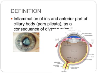
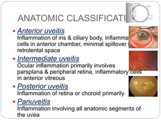
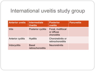
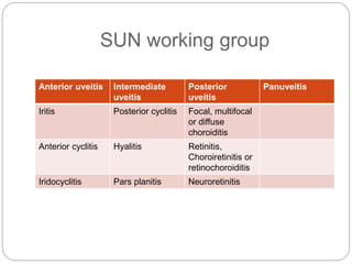
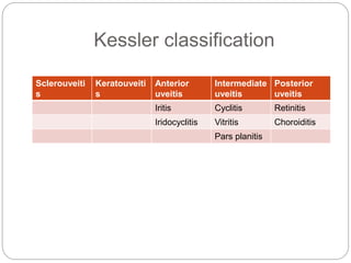
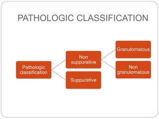
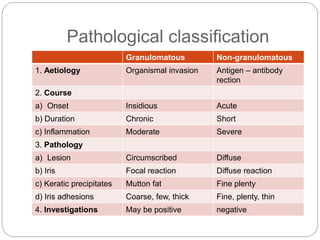
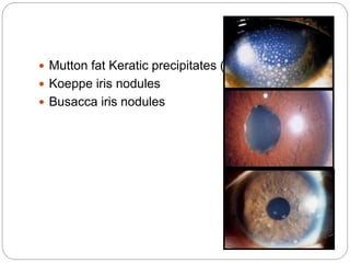
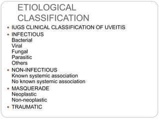
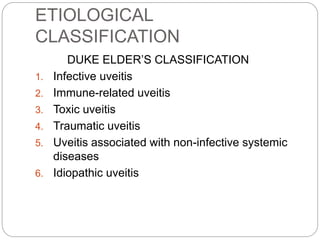
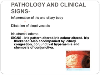
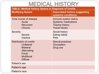
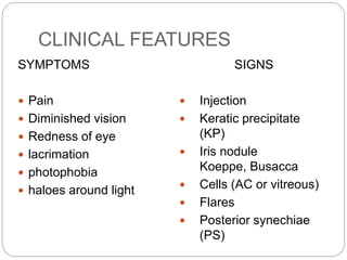
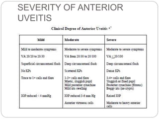
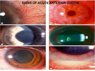
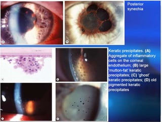
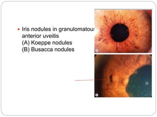
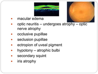
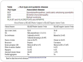
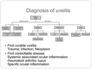
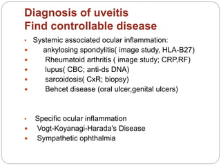
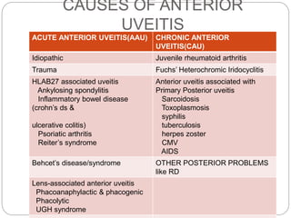
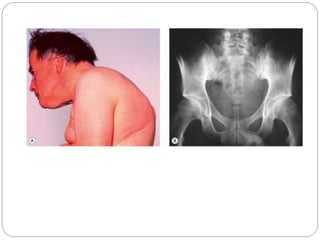
Dr. TP Chhangte discusses anterior uveitis, including its definition, epidemiology, classification, pathology, clinical features, complications, and differential diagnosis. Some key points:
- Anterior uveitis is inflammation of the iris and anterior part of the ciliary body. Its prevalence has been increasing and it commonly affects people aged 20-50.
- Uveitis can be classified anatomically, clinically, etiologically, and pathologically. The anatomical classification divides uveitis into anterior, intermediate, posterior, and panuveitis based on location of inflammation.
- Clinical features of anterior uveitis include eye pain, redness, photophobia, and decreased vision. On examination
This presentation describes the background of the cornea and the corneal diseases in general, also it describes in detailed manner how to manage the corneal ulcer with its different causes
Uveitis: Pathogenesis, Clinical presentations and Treatmentiosrphr_editor
The IOSR Journal of Pharmacy (IOSRPHR) is an open access online & offline peer reviewed international journal, which publishes innovative research papers, reviews, mini-reviews, short communications and notes dealing with Pharmaceutical Sciences( Pharmaceutical Technology, Pharmaceutics, Biopharmaceutics, Pharmacokinetics, Pharmaceutical/Medicinal Chemistry, Computational Chemistry and Molecular Drug Design, Pharmacognosy & Phytochemistry, Pharmacology, Pharmaceutical Analysis, Pharmacy Practice, Clinical and Hospital Pharmacy, Cell Biology, Genomics and Proteomics, Pharmacogenomics, Bioinformatics and Biotechnology of Pharmaceutical Interest........more details on Aim & Scope).
Uvea consists of the middle, pigmented, vascular structures of the eye and includes the iris, ciliary body, and choroid
Uveitis is broadly defined as inflammation (ie, -itis)of the uvea (from the Latin uva, meaning "grape")
It emphasizes the close relationship between the anatomically distinct parts of the uveal tract, for inflammatory processes tend to involve the uvea as a whole and are generally not limited to a single region
The corneal diseases are one of the leading causes of blindness in the world. in most cases, these infections are preventable or treatable.
This seminar provides an overview of the anatomy and physiology of the cornea, as well as an overview of common conditions.
www.ophthalclass.blogspot.com has the complete class and MCQs on uveitis for undergraduate medical students. Class 5 in the series of classes on uveitis deals with the common causes of panuveitis and briefly discusses their management. The clinical feature of each of the disease entities is explained with the help of case studies.
Report Back from SGO 2024: What’s the Latest in Cervical Cancer?bkling
Are you curious about what’s new in cervical cancer research or unsure what the findings mean? Join Dr. Emily Ko, a gynecologic oncologist at Penn Medicine, to learn about the latest updates from the Society of Gynecologic Oncology (SGO) 2024 Annual Meeting on Women’s Cancer. Dr. Ko will discuss what the research presented at the conference means for you and answer your questions about the new developments.
Flu Vaccine Alert in Bangalore Karnatakaaddon Scans
As flu season approaches, health officials in Bangalore, Karnataka, are urging residents to get their flu vaccinations. The seasonal flu, while common, can lead to severe health complications, particularly for vulnerable populations such as young children, the elderly, and those with underlying health conditions.
Dr. Vidisha Kumari, a leading epidemiologist in Bangalore, emphasizes the importance of getting vaccinated. "The flu vaccine is our best defense against the influenza virus. It not only protects individuals but also helps prevent the spread of the virus in our communities," he says.
This year, the flu season is expected to coincide with a potential increase in other respiratory illnesses. The Karnataka Health Department has launched an awareness campaign highlighting the significance of flu vaccinations. They have set up multiple vaccination centers across Bangalore, making it convenient for residents to receive their shots.
To encourage widespread vaccination, the government is also collaborating with local schools, workplaces, and community centers to facilitate vaccination drives. Special attention is being given to ensuring that the vaccine is accessible to all, including marginalized communities who may have limited access to healthcare.
Residents are reminded that the flu vaccine is safe and effective. Common side effects are mild and may include soreness at the injection site, mild fever, or muscle aches. These side effects are generally short-lived and far less severe than the flu itself.
Healthcare providers are also stressing the importance of continuing COVID-19 precautions. Wearing masks, practicing good hand hygiene, and maintaining social distancing are still crucial, especially in crowded places.
Protect yourself and your loved ones by getting vaccinated. Together, we can help keep Bangalore healthy and safe this flu season. For more information on vaccination centers and schedules, residents can visit the Karnataka Health Department’s official website or follow their social media pages.
Stay informed, stay safe, and get your flu shot today!
Ethanol (CH3CH2OH), or beverage alcohol, is a two-carbon alcohol
that is rapidly distributed in the body and brain. Ethanol alters many
neurochemical systems and has rewarding and addictive properties. It
is the oldest recreational drug and likely contributes to more morbidity,
mortality, and public health costs than all illicit drugs combined. The
5th edition of the Diagnostic and Statistical Manual of Mental Disorders
(DSM-5) integrates alcohol abuse and alcohol dependence into a single
disorder called alcohol use disorder (AUD), with mild, moderate,
and severe subclassifications (American Psychiatric Association, 2013).
In the DSM-5, all types of substance abuse and dependence have been
combined into a single substance use disorder (SUD) on a continuum
from mild to severe. A diagnosis of AUD requires that at least two of
the 11 DSM-5 behaviors be present within a 12-month period (mild
AUD: 2–3 criteria; moderate AUD: 4–5 criteria; severe AUD: 6–11 criteria).
The four main behavioral effects of AUD are impaired control over
drinking, negative social consequences, risky use, and altered physiological
effects (tolerance, withdrawal). This chapter presents an overview
of the prevalence and harmful consequences of AUD in the U.S.,
the systemic nature of the disease, neurocircuitry and stages of AUD,
comorbidities, fetal alcohol spectrum disorders, genetic risk factors, and
pharmacotherapies for AUD.
The prostate is an exocrine gland of the male mammalian reproductive system
It is a walnut-sized gland that forms part of the male reproductive system and is located in front of the rectum and just below the urinary bladder
Function is to store and secrete a clear, slightly alkaline fluid that constitutes 10-30% of the volume of the seminal fluid that along with the spermatozoa, constitutes semen
A healthy human prostate measures (4cm-vertical, by 3cm-horizontal, 2cm ant-post ).
It surrounds the urethra just below the urinary bladder. It has anterior, median, posterior and two lateral lobes
It’s work is regulated by androgens which are responsible for male sex characteristics
Generalised disease of the prostate due to hormonal derangement which leads to non malignant enlargement of the gland (increase in the number of epithelial cells and stromal tissue)to cause compression of the urethra leading to symptoms (LUTS
Title: Sense of Smell
Presenter: Dr. Faiza, Assistant Professor of Physiology
Qualifications:
MBBS (Best Graduate, AIMC Lahore)
FCPS Physiology
ICMT, CHPE, DHPE (STMU)
MPH (GC University, Faisalabad)
MBA (Virtual University of Pakistan)
Learning Objectives:
Describe the primary categories of smells and the concept of odor blindness.
Explain the structure and location of the olfactory membrane and mucosa, including the types and roles of cells involved in olfaction.
Describe the pathway and mechanisms of olfactory signal transmission from the olfactory receptors to the brain.
Illustrate the biochemical cascade triggered by odorant binding to olfactory receptors, including the role of G-proteins and second messengers in generating an action potential.
Identify different types of olfactory disorders such as anosmia, hyposmia, hyperosmia, and dysosmia, including their potential causes.
Key Topics:
Olfactory Genes:
3% of the human genome accounts for olfactory genes.
400 genes for odorant receptors.
Olfactory Membrane:
Located in the superior part of the nasal cavity.
Medially: Folds downward along the superior septum.
Laterally: Folds over the superior turbinate and upper surface of the middle turbinate.
Total surface area: 5-10 square centimeters.
Olfactory Mucosa:
Olfactory Cells: Bipolar nerve cells derived from the CNS (100 million), with 4-25 olfactory cilia per cell.
Sustentacular Cells: Produce mucus and maintain ionic and molecular environment.
Basal Cells: Replace worn-out olfactory cells with an average lifespan of 1-2 months.
Bowman’s Gland: Secretes mucus.
Stimulation of Olfactory Cells:
Odorant dissolves in mucus and attaches to receptors on olfactory cilia.
Involves a cascade effect through G-proteins and second messengers, leading to depolarization and action potential generation in the olfactory nerve.
Quality of a Good Odorant:
Small (3-20 Carbon atoms), volatile, water-soluble, and lipid-soluble.
Facilitated by odorant-binding proteins in mucus.
Membrane Potential and Action Potential:
Resting membrane potential: -55mV.
Action potential frequency in the olfactory nerve increases with odorant strength.
Adaptation Towards the Sense of Smell:
Rapid adaptation within the first second, with further slow adaptation.
Psychological adaptation greater than receptor adaptation, involving feedback inhibition from the central nervous system.
Primary Sensations of Smell:
Camphoraceous, Musky, Floral, Pepperminty, Ethereal, Pungent, Putrid.
Odor Detection Threshold:
Examples: Hydrogen sulfide (0.0005 ppm), Methyl-mercaptan (0.002 ppm).
Some toxic substances are odorless at lethal concentrations.
Characteristics of Smell:
Odor blindness for single substances due to lack of appropriate receptor protein.
Behavioral and emotional influences of smell.
Transmission of Olfactory Signals:
From olfactory cells to glomeruli in the olfactory bulb, involving lateral inhibition.
Primitive, less old, and new olfactory systems with different path
These simplified slides by Dr. Sidra Arshad present an overview of the non-respiratory functions of the respiratory tract.
Learning objectives:
1. Enlist the non-respiratory functions of the respiratory tract
2. Briefly explain how these functions are carried out
3. Discuss the significance of dead space
4. Differentiate between minute ventilation and alveolar ventilation
5. Describe the cough and sneeze reflexes
Study Resources:
1. Chapter 39, Guyton and Hall Textbook of Medical Physiology, 14th edition
2. Chapter 34, Ganong’s Review of Medical Physiology, 26th edition
3. Chapter 17, Human Physiology by Lauralee Sherwood, 9th edition
4. Non-respiratory functions of the lungs https://academic.oup.com/bjaed/article/13/3/98/278874
These lecture slides, by Dr Sidra Arshad, offer a quick overview of physiological basis of a normal electrocardiogram.
Learning objectives:
1. Define an electrocardiogram (ECG) and electrocardiography
2. Describe how dipoles generated by the heart produce the waveforms of the ECG
3. Describe the components of a normal electrocardiogram of a typical bipolar leads (limb II)
4. Differentiate between intervals and segments
5. Enlist some common indications for obtaining an ECG
Study Resources:
1. Chapter 11, Guyton and Hall Textbook of Medical Physiology, 14th edition
2. Chapter 9, Human Physiology - From Cells to Systems, Lauralee Sherwood, 9th edition
3. Chapter 29, Ganong’s Review of Medical Physiology, 26th edition
4. Electrocardiogram, StatPearls - https://www.ncbi.nlm.nih.gov/books/NBK549803/
5. ECG in Medical Practice by ABM Abdullah, 4th edition
6. ECG Basics, http://www.nataliescasebook.com/tag/e-c-g-basics
Ozempic: Preoperative Management of Patients on GLP-1 Receptor Agonists Saeid Safari
Preoperative Management of Patients on GLP-1 Receptor Agonists like Ozempic and Semiglutide
ASA GUIDELINE
NYSORA Guideline
2 Case Reports of Gastric Ultrasound
Couples presenting to the infertility clinic- Do they really have infertility...Sujoy Dasgupta
Dr Sujoy Dasgupta presented the study on "Couples presenting to the infertility clinic- Do they really have infertility? – The unexplored stories of non-consummation" in the 13th Congress of the Asia Pacific Initiative on Reproduction (ASPIRE 2024) at Manila on 24 May, 2024.
Factory Supply Best Quality Pmk Oil CAS 28578–16–7 PMK Powder in Stockrebeccabio
Factory Supply Best Quality Pmk Oil CAS 28578–16–7 PMK Powder in Stock
Telegram: bmksupplier
signal: +85264872720
threema: TUD4A6YC
You can contact me on Telegram or Threema
Communicate promptly and reply
Free of customs clearance, Double Clearance 100% pass delivery to USA, Canada, Spain, Germany, Netherland, Poland, Italy, Sweden, UK, Czech Republic, Australia, Mexico, Russia, Ukraine, Kazakhstan.Door to door service
Hot Selling Organic intermediates
Tom Selleck Health: A Comprehensive Look at the Iconic Actor’s Wellness Journeygreendigital
Tom Selleck, an enduring figure in Hollywood. has captivated audiences for decades with his rugged charm, iconic moustache. and memorable roles in television and film. From his breakout role as Thomas Magnum in Magnum P.I. to his current portrayal of Frank Reagan in Blue Bloods. Selleck's career has spanned over 50 years. But beyond his professional achievements. fans have often been curious about Tom Selleck Health. especially as he has aged in the public eye.
Follow us on: Pinterest
Introduction
Many have been interested in Tom Selleck health. not only because of his enduring presence on screen but also because of the challenges. and lifestyle choices he has faced and made over the years. This article delves into the various aspects of Tom Selleck health. exploring his fitness regimen, diet, mental health. and the challenges he has encountered as he ages. We'll look at how he maintains his well-being. the health issues he has faced, and his approach to ageing .
Early Life and Career
Childhood and Athletic Beginnings
Tom Selleck was born on January 29, 1945, in Detroit, Michigan, and grew up in Sherman Oaks, California. From an early age, he was involved in sports, particularly basketball. which played a significant role in his physical development. His athletic pursuits continued into college. where he attended the University of Southern California (USC) on a basketball scholarship. This early involvement in sports laid a strong foundation for his physical health and disciplined lifestyle.
Transition to Acting
Selleck's transition from an athlete to an actor came with its physical demands. His first significant role in "Magnum P.I." required him to perform various stunts and maintain a fit appearance. This role, which he played from 1980 to 1988. necessitated a rigorous fitness routine to meet the show's demands. setting the stage for his long-term commitment to health and wellness.
Fitness Regimen
Workout Routine
Tom Selleck health and fitness regimen has evolved. adapting to his changing roles and age. During his "Magnum, P.I." days. Selleck's workouts were intense and focused on building and maintaining muscle mass. His routine included weightlifting, cardiovascular exercises. and specific training for the stunts he performed on the show.
Selleck adjusted his fitness routine as he aged to suit his body's needs. Today, his workouts focus on maintaining flexibility, strength, and cardiovascular health. He incorporates low-impact exercises such as swimming, walking, and light weightlifting. This balanced approach helps him stay fit without putting undue strain on his joints and muscles.
Importance of Flexibility and Mobility
In recent years, Selleck has emphasized the importance of flexibility and mobility in his fitness regimen. Understanding the natural decline in muscle mass and joint flexibility with age. he includes stretching and yoga in his routine. These practices help prevent injuries, improve posture, and maintain mobilit
Pulmonary Thromboembolism - etilogy, types, medical- Surgical and nursing man...VarunMahajani
Disruption of blood supply to lung alveoli due to blockage of one or more pulmonary blood vessels is called as Pulmonary thromboembolism. In this presentation we will discuss its causes, types and its management in depth.
2. DEFINITION
Inflammation of iris and anterior part of
ciliary body (pars plicata), as a
consequence of diverse stimuli.
3. EPIDEMIOLOGY
The prevalence of uveitis which was estimated to be
around 17.4 per 100,000 population in 1960’s, in a
more recent investigation has been reported to be
52.4 per 100,000 people and the incidence seems
upraising
It usually affects people 20-50 years of age and
account for 10-15% of cases of legal blindness in
developed countries. It is often combined with
autoimmune diseases and characterised by its
severe complications and recurrencythe
most common presentation for uveitis is undoubtedly
acute anterior uveitis or AAU .AMERICAL UVEITIS
SOCIETY
HLAB27 antigen is the commonest factor responsible
for uveitis in caucasians; in the afro-caribbean
community, sarcoidosis is common while Behcet’s
disease affects those of asian and middle eastern
4. CLASSIFICATION
WHY TO CLASSIFY?
Uvea consist of three continuous but distinct parts
Uveitis may be caused by vast number of highly
variable conditions
Uveitis may be one of the features of a serious or
life threatening systemic disease
Uveitis is an entity for which no causative agent
may be found, despite most thorough diagnostic
investigations, in a number of cases
Proper classification is essential if one is to avoid
confusion & misinterpretation
6. ANATOMIC CLASSIFICATION
Anterior uveitis
Inflammation of iris & ciliary body, inflammatory
cells in anterior chamber, minimal spillover into
retrolental space
Intermediate uveitis
Ocular inflammation primarily involves
parsplana & peripheral retina, inflammatory cells
in anterior vitreous
Posterior uveitis
Inflammation of retina or choroid primarily
Panuveitis
Inflammation involving all anatomic segments of
the uvea
7. International uveitis study group
Anterior uvetis Intermediate
Uveitis
Posterior
uveitis
Panuveitis
Iritis Posterior cyclitis Focal, multifocal
or diffuse
choroiditis
Anterior cyclitis Hyalitis Choroiretinitis or
retinochoroiditis
Iridocyclitis Basal
retinochoroiditis
Neuroretinitis
8. SUN working group
Anterior uveitis Intermediate
uveitis
Posterior
uveitis
Panuveitis
Iritis Posterior cyclitis Focal, multifocal
or diffuse
choroiditis
Anterior cyclitis Hyalitis Retinitis,
Choroiretinitis or
retinochoroiditis
Iridocyclitis Pars planitis Neuroretinitis
9. Immunology & uveitis service,
Massachusetts eye & ear infirmary
Harvard Medical school
Anterio
r uveitis
Intermediat
e
uveitis
Posterior
uveitis
Panuveit
is
Sclerouveiti
s
Keratouveiti
s
Iritis Iridocyclitis Focal,
multifocal,
or diffuse
choroiditis
Uveitis &
scleritis
Uveitis &
keratitis
Cyclitis chorioretiniti
s
Fuch’s
heterochromi
c
iridocyclitis
Retinochoro
iditis
Phacogenic
uveitis
Retinal
vasculitis
Pars planitis Neuroretiniti
s
13. Granulomatous inflammation
Granulomatous inflammation
is a distinctive type of chronic
inflammatory reaction in which
the predominant cell type is an
activated macrophage with a
modified epithelial-like
appearance
Granuloma is a focal area of
granulomatous inflammation
consisting of an aggregation of
macrophages, which may or may
not be surrounded by mononu
clear leukocytes
14. Pathological classification
Granulomatous Non-granulomatous
1. Aetiology Organismal invasion Antigen – antibody
rection
2. Course
a) Onset Insidious Acute
b) Duration Chronic Short
c) Inflammation Moderate Severe
3. Pathology
a) Lesion Circumscribed Diffuse
b) Iris Focal reaction Diffuse reaction
c) Keratic precipitates Mutton fat Fine plenty
d) Iris adhesions Coarse, few, thick Fine, plenty, thin
4. Investigations May be positive negative
18. ETIOLOGICAL
CLASSIFICATION
1)Exogenous –
Introduction of organism into the eye through a
perforating wound or ulcer acute iridocyclitis of
suppurative type, pan ophthalmitis
2) Secondary infection –
Due to direct spread from adjoining structures –
CORNEA
SCLERA
RETINA
19. 3)Endogenous
Bacterial e.g. TB, syphilis, gonorrhea
Viral e.g., Mumps, small pox, influenza
Protozoal e.g., toxoplasmosis
4) Antigen-antibody reaction
Result of an antigen-antibody reaction occuring in
the eye due to previous sensitization of uveal
tissue to some allergen. The allergen is a foreign
protein
Most of the cases of iridocyclitis do not have
any specific cause and are probably allergic in
nature
20. 5)Auto-immune -
a)Immune disorders affecting the body as a
whole have ocular manifestations in the
form of iridocyclitis.
e.g. rheumatoid arthritis, SLE, ankylosing
spondylitis, Reiter’s syndrome, Behcet’s
Syndrome.
b) response to antigenic stimuli in other part of
the eye. Iridocyclitis is a common
accompaniment of severe corneal infection
and choroiditis of retinal inflammation.
HLA antigenic involvement
Disproportionately high percentage of
patients of B-27 antigenic group develop
21. SPECIFIC CLINICO-ETIOLOGICAL TYPES
OF NON-SUPPURATIVE UVEITIS
I. UVEITIS ASSOCIATED WITH CHRONIC
SYSTEMIC BACTERIAL INFECTIONS:
1. Tubercular uveitis
2. Syphilitic uveitis
3. Leprotic uveitis
II. UVEITIS ASSOCIATED WITH NONINFECTIOUS
SYSTEMIC DISEASES
1. Uveitis in sarcoidosis
2. Behcet’s disease.
III. UVEITIS ASSOCIATED WITH ARTHRITIS
1. Uveitis with Ankylosing spondylitis
2. Reiter’s syndrome
3. Still’s disease
IV. PARASITIC UVEITIS
1. Toxoplasmosis
2. Toxocariasis
3. Onchocerciasis
4. Amoebiasis
V. FUNGAL UVEITIS
1. Presumed ocular histoplasmosis
syndrome
2. Candidiasis
VI. VIRAL UVEITIS
1. Herpes simplex uveitis
2. Herpes zoster uveitis
3. Acquired cytomegalovirus uveitis
4. Uveitis in acquired immune deficiency
syndrome (AIDS)
VII. LENS INDUCED UVEITIS
1. Phacotoxic uveitis
2. Phacoanaphylactic endophthalmitis
VIII. TRAUMATIC UVEITIS
IX. UVEITIS ASSOCIATED WITH MALIGNANT
INTRAOCULAR TUMOURS
X. IDIOPATHIC SPECIFIC UVEITIS
SYNDROMES
1. Fuchs’ uveitis syndrome
2. Intermediate uveitis (pars planitis)
3. Sympathetic ophthalmitis (see page 413-
414)
4. Glaucomatocyclitic crisis.
5. Vogt-Koyanagi-Harada’s syndrome.
6. Bird shot retinochoroidopathy.
7. Acute multifocal placoid pigment epitheliopathy
(AMPPE)
8. Serpiginous choroidopathy.
22. PATHOLOGY AND CLINICAL
SIGNS-
Inflammation of iris and ciliary body
Dilatation of blood vessels
Iris stromal edema.
SIGNS - Iris pattern altered.Iris colour altered. Iris
thickened.Also accompanied by, ciliary
congestion, conjunctival hyperaemia and
chemosis of conjunctiva.
23. Exudation of fibrin-rich fluid and inflammatory
cells in the tissues
Exudates escape into anterior chamber
Plasmoid aqueous
SIGNS - Aqueous flare (like the beam of projector in
smokey theatre) and Cells
24. Nutrition of corneal endothelium is affected due
to toxins
Corneal endothelium becomes sticky and
edematous
Cells desquamated at places
Inflammatory cells stick to endothelial layer as
cellular deposits
SIGN – Keratic precipitates
25. In very intense cases, polymorphs pour out to
sink to bottom of anterior chamber
SIGN – Hypopyon
26. Exudates cover the iris as a thin film and spread over
pupillary area
SIGN – Irritation of iris musculature constrictor
being more powerful than dilator, spasm results in
miosis.
If exudate is profuse
SIGN – Plastic iritis
Blockage of pupil
SIGN – impairment of sight
27. In early stages, there is adhesion of iris to lens capsule
(Atropine may free the iris)
SIGN – Spots of exudate or pigment derived from
posterior layer of iris left permanently upon
anterior capsule of lens (valuable evidence of
previous iritis)
Later on, the organization of the adhesion leads to
formation of fibrous bands between pupillary margin
of iris and lens capsule (atropine cannot rupture them)
SIGN – Posterior synechiae (more in lower part of
pupil due to effect of gravity)
28. When adhesions are localized and a
mydriatic is instilled, it causes
intervening portions of circle of pupil to
dilate.
29. Pigment epithelium on posterior surface is
pulled around pupillary margin so that
patches of pigment on anterior surface
of iris are seen.
SIGN – Ectropion of uveal pigment
(due to contraction of organizing
exudates upon iris)
With recurrent attacks or severe cases,
the whole circle of pupillary margin gets
tied to lens capsule.
SIGNS – Annular or ring synechiae
or Seclusio pupillae
30. Collection of aqueous behind iris since aqueous
drainage is hampered.
Iris is hence bowed forwards like sail.
SIGN – Iris Bombe (anterior chamber is
funnel shaped i.e. deepest in centre,
shallowest at periphery)
31. As iris bulges forward and comes into contact
with cornea
Adhesions of iris to cornea at periphery
develop
SIGNS – Peripheral anterior synechiae
Obliteration of filtration angle (Hypertensive
iridocyclitis)
SIGNS – Rise in IOT (secondary glaucoma)
32. When exudate is more extensive
Organization of exudate across entire pupillary
area
Film of opaque fibrous tissue in pupillary area
SIGNS – Occlusio pupillae or Blocked pupil
Exudates fill up posterior chamber if there is
much of cyclitis
When these adhesions organize, the iris
adheres to lens capsule.
SIGNS – Total posterior synechiae
33. When these adhesions organize, the iris
adheres to lens capsule.
SIGNS – Total posterior synechiae
Retraction of peripheral part of iris
Anterior chamber is abnormally deep at
periphery
In worst cases of plastic iridocyclitis
34. Cyclitic membrane formed
behind lens
Finally, degenerative
changes in ciliary body
Vitreous becomes fluid
Nutrition of lens impaired
SIGNS – Complicated
cataract
35. In final stages, there is
interference with
secretion of aqueous
Fall in IOT
Eye shrinks
(development of soft
eye is an ominous
sign)
SIGNS – Phthisis bulbi
36. MEDICAL HISTORY
TABLE: Medical history factors in Diagnosis of Uveitis
Modifying factors Associated factors suggesting
systemic conditions
Time course of disease
Acute
Recurrent
Chronic
Immune system status
Systemic medications
Trauma history
Travel hisotry
Severity
Severe
Inactive
Social history
Eating habits
Pets
Distribution of uveitis
Unilateral
Bilateral
Alternating
Focal
Multifocal
Diffuse
Sexual practices
Occupation
Drug use
Patient’s sex
Patient’s age
Patient’s race
44. DIFFERENTIAL DIAGNOSIS
Character Conjunctivitis Iridocyclitis ACG
Infection Superficial Deep ----
Secretion Mucopurulent Watery Watery
Pupil Normal Small, irregular Large, oval
Media Clear Sometimes pupil
opaque
Corneal oedema
Tension Normal Usually normal High
Pain Mild Moderate
with first division
of trigeminal
Severe and
entire trigeminal
Tenderness Absent Marked Marked
Vision Good Fair Poor
Onset Gradual Usually gradual Sudden
Systemic
complications
Absent Little Prostration
& vomiting
45. COMPLICATIONS OF UVEITIS
Hypertensive uveitis – Secondary glaucoma
Endothelial opacities in cornea due to
formation of keratic precipitates
Hypopyon and hyphaema
suppurative uveitis may progress to end-
ophthalmitis or pan-ophthalmitis
toxic matter goes into lens – complicated
cataract.
Post inflammatory atrophy of zonules –
subluxation of lens
vitreous – opacification of vitreous,
liquification of gel, shrinkage of gel, retinal
detachment
47. Uveitis and Systemic Disease – avoid a shotgun
approach to investigation!!
Do not wade in like John Wayne!!
48. When to investigate?
One of the most pressing questions that arises in
the mind of every ophthalmologist who sees a
new case of uveitis is “what is the cause of this
disease?” In evaluating patients with uveitis, the
ophthalmologist must consider that a lengthy list
of infections, autoimmune systemic diseases,
distinctive inflammatory conditions and
masquerade syndromes may all cause uveal
inflammation. Despite this array of potential
diagnoses, the vast majority of patients have
disease that defies categorisation
49. SPECIAL INVESTIGATIONS
INDICATIONS
Granulomatous inflammation.
Recurrent uveitis.
Bilateral disease.
Systemic manifestations without a specific
diagnosis.
Confirmation of a suspected ocular picture
which depends on the test result as part of
the criteria for diagnosis such as HLA-A29
testing for birdshot chorioretinopathy
50. INVESTIGATIONS
Local
vision, refraction, fundus examination
IOT by Schiotz Tonometer
Slit Lamp examination
Focal –
ENT, Dental, Genito-urinatory examination for
septic focus.
51. SKIN TEST
(A) Positive tuberculin skin reaction
(B) Strongly positive tuberculin skin react
(C) Positive pathergy test in Behcet synd
52. For associated systemic disorders –
CBC, ESR, MT, X-ray chest – Tuberculosis
Urine, Blood examination-Diabetes
VDRL, Kahn Test – syphilis
Urethral smear – gonorrhoeae
Urine culture – for UTI
Blood culture – Septicemia
ASLO Titre, C-reactive protein – for rheumatic
disorders
Screening test for auto immune disorders
Imaging :- FFA, ICGA, OCT, Sacroiliac joint x
ray, CT and MRI
58. TRAUMATIC ANTERIOR
UVEITIS
Trauma is one of the most common causes of
anterior uveitis
There is usually a h/o blunt trauma to the eye or
adnexa
Other injuries such as ocular burns, foreign
bodies or corneal abrasions may also result in
uveitis
VA and IOP may be affected
There may be hyphaema
59. IDIOPATHIC ANTERIOR
UVEITIS
The term ‘Idiopathic’ applies to anterior uveitis
with no obvious systemic or tramatic etiology
Diagnosis is established after exclusion of other
causes by history and examination
60. Ankylosing Spondylitis
Characterized by inflammation, calcification and
finally ossification of ligaments and capsules of
joints with resultant bony ankylosis of the axial
skeleton
Typically affects males, of whom 90% are HLA-
B27 positive
3-4th decade, with insidious onset of pain and
stiffness in the lower back or buttock
SIGNS: a) Spondyloarhritis b) enthesopathy
RADIOLOGY: bamboo spine
AAU occurs in 25% of pts with AS; conversely
25% of males with AAU will have AS
61.
62. Reiter Syndrome
Young adult male (3rd decade of life)
Episode of arthritis of more than 1 month duration
About 85% are + for HLA-B27
Classic triad
Non-gonococcal Urethritis
Seronegative Polyarthritis(mostly knee, ankles
and toes)
Conjunctivitis (the most common) or acute iritis
(12%)
Ocular features
- Bilateral mucopurulent conjunctivitis
- Acute anterior iritis
- Punctate epithelial keratitis
Keratoderma blenorhagica
Circinate balanitis
63. Psoriatic arthritis
About 7% of pts with psoriasis develop arthritis;
3-4th decade
HLA-B27, HLA-B17
SIGNS: SKIN (plaque & flexural); NAIL
(pitting+transverse depression and onycholysis);
ARTHRITIS (asymmetrical and distal IPJ)
Ocular features:
- Conjunctivitis
- Acute iritis(seen in 7%)
- Keratitis
- Secondary
sjorgen’s syndrome
68. Glaucomatocyclitic Crisis
(Posner- Schlossman Syndrome)
a disease typified by acute, unilateral, recurrent attacks of
elevated intraocular pressure (IOP) accompanied by mild
anterior chamber inflammation.
S/S: KP, IOP elevation
Eti: still unknown; autoimmune to infectious
; prostaglandin induced trabeculitis
Dx: clinical criteria
1. Fine KP 2. trabeculitis
3. IOP elevation
TX: 1. Steroid
2 atropine
3 control IOP
4 avoid miotic
5 avoid prostagradin
69. Lens-Associated Uveitis
Phacoantigenic glaucoma and phacoanaphylactic
glaucoma
Lens capsule injury (immune response to
lens protein)
Mutton-fat KP, dense cells & flare, PS
Phacolytic glaucoma
Intact capsule, hypermature cataract
Acute increase of IOP (clogging of TM by
lens protein & engorged macrophages)
Lack of KP, PS
UGH syndrome
70. Juvenile Idiopathic arthritis
Idiopathic, inflammatory arthritis of at least 6 wks duration
before 16 yrs of age
M:F 2:3
Seronegative for IgM rheumatoid factor
Presentation:
- Systemic onset(20%): transient maculopapular
rash, genelarized lymphadenopathy, hepato
splenomegaly and serositis
- Pauciarticular onset (60%)
- Polyarticular onset (20%)
Ocular findings:- asymptomatic, uninjected white eye even
in the presence of severe uveitis, KPs are usually small to
medium- Posterior synechiae
Complications: cataract (84%), band keratopathy (70%),
macular edema, vitreous debris, glaucoma, phthisis
Treatment: topical steroids
71. Fuchs heterochromic iridocyclitis
Chronic u/l uveitis
TRIAD:
- heterochromia
- predisposition to cataract and glaucoma
- KPs
Symptoms vary from none to mild blurring or
discomfort
ETIOLOGY: NO SINGLE THEORY; Toxoplasma
gondii, HSV, Rubella, neurogenic causes and
autoimmune
Complications: glaucoma, cataract, decreased
VA, iris atrophy
74. MYDRIATICS
PREPARATIONS
1. Short-acting
Tropicamide (0.5 and 1%) has a duration of 6
hours
Cyclopentolate (0.5% and 1%) has a duration of
24 hours
Phenylephrine (2.5% and 10%) has a duration of
3 hours but no cycloplegic effects
2. Long-acting:
Homatripine 2% has a duration of 2 days
Atropine 1% is the most powerful cycloplegic and
mydriactic with a duration of action lasting upto 2
weeks. Use of atropine may lead to PAC (SEC
75. Mydriatics
Acts in 3 ways
by keeping the iris and ciliary body at rest
by diminishing hyperaemia
by preventing formation of posterior synechiae
and breaking down any already formed
76. Essentials of treatment of anterior
uveitis
Dilatation of pupil with atropine
Hot application
Control of acute phase of inflammation with
steroids
77. Method of administration and
dose
Atropine may be used in form of drops or ointment
(1%) ,every four hours is usually sufficient.
When pupil is well dilated, twice a day suffices.
If atropine irritation ensues, one or the other
substitutes for this drug may be used.
e.g. Homatropine, Cyclopentolate.
Mydriasis -the sub-conjunctival injection of 0.3 ml. of
mydricaine, a mixture of atropine, procaine and
adrenaline.
To avoid relapse-Atropine, or its equivalent -continued
for at least 10 days to a fortnight after the eye
appears to be quiet
78. Hot application
extremely soothing to patient by diminishing the
pain.
of therapeutic service in increasing the
circulation.
79. Corticosteroids
Mainstay of treatment
Dexamethasone, Bethamethasone, prednisolone
Administered as drops or ointment, or more
effectively as subconjunctival injections are of great
value in controlling the inflammation in the acute
phase.
Occasionally, results are dramatic and eye
becomes white with great rapidity.
Minimize damages of antigen antibody reaction.
80. Corticosteroids
INDICATIONS OF SYSTEMIC ADMINISTRATION
Severe acute anterior uveitis, especially in
patients with ankylosing spondylitis with a marked
fibrinous exudate in the anterior chamber or
hypopyon
As an adjunct to topical or systemic therapy in
resistant chronic anterior uveitis
Poor patient compliance with topical or systemic
medication
At time of surgery in eyes with uveitis
81. Aspirin
Is very useful in relieving pain but if it is intense,
stronger preparation are required
82. IMMUNOSUPPRESSIVE
AGENTS
Immunosuppressive agents used in the
treatment of uveitis are (a) antimetabolites
(Cytotoxics) and (b)
T-cell inhibitors
Indications
1. Sight threatening uveitis : which is
usually b/l, non infectious, reversible and
has failed to respond to adequate steroid
therapy
2. Steroid sparing therapy:in patients with
intolerable side effects from systemic
84. Treatment of complications and
sequelae-
Secondary glaucoma-
Before formation of posterior or peripheral
synechiae,- intensify atropinisation in order to
allay the inflammatory congestion.
Corticosteroids - topically and acetazolamide -
systematically are very useful in such cases..
85. Annular synechiae-
Iridectomy ‘
( No operative procedure of this kind must
be undertaken during an acute attack of
iritis if it can be avoided. Reason –
operation will set up a traumatic iritis which
will result in the opening getting filled with
exudates.)
preventive iridectomy- Since ring
synechiae is the result of recurrent attacks,
iridectomy can be performed during
quiescent interval.
Difficulty – iris is atrophied, friable.
Haemorrhage is common. Synechiae can
86. Hypopyon and Hyphaema may need
evacuation and A.C. Wash.
End-ophthalmitis – intravitreal injection of
Decadron and Gentamicin
Pan ophthalmitis – Evisceration
Iris Bombe
Medical – 1. Atropine 2. Diamox
Surgical – 1. 4-dot Iridotomy
using von Graefe’s knife
YAG Laser for breaking posterior synechiae