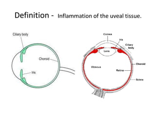
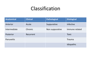
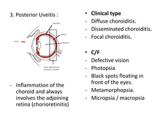
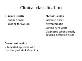
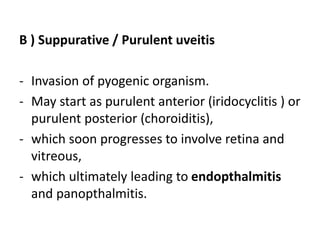
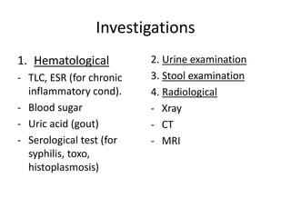
Najihah Binti Mohd Yusri (082012100064)
Definition - Inflammation of the uveal tissue.
Classification
Anatomical Clinical Pathological Etiological
Anterior Acute Suppurative Infective
Intermediate Chronic Non suppurative Immune related
Posterior Recurrent Toxic
Panuveitis Trauma
Idiopathic
Anatomical classification
1. Anterior uveitis :
• Inflammation from iris up to
pars plicata of ciliary body.
• Subdivision :
- Iritis
- Cyclitis ( inflammation of
ciliary body)
- Iridocyclitis.
• C/F
- Pain (dull aching throbbing
worse at night).
- Redness
- Photophobia
- Lacrimation
- Defective vision
• Signs
(slit lamp biomicroscopic
examination)
- Lid edema
- Circumcorneal
congestion.
- corneal sign.
- Anterior chamber sign.
- Iris sign
Loss of normal pattern,
and muddy in color,
synechiae,
• Complication
- Cataract
- Secondary glaucoma
- Cyclitic membrane
(fibrosis behind lens)
- Choroiditis
- Retinal complication
(scar, fibrosis)
• Ddx
- Acute red eye.
- Granulomatous / non
granulomatous uveitis.
TREATMENT OF ANTERIOR UVEITIS
LOCAL SYSTEMIC TREATMENT OF
COMPLICATION
• Hot compress
• Dark glassess
• Atropine 1%
eyedrop
• Corticosteroid
• Corticosteroid
(dexamethason
e)
• Analgesics
(NSAID)
• Immunosupress
ant
• Cataract – lens
extraction
• Secondary
glaucoma
(timolol,
acetazolamide
tab)
• Annular
synechiae
laser
iridotomy
2. Intermediate uveitis
- Inflammation involving
pars plana, peripheral
retina, choroid and
vitreous base.
• 80% bilateral
• Age : 2-4 decade
• 85% idiopathic
• 15%- tb, syphilis
• C/F
- Usually asymptomatic
- Later- blurry and
decrease vision.
• Signs
- Anterior segment appears
normal or little KPs, flare.
- Posterior segment shows
snowball / cotton ball
opacities (vitreous
condensation)
• Prognosis
- Good
• Complication
- 42% cataract
- Secondary glaucoma
- Vitreous hemorrhage
- Retinal detachment
• Rx
- Tropical steroid
- Immunosupressive drugs.
- Cryotherapy or indirect
laser photocoagulation
for snowbanking.
3. Posterior Uveitis :
- Inflammation of the
choroid and always
involves the adjoining
retina (chorioretinitis)
• Clinical type
- Diffuse choroiditis.
- Disseminated choroiditis.
- Focal choroiditis.
• C/F
- Defective vision
- Photopsia
- Black spots floating in
front of the eyes.
- Metamorphopsia.
- Micropsia / macropsia
• Signs
- Fundus examination
shows features of a
patch of choroiditis.
- active patch of
choriditis :
pale yellow / dirty white
raised area.
• Complication
- complicated cataract
- Vitreus degeneration
- retinal detachment
• Rx
- Corticosteroid
- Immunosupressive
- Specific treatment for
tb, syphilis
Clinical classification
• Acute uveitis
- Sudden onset
- Lasting for 6w-3m
*recurrent uveitis :
- Repeated episodes with
inactive period of >3m of rx
• Chronic uveitis
- Insidious onset
- Asymptomatic
- Lasting >3m-years
- Diagnosed when already
develop defective vision
Pathological classification
o Non suppurative
o Suppurative
A) Non-suppurative uveitis
B ) Suppurative / Purulent uveitis
- Invasion of pyogenic organism.
- May start as purulent anterior (iridocyclitis ) or
purulent posterior (choroiditis),
- which soon progresses to involve retina and
vitreous,
- which ultimately leading to endopthalmitis
and panopthalmitis.
• Endopthalmitis –
inflammation of inner
structure of eyeball
- Etiology :
 GP cocci, streptococci,
pseudomonas,
pneumococci.
 Fungal (rare)-
Aspergilus, Fusarium,
Candida.
• Panopthalmitis –
intense purulent
inflammation of whole
eyeball.
- Etiology :
 Same
• Endopthalmitis
- Modes of infection :
 Exogenous following
perforating injury,
perforated corneal ulcer,
post eye surgery.
 Endogenous tru blood
stream from caries teeth,
septicemia.
 Secondary infection
following orbital cellulitis,
thrombophlebitis,
infected corneal ulcer.
• Panopthalmitis
- Modes of infection :
 Same
• Endopthalmitis
- c/f :
 Severe ocular pain,
redness
 lacrimation,
 photophobia,
 loss of vision
• Panopthalmitis
- c/f :
 Severe ocular pain,
headache,
 complete loss of vision,
 profuse watering,
 purulent discharge,
 marked swelling and
redness of eye,
 associated fever and
malaise.
• Endopthalmitis
- Swollen lid, conjunctiva
chemosis, edematous
cornea, pupil yellow
reflex, increase iop.
• Panopthalmitis
- Lid edema and
hyperemia, eyeball
slightly proptosed, limited
ocular movement,
conjuctival congestion,
claudy cornea, absent
perception of light, raised
iop.
• Endopthalmitis
- Rx :
Antibiotic intavitreal
injection, tropical and
systemic.
Steroid
Antiglaucoma
Vitrectomy
• Panopthalmitis
- Rx :
Antiinflammatory,
analgesic
Broad spectrum antibiotic
Evisceration
- Avoid complication of
intracranial dissemination
- Remove content of
eyeball leving behind
sclera.
Etiological (Duke Elder’s) classification
1. Infective uveitis
- Bacterial (granulomatous
and non granulomatous)
- Viral (herpes simplex,
herpes zoster,CMV)
- Fungal (aspergillosis,
candidiasis, blastomycosis)
- Parasitic (toxoplasmosis,
toxocariasis, amoebiasis)
- Rickettsial (scrub typhus,
epidemic typhus)
2. Immune mediated uveitis
- Anaphylactic uveitis
- Atopic uveitis (pollen)
- Autoimmune (RA, SLE)
- HLA-associated uveitis
3. Toxic Uveitis
- Endotoxin (microbial
organism)
- Endocular toxin (from
ocular tissue)
- Exogenous toxin (certain
drugs, inorganic chemical
substance)
4. Traumatic uveitis
- Accidental / operative
injury of uveal tissue.
- Direct mechanical
trauma.
- Foreign body
5. Idiopathic
Investigations
1. Hematological
- TLC, ESR (for chronic
inflammatory cond).
- Blood sugar
- Uric acid (gout)
- Serological test (for
syphilis, toxo,
histoplasmosis)
2. Urine examination
3. Stool examination
4. Radiological
- Xray
- CT
- MRI
Reference
• AK Khurana, Review of Opthalmology, 6th ed.
Thank you

Uveitis

  • 1.
    Najihah Binti MohdYusri (082012100064)
  • 3.
    Definition - Inflammationof the uveal tissue.
  • 4.
    Classification Anatomical Clinical PathologicalEtiological Anterior Acute Suppurative Infective Intermediate Chronic Non suppurative Immune related Posterior Recurrent Toxic Panuveitis Trauma Idiopathic
  • 5.
    Anatomical classification 1. Anterioruveitis : • Inflammation from iris up to pars plicata of ciliary body. • Subdivision : - Iritis - Cyclitis ( inflammation of ciliary body) - Iridocyclitis. • C/F - Pain (dull aching throbbing worse at night). - Redness - Photophobia - Lacrimation - Defective vision
  • 6.
    • Signs (slit lampbiomicroscopic examination) - Lid edema - Circumcorneal congestion. - corneal sign. - Anterior chamber sign. - Iris sign Loss of normal pattern, and muddy in color, synechiae,
  • 7.
    • Complication - Cataract -Secondary glaucoma - Cyclitic membrane (fibrosis behind lens) - Choroiditis - Retinal complication (scar, fibrosis) • Ddx - Acute red eye. - Granulomatous / non granulomatous uveitis.
  • 8.
    TREATMENT OF ANTERIORUVEITIS LOCAL SYSTEMIC TREATMENT OF COMPLICATION • Hot compress • Dark glassess • Atropine 1% eyedrop • Corticosteroid • Corticosteroid (dexamethason e) • Analgesics (NSAID) • Immunosupress ant • Cataract – lens extraction • Secondary glaucoma (timolol, acetazolamide tab) • Annular synechiae laser iridotomy
  • 9.
    2. Intermediate uveitis -Inflammation involving pars plana, peripheral retina, choroid and vitreous base. • 80% bilateral • Age : 2-4 decade • 85% idiopathic • 15%- tb, syphilis • C/F - Usually asymptomatic - Later- blurry and decrease vision.
  • 10.
    • Signs - Anteriorsegment appears normal or little KPs, flare. - Posterior segment shows snowball / cotton ball opacities (vitreous condensation) • Prognosis - Good • Complication - 42% cataract - Secondary glaucoma - Vitreous hemorrhage - Retinal detachment • Rx - Tropical steroid - Immunosupressive drugs. - Cryotherapy or indirect laser photocoagulation for snowbanking.
  • 11.
    3. Posterior Uveitis: - Inflammation of the choroid and always involves the adjoining retina (chorioretinitis) • Clinical type - Diffuse choroiditis. - Disseminated choroiditis. - Focal choroiditis. • C/F - Defective vision - Photopsia - Black spots floating in front of the eyes. - Metamorphopsia. - Micropsia / macropsia
  • 12.
    • Signs - Fundusexamination shows features of a patch of choroiditis. - active patch of choriditis : pale yellow / dirty white raised area. • Complication - complicated cataract - Vitreus degeneration - retinal detachment • Rx - Corticosteroid - Immunosupressive - Specific treatment for tb, syphilis
  • 13.
    Clinical classification • Acuteuveitis - Sudden onset - Lasting for 6w-3m *recurrent uveitis : - Repeated episodes with inactive period of >3m of rx • Chronic uveitis - Insidious onset - Asymptomatic - Lasting >3m-years - Diagnosed when already develop defective vision
  • 14.
    Pathological classification o Nonsuppurative o Suppurative
  • 15.
  • 16.
    B ) Suppurative/ Purulent uveitis - Invasion of pyogenic organism. - May start as purulent anterior (iridocyclitis ) or purulent posterior (choroiditis), - which soon progresses to involve retina and vitreous, - which ultimately leading to endopthalmitis and panopthalmitis.
  • 17.
    • Endopthalmitis – inflammationof inner structure of eyeball - Etiology :  GP cocci, streptococci, pseudomonas, pneumococci.  Fungal (rare)- Aspergilus, Fusarium, Candida. • Panopthalmitis – intense purulent inflammation of whole eyeball. - Etiology :  Same
  • 18.
    • Endopthalmitis - Modesof infection :  Exogenous following perforating injury, perforated corneal ulcer, post eye surgery.  Endogenous tru blood stream from caries teeth, septicemia.  Secondary infection following orbital cellulitis, thrombophlebitis, infected corneal ulcer. • Panopthalmitis - Modes of infection :  Same
  • 19.
    • Endopthalmitis - c/f:  Severe ocular pain, redness  lacrimation,  photophobia,  loss of vision • Panopthalmitis - c/f :  Severe ocular pain, headache,  complete loss of vision,  profuse watering,  purulent discharge,  marked swelling and redness of eye,  associated fever and malaise.
  • 20.
    • Endopthalmitis - Swollenlid, conjunctiva chemosis, edematous cornea, pupil yellow reflex, increase iop. • Panopthalmitis - Lid edema and hyperemia, eyeball slightly proptosed, limited ocular movement, conjuctival congestion, claudy cornea, absent perception of light, raised iop.
  • 21.
    • Endopthalmitis - Rx: Antibiotic intavitreal injection, tropical and systemic. Steroid Antiglaucoma Vitrectomy • Panopthalmitis - Rx : Antiinflammatory, analgesic Broad spectrum antibiotic Evisceration - Avoid complication of intracranial dissemination - Remove content of eyeball leving behind sclera.
  • 22.
    Etiological (Duke Elder’s)classification 1. Infective uveitis - Bacterial (granulomatous and non granulomatous) - Viral (herpes simplex, herpes zoster,CMV) - Fungal (aspergillosis, candidiasis, blastomycosis) - Parasitic (toxoplasmosis, toxocariasis, amoebiasis) - Rickettsial (scrub typhus, epidemic typhus) 2. Immune mediated uveitis - Anaphylactic uveitis - Atopic uveitis (pollen) - Autoimmune (RA, SLE) - HLA-associated uveitis 3. Toxic Uveitis - Endotoxin (microbial organism) - Endocular toxin (from ocular tissue) - Exogenous toxin (certain drugs, inorganic chemical substance)
  • 23.
    4. Traumatic uveitis -Accidental / operative injury of uveal tissue. - Direct mechanical trauma. - Foreign body 5. Idiopathic
  • 24.
    Investigations 1. Hematological - TLC,ESR (for chronic inflammatory cond). - Blood sugar - Uric acid (gout) - Serological test (for syphilis, toxo, histoplasmosis) 2. Urine examination 3. Stool examination 4. Radiological - Xray - CT - MRI
  • 25.
    Reference • AK Khurana,Review of Opthalmology, 6th ed. Thank you

Editor's Notes

  • #7 Kp - Epitheloid cell and macrophages Flare – leakage of protein particles into aq humour from damaged BV Synechiae – adhere of iris and lens
  • #9 Hot compress- increase circulation, reduce stasis Dark glass – reduce photophobia, lacrimation
  • #12 Floating – large exudative clump in vitreous. Metamorphopsia – distorted images of the object. Micropsia – sepration of visual cells, macropsia – crowding together of rods and cones.
  • #17 Endopthalmitis – inf of inner structure of eyeball
  • #18 GPC – staph aureus