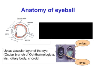
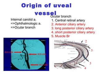
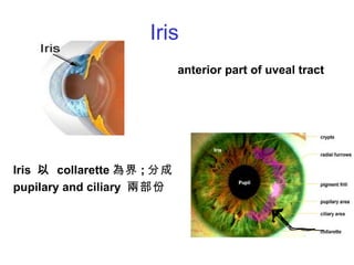
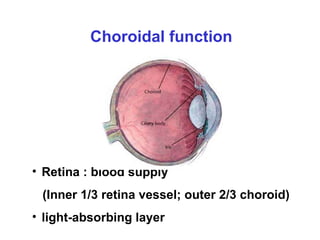
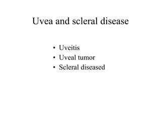
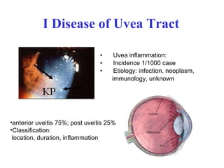
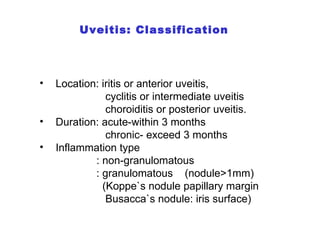
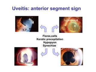
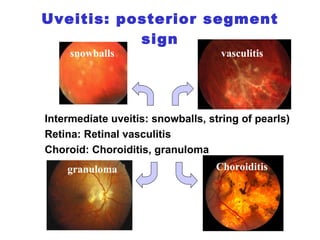
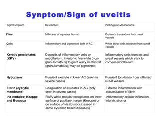
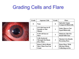
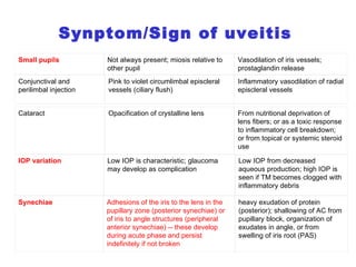
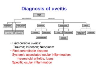
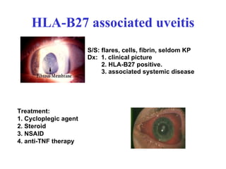
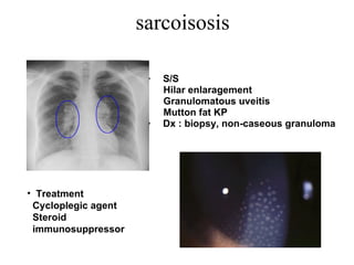
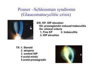
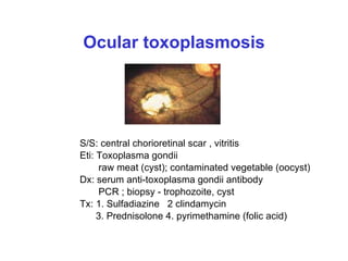
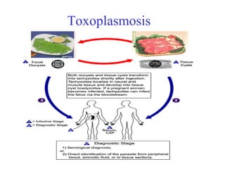
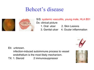
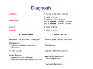
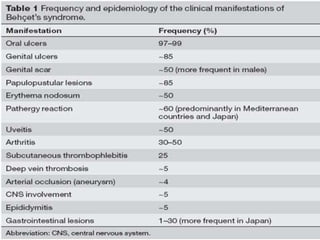
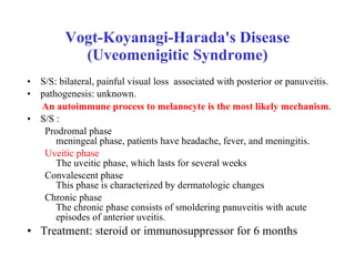
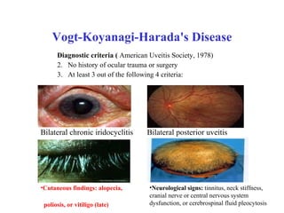
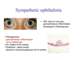
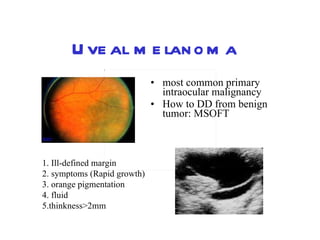
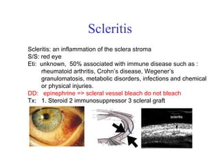
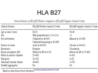
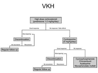
三軍總醫院  蔡明霖醫師  M.D. Ph.D.  Uveitis and Sclera
Anatomy of eyeball Uvea: vascular layer of the eye ( Ocular branch of Ophthalmologic a.) iris,  ciliary body, choroid. uvea sclera Eyeball: outer coat (cornea, sclera),  middle vascular layer (uveal tract)  inner sensory layer ( retina)
Origin of uveal vessel   Ocular branch  1. Central retinal artery  2. Anterior ciliary artery  3. long posterior ciliary artery 4. short posterior ciliary artery 5. Muscle Br   Internal carotid a. =>Ophthalmologic a. =>Ocular branch  4 3 5 2
Iris anterior part of uveal tract Iris  以  collarette 為界 ; 分成 pupilary and ciliary  兩部份
Function of iris pupil open  Iris dilator muscle  cervical sympathetic trunk pupil close Iris sphincter muscle  short ciliary nerve   (brainstem: E-W Nu )
散瞳劑 副交感神經阻斷劑 : Atropine(1%) :2 weeks 。 Scopolamine:1-3 days Homatropine:1-3 days cyclopentolate(0.5%):24 hours tropicamide (0.5%): 6 hours 。
Ciliary body middle part of uvea Function:  aqueous production accommodation
Histology Stroma : ciliary  muscle (accommodation ) circular muscle=>near ( lens thickening) radial muscle=>far (lens thinning) Epithelium: pigmented epithelium  non-pigmented epithelium (aqueous formation)
Accommodation near lens thickening  parasympathetic fiber  ciliary circular M. contraction zonular relax   Far lens thinning sympathetic fiber ciliary radial M. contraction zonular contraction
睫狀肌鬆弛劑 副交感神經阻斷劑 : Atropine(1%) :2 weeks 。 Scopolamine:1-3 days Homatropine:1-3 days cyclopentolate(0.5%):24 hours tropicamide (0.5%):6 hours 。
Choroidal function  Retina : blood supply (Inner 1/3 retina vessel; outer 2/3 choroid) light-absorbing layer
Uvea and scleral disease Uveitis Uveal tumor Scleral diseased
I Disease of Uvea Tract Uvea inflammation: Incidence 1/1000 case  Etiology: infection, neoplasm,  immunology, unknown anterior uveitis 75%; post uveitis 25% Classification: location, duration, inflammation
Uveitis: Classification Location: iritis or anterior uveitis, cyclitis or intermediate uveitis choroiditis or posterior uveitis. Duration: acute-within 3 months chronic- exceed 3 months Inflammation type  : non-granulomatous : granulomatous  (nodule>1mm)  (Koppe`s nodule papillary margin Busacca`s nodule: iris surface)
Uveitis: anterior segment sign hypopyron synethiae Flares,cells Keratic precepitation Hypopyon Synechiae   KP
Uveitis: posterior segment sign hypopyron synethiae Intermediate uveitis: snowballs, string of pearls) Retina: Retinal vasculitis Choroid: Choroiditis, granuloma snowballs vasculitis Choroiditis granuloma
Synptom/Sign of uveitis   Sign/Symptom Description Pathogenic Mechanisms Flare Milkiness of aqueous humor Protein is transudate from uveal vessels Cells Inflammatory and pigmented cells in AC White blood cells released from uveal vessels Fibrin (cyclytic membrane) Coagulation of exudates in AC (only seen in severe cases) Extreme inflammation with accumulation of fibrin  Iris nodules: Koeppe and Busacca Fluffy white nodular precipitates on inner surface of pupillary margin (Koeppe) or on surface of iris (Busacca) (seen in some systemic based diseases) Inflammatory cellular infiltration into iris stroma Keratic precipitates (KP's) Deposits of inflammatory cells on endothelium. inferiorly; fine white (non-granulomatous) to giant waxy mutton fat (granulomatous); may be pigmented Inflammatory cells from iris and uveal vessels which stick to corneal endothelium Hypopyon Purulent exudate in lower AC (seen in severe cases) Purulent Exudation from inflamed uveal vessels
Grading Cells and Flare  Dense Haze: With Obvious Fibrin Collecting on Iris 4 Dense Cells in Beam, More Than You Can Count 4 Marked: Iris Details Becoming Hazy 3 Cells Scattered Through Out Beam 20 or More 3 Moderate: But Iris Detail Still Clear 2 5-10 Cell Seen at Once 2 Faint: Haze or Not Equal Bilaterally 1 2-5 Cells Seen in 45 Seconds or One Minute 1 Optically Empty Compared Bilaterally 0 None 0 Flare Grade Aqueous Cells Grade
Synptom/Sign of uveitis   IOP variation Low IOP is characteristic; glaucoma may develop as complication Low IOP from decreased aqueous production; high IOP is seen if TM becomes clogged with inflammatory debris Synechiae Adhesions of the iris to the lens in the pupillary zone (posterior synechiae) or of iris to angle structures (peripheral anterior synechiae) -- these develop during acute phase and persist indefinitely if not broken heavy exudation of protein (posterior); shallowing of AC from pupillary block, organization of exudates in angle, or from swelling of iris root (PAS) Conjunctival and perilimbal injection Pink to violet circumlimbal episcleral vessels (ciliary flush) Inflammatory vasodilation of radial episcleral vessels Cataract Opacification of crystalline lens From nutritional deprivation of lens fibers; or as a toxic response to inflammatory cell breakdown; or from topical or systemic steroid use Small pupils Not always present; miosis relative to other pupil Vasodilation of iris vessels; prostaglandin release
Diagnosis of uveitis Find curable uveitis: Trauma; Infection; Neoplasm Find controllable disease Systemic associated ocular inflammation:  rheumatoid arthritis; lupus Specific ocular inflammation
Diagnosis of uveitis Find curable uveitis Trauma : Hx Infection:  Bacteria (complete blood count; culture) Fungus (complete blood count; culture, culture) Parasite (complete blood count; culture, eosinophil) Virus: PCR; Serology: VDRL, HIV, Torch marker  Neoplasm: CBC, image study( PET)
Diagnosis of uveitis Find controllable disease Systemic associated ocular inflammation:  ankylosing spondylitis( image study, HLA-B27) Rheumatoid arthritis ( image study; CRP) lupus( CBC; anti-ds DNA) sarcoidosis( CxR; biopsy) Behcet disease (oral ulcer) Specific ocular inflammation Vogt-Koyanagi-Harada's Disease Sympathetic ophthalmia
Uveitis treatment  Mandatory :   1. Treatment of underlying disease, if known  ( trauma, infection, n eoplasm) 2. Cycloplegic agent  3. topical steroid   Optional :   1. Periocular or systemic steroids: unresponsive  to topical steroid 2. Immunosuppressors, anti-TNF therapy sight-threatening inflammation which has been unresponsive to steroids   3. control elevated intraocular pressure
Specific ocular  uveitis Anterior uveitis HLA-B27 associated uveitis(20-40%) Sarcoidosis (15%)  Posner-Schlossman Syndrome  (1-5%) Posterior uveitis Ocular toxoplasmosis(8-40%) Behcet disease (5%) Vogt-Koyanagi-Harada's Disease Sympathetic ophthalmia
HLA-B27 associated uveitis S/S: flares, cells, fibrin, seldom KP Dx:  1. clinical picture  2. HLA-B27 positive. 3. associated   systemic disease Treatment:  1. Cycloplegic agent  2. Steroid 3. NSAID 4. anti-TNF therapy
HLA-B27 associated system disease   Treatment: control uveitis, consult: CV, RIA , REH AS :systemic anti-inflammatory agent CV consult(heart block, aortic insufficiency) Reiter syndrome: systemic antibiotic Psoriasis: analgesics,  immunosuppressive AS (55-90%) Psoriasis 3-4% Reiter syndrome (8-21% )
sarcoisosis  S/S  Hilar enlaragement  Granulomatous uveitis Mutton fat KP Dx : biopsy, non-caseous granuloma Treatment Cycloplegic agent  Steroid  immunosuppressor
Posner –Schlossman syndromn (Glaucomatocyclitic crisis) S/S: KP, IOP elevation  Eti: prostaglandin induced trabeculitis Dx: clinical criteria  1. Fine KP  2. trabeculitis  3. IOP elevation TX: 1. Steroid  2  atropine 3  control IOP  4 avoid miotic 5 avoid prostagradin
Ocular toxoplasmosis S/S: central chorioretinal scar , vitritis Eti: Toxoplasma gondii  raw meat (cyst); contaminated vegetable (oocyst) Dx: serum anti-toxoplasma gondii antibody  PCR ; biopsy - trophozoite, cyst Tx: 1. Sulfadiazine  2 clindamycin 3. Prednisolone 4. pyrimethamine (folic acid)
Toxoplasmosis
Behcet’s disease  S/S:  systemic vasculitis, young male, HLA B51   Dx: clinical picture 1. Oral  ulcer  2. Skin Lesions  3. Genital ulcer  4. Ocular inflammation  Eti:  unknown.  infection-induced autoimmune process to vessel  endothelium is the most likely mechanism.  TX:  1. Steroid  2 immunosuppressor
Diagnosis MAJOR CRITERIA   MINOR CRITERIA   Recurrent Oral Aphthous Ulcers (pain) Arthritis (heat, effuse, limitation) Skin Lesions: **Erythema nodosum-like lesions **Folliculitis Epididymitis Genital Ulcers( Gastrointestinal Involvement Ocular  Disease : **Iridocyclitis with hypopyon **Posterior Uveitis with retinal vasculitis Vascular Involevement: **Thrombophlebitis Neurologic Symptoms Complete   Presence of all major criteria Incomplete 3 major criteria 2 major + 2 minor criteria ocular  disease  + 1 major criterion ocular  disease  + 2 minor criteria Suspect 2 major criteria Possible 1 major criterion
 
Vogt-Koyanagi-Harada's Disease (Uveomenigitic Syndrome) S/S: bilateral, painful visual loss  associated with posterior or panuveitis.  pathogenesis: unknown. An autoimmune process to melanocyte is the most likely mechanism .   S/S : Prodromal phase meningeal phase, patients have headache, fever, and meningitis.  Uveitic phase The uveitic phase, which lasts for several weeks Convalescent phase This phase is characterized by dermatologic changes  Chronic phase The chronic phase consists of smoldering panuveitis with acute episodes of anterior uveitis.  Treatment: steroid or immunosuppressor for 6 months
Vogt-Koyanagi-Harada's Disease Diagnostic criteria (  American Uveitis Society, 1978) No history of ocular trauma or surgery  At least 3 out of the following 4 criteria: Bilateral chronic iridocyclitis  Bilateral posterior uveitis  Cutaneous findings: alopecia,  poliosis, or vitiligo (late) Neurological signs:  tinnitus, neck stiffness, cranial nerve or central nervous system dysfunction, or cerebrospinal fluid pleocytosis
Sympathetic ophthalmia S/S: injury in one eye , granulomatous inflammation developed in following eye   Pathogenesis:  granulomatous inflammation  to to melanocyte   Dx: Dallen-Fuch nodule Treatment : repair would  steroid or immunosuppressor for 6 months
Uveal melanoma most common primary intraocular malignancy How to DD from benign tumor: MSOFT  1. Ill-defined margin  2. symptoms (Rapid growth)  3. orange pigmentation  4. fluid  5.thinkness>2mm
II  Uveal tumor Uveal melanoma 最常見的原發性眼內惡性腫瘤 眼內最常見的腫瘤為 metastatic uveal tumor 女性最常見的的轉移性腫瘤 , 來源為乳癌 男性最常見的的轉移性腫瘤 , 來源為肺癌
Uveal melanoma Pathology:spindle cell A, spindle cell B, mixed type Treatment  Photocoagulation, brachytherapy, enucleation, cyberknife
III Sclera and scleral disease  Sclera histology : two layer Episclera,  Scleral stroma( 越後面越厚 ) Sclera: white outer covering of the eye Muscle insertion :  danger site of eyeball rupture MR 5.5; IR 6.5; LR 7.0; SR 7.8mm
Episcleritis: episcleral tissue inflammation S/S:  red eye  episcleral vessel engorgement (between the conjunctiva and the sclera) Eti: allergy, unknown,… DD: epinephrine => bleach  Tx:  steroid
Scleritis Scleritis: an inflammation of the sclera stroma S/S: red eye  Eti:  unknown,  50% associated with immune disease such as :  rheumatoid arthritis, Crohn’s disease, Wegener’s  granulomatosis, metabolic disorders, infections and chemical  or physical injuries. DD:  epinephrine => scleral vessel bleach do not bleach   Tx:  1. Steroid  2 immunosuppressor 3 scleral graft
Video for uveitis Biopsy
THANKS
Find systemic disease Diagnostic imaging  Chest x-ray (TB, sarcoidosis, histoplasmosis, tumor)  Sacroiliac films (HLA-B27, Reiter, ankylosing spondylitis)  Orbital image (tumor, foreign body, thyroid, scleritis)  Skull films (congenital toxoplasmosis)  Joint films (rheumatoid, HLA-B27, JRA, lupus, gonorrhea)  Gallium scan (sarcoidosis, infection, metastasis)
HLA B27
VKH
VKH minimum of 6 months, has been shown to improve the prognosis by reducing the length of disease, increasing the incidence of a convalescent phase, and decreasing the extraocular manifestations.
SO:  須找出傷口


Sarcoidosis The initial dose is 30 to 60 mg of prednisone daily for 8 to 12 weeks, followed by slow tapering over 6 or 12 weeks to establish the minimal effective dose.5,6 Pulsed intravenous methylprednisolone might be necessary in some cases. A maintenance dose of oral corticosteroid at 10 to 15 mg per day in alternate days should be given at least for 3 months.
HLA-B27 associated system disease   Treatment: control uveitis, consult: CV, RIA , REH AS :systemic anti-inflammatory agent CV consult(heart block, aortic insufficiency) Reiter syndrome: systemic antibiotic Psoriasis: analgesics,  immunosuppressive AS (55-90%) Psoriasis 3-4% Reiter syndrome (8-21% )
Find systemic disease Diagnostic imaging  Chest x-ray (TB, sarcoidosis, histoplasmosis, tumor)  Sacroiliac films (HLA-B27, Reiter, ankylosing spondylitis)  Orbital image (tumor, foreign body, thyroid, scleritis)  Skull films (congenital toxoplasmosis)  Joint films (rheumatoid, HLA-B27, JRA, lupus, gonorrhea)  Gallium scan (sarcoidosis, infection, metastasis)

Uvea sclera 990829

  • 1.
    三軍總醫院 蔡明霖醫師 M.D. Ph.D. Uveitis and Sclera
  • 2.
    Anatomy of eyeballUvea: vascular layer of the eye ( Ocular branch of Ophthalmologic a.) iris, ciliary body, choroid. uvea sclera Eyeball: outer coat (cornea, sclera), middle vascular layer (uveal tract) inner sensory layer ( retina)
  • 3.
    Origin of uvealvessel Ocular branch 1. Central retinal artery 2. Anterior ciliary artery 3. long posterior ciliary artery 4. short posterior ciliary artery 5. Muscle Br Internal carotid a. =>Ophthalmologic a. =>Ocular branch 4 3 5 2
  • 4.
    Iris anterior partof uveal tract Iris 以 collarette 為界 ; 分成 pupilary and ciliary 兩部份
  • 5.
    Function of irispupil open Iris dilator muscle cervical sympathetic trunk pupil close Iris sphincter muscle short ciliary nerve (brainstem: E-W Nu )
  • 6.
    散瞳劑 副交感神經阻斷劑 :Atropine(1%) :2 weeks 。 Scopolamine:1-3 days Homatropine:1-3 days cyclopentolate(0.5%):24 hours tropicamide (0.5%): 6 hours 。
  • 7.
    Ciliary body middlepart of uvea Function: aqueous production accommodation
  • 8.
    Histology Stroma :ciliary muscle (accommodation ) circular muscle=>near ( lens thickening) radial muscle=>far (lens thinning) Epithelium: pigmented epithelium non-pigmented epithelium (aqueous formation)
  • 9.
    Accommodation near lensthickening parasympathetic fiber ciliary circular M. contraction zonular relax Far lens thinning sympathetic fiber ciliary radial M. contraction zonular contraction
  • 10.
    睫狀肌鬆弛劑 副交感神經阻斷劑 :Atropine(1%) :2 weeks 。 Scopolamine:1-3 days Homatropine:1-3 days cyclopentolate(0.5%):24 hours tropicamide (0.5%):6 hours 。
  • 11.
    Choroidal function Retina : blood supply (Inner 1/3 retina vessel; outer 2/3 choroid) light-absorbing layer
  • 12.
    Uvea and scleraldisease Uveitis Uveal tumor Scleral diseased
  • 13.
    I Disease ofUvea Tract Uvea inflammation: Incidence 1/1000 case Etiology: infection, neoplasm, immunology, unknown anterior uveitis 75%; post uveitis 25% Classification: location, duration, inflammation
  • 14.
    Uveitis: Classification Location:iritis or anterior uveitis, cyclitis or intermediate uveitis choroiditis or posterior uveitis. Duration: acute-within 3 months chronic- exceed 3 months Inflammation type : non-granulomatous : granulomatous (nodule>1mm) (Koppe`s nodule papillary margin Busacca`s nodule: iris surface)
  • 15.
    Uveitis: anterior segmentsign hypopyron synethiae Flares,cells Keratic precepitation Hypopyon Synechiae KP
  • 16.
    Uveitis: posterior segmentsign hypopyron synethiae Intermediate uveitis: snowballs, string of pearls) Retina: Retinal vasculitis Choroid: Choroiditis, granuloma snowballs vasculitis Choroiditis granuloma
  • 17.
    Synptom/Sign of uveitis Sign/Symptom Description Pathogenic Mechanisms Flare Milkiness of aqueous humor Protein is transudate from uveal vessels Cells Inflammatory and pigmented cells in AC White blood cells released from uveal vessels Fibrin (cyclytic membrane) Coagulation of exudates in AC (only seen in severe cases) Extreme inflammation with accumulation of fibrin  Iris nodules: Koeppe and Busacca Fluffy white nodular precipitates on inner surface of pupillary margin (Koeppe) or on surface of iris (Busacca) (seen in some systemic based diseases) Inflammatory cellular infiltration into iris stroma Keratic precipitates (KP's) Deposits of inflammatory cells on endothelium. inferiorly; fine white (non-granulomatous) to giant waxy mutton fat (granulomatous); may be pigmented Inflammatory cells from iris and uveal vessels which stick to corneal endothelium Hypopyon Purulent exudate in lower AC (seen in severe cases) Purulent Exudation from inflamed uveal vessels
  • 18.
    Grading Cells andFlare Dense Haze: With Obvious Fibrin Collecting on Iris 4 Dense Cells in Beam, More Than You Can Count 4 Marked: Iris Details Becoming Hazy 3 Cells Scattered Through Out Beam 20 or More 3 Moderate: But Iris Detail Still Clear 2 5-10 Cell Seen at Once 2 Faint: Haze or Not Equal Bilaterally 1 2-5 Cells Seen in 45 Seconds or One Minute 1 Optically Empty Compared Bilaterally 0 None 0 Flare Grade Aqueous Cells Grade
  • 19.
    Synptom/Sign of uveitis IOP variation Low IOP is characteristic; glaucoma may develop as complication Low IOP from decreased aqueous production; high IOP is seen if TM becomes clogged with inflammatory debris Synechiae Adhesions of the iris to the lens in the pupillary zone (posterior synechiae) or of iris to angle structures (peripheral anterior synechiae) -- these develop during acute phase and persist indefinitely if not broken heavy exudation of protein (posterior); shallowing of AC from pupillary block, organization of exudates in angle, or from swelling of iris root (PAS) Conjunctival and perilimbal injection Pink to violet circumlimbal episcleral vessels (ciliary flush) Inflammatory vasodilation of radial episcleral vessels Cataract Opacification of crystalline lens From nutritional deprivation of lens fibers; or as a toxic response to inflammatory cell breakdown; or from topical or systemic steroid use Small pupils Not always present; miosis relative to other pupil Vasodilation of iris vessels; prostaglandin release
  • 20.
    Diagnosis of uveitisFind curable uveitis: Trauma; Infection; Neoplasm Find controllable disease Systemic associated ocular inflammation: rheumatoid arthritis; lupus Specific ocular inflammation
  • 21.
    Diagnosis of uveitisFind curable uveitis Trauma : Hx Infection: Bacteria (complete blood count; culture) Fungus (complete blood count; culture, culture) Parasite (complete blood count; culture, eosinophil) Virus: PCR; Serology: VDRL, HIV, Torch marker Neoplasm: CBC, image study( PET)
  • 22.
    Diagnosis of uveitisFind controllable disease Systemic associated ocular inflammation: ankylosing spondylitis( image study, HLA-B27) Rheumatoid arthritis ( image study; CRP) lupus( CBC; anti-ds DNA) sarcoidosis( CxR; biopsy) Behcet disease (oral ulcer) Specific ocular inflammation Vogt-Koyanagi-Harada's Disease Sympathetic ophthalmia
  • 23.
    Uveitis treatment Mandatory : 1. Treatment of underlying disease, if known ( trauma, infection, n eoplasm) 2. Cycloplegic agent 3. topical steroid Optional : 1. Periocular or systemic steroids: unresponsive to topical steroid 2. Immunosuppressors, anti-TNF therapy sight-threatening inflammation which has been unresponsive to steroids 3. control elevated intraocular pressure
  • 24.
    Specific ocular uveitis Anterior uveitis HLA-B27 associated uveitis(20-40%) Sarcoidosis (15%) Posner-Schlossman Syndrome (1-5%) Posterior uveitis Ocular toxoplasmosis(8-40%) Behcet disease (5%) Vogt-Koyanagi-Harada's Disease Sympathetic ophthalmia
  • 25.
    HLA-B27 associated uveitisS/S: flares, cells, fibrin, seldom KP Dx: 1. clinical picture 2. HLA-B27 positive. 3. associated systemic disease Treatment: 1. Cycloplegic agent 2. Steroid 3. NSAID 4. anti-TNF therapy
  • 26.
    HLA-B27 associated systemdisease Treatment: control uveitis, consult: CV, RIA , REH AS :systemic anti-inflammatory agent CV consult(heart block, aortic insufficiency) Reiter syndrome: systemic antibiotic Psoriasis: analgesics, immunosuppressive AS (55-90%) Psoriasis 3-4% Reiter syndrome (8-21% )
  • 27.
    sarcoisosis S/S Hilar enlaragement Granulomatous uveitis Mutton fat KP Dx : biopsy, non-caseous granuloma Treatment Cycloplegic agent Steroid immunosuppressor
  • 28.
    Posner –Schlossman syndromn(Glaucomatocyclitic crisis) S/S: KP, IOP elevation Eti: prostaglandin induced trabeculitis Dx: clinical criteria 1. Fine KP 2. trabeculitis 3. IOP elevation TX: 1. Steroid 2 atropine 3 control IOP 4 avoid miotic 5 avoid prostagradin
  • 29.
    Ocular toxoplasmosis S/S:central chorioretinal scar , vitritis Eti: Toxoplasma gondii raw meat (cyst); contaminated vegetable (oocyst) Dx: serum anti-toxoplasma gondii antibody PCR ; biopsy - trophozoite, cyst Tx: 1. Sulfadiazine 2 clindamycin 3. Prednisolone 4. pyrimethamine (folic acid)
  • 30.
  • 31.
    Behcet’s disease S/S: systemic vasculitis, young male, HLA B51 Dx: clinical picture 1. Oral ulcer 2. Skin Lesions 3. Genital ulcer 4. Ocular inflammation Eti: unknown. infection-induced autoimmune process to vessel endothelium is the most likely mechanism. TX: 1. Steroid 2 immunosuppressor
  • 32.
    Diagnosis MAJOR CRITERIA MINOR CRITERIA Recurrent Oral Aphthous Ulcers (pain) Arthritis (heat, effuse, limitation) Skin Lesions: **Erythema nodosum-like lesions **Folliculitis Epididymitis Genital Ulcers( Gastrointestinal Involvement Ocular Disease : **Iridocyclitis with hypopyon **Posterior Uveitis with retinal vasculitis Vascular Involevement: **Thrombophlebitis Neurologic Symptoms Complete   Presence of all major criteria Incomplete 3 major criteria 2 major + 2 minor criteria ocular disease + 1 major criterion ocular disease + 2 minor criteria Suspect 2 major criteria Possible 1 major criterion
  • 33.
  • 34.
    Vogt-Koyanagi-Harada's Disease (UveomenigiticSyndrome) S/S: bilateral, painful visual loss associated with posterior or panuveitis. pathogenesis: unknown. An autoimmune process to melanocyte is the most likely mechanism . S/S : Prodromal phase meningeal phase, patients have headache, fever, and meningitis. Uveitic phase The uveitic phase, which lasts for several weeks Convalescent phase This phase is characterized by dermatologic changes Chronic phase The chronic phase consists of smoldering panuveitis with acute episodes of anterior uveitis. Treatment: steroid or immunosuppressor for 6 months
  • 35.
    Vogt-Koyanagi-Harada's Disease Diagnosticcriteria ( American Uveitis Society, 1978) No history of ocular trauma or surgery At least 3 out of the following 4 criteria: Bilateral chronic iridocyclitis Bilateral posterior uveitis Cutaneous findings: alopecia, poliosis, or vitiligo (late) Neurological signs: tinnitus, neck stiffness, cranial nerve or central nervous system dysfunction, or cerebrospinal fluid pleocytosis
  • 36.
    Sympathetic ophthalmia S/S:injury in one eye , granulomatous inflammation developed in following eye Pathogenesis: granulomatous inflammation to to melanocyte Dx: Dallen-Fuch nodule Treatment : repair would steroid or immunosuppressor for 6 months
  • 37.
    Uveal melanoma mostcommon primary intraocular malignancy How to DD from benign tumor: MSOFT 1. Ill-defined margin 2. symptoms (Rapid growth) 3. orange pigmentation 4. fluid 5.thinkness>2mm
  • 38.
    II Uvealtumor Uveal melanoma 最常見的原發性眼內惡性腫瘤 眼內最常見的腫瘤為 metastatic uveal tumor 女性最常見的的轉移性腫瘤 , 來源為乳癌 男性最常見的的轉移性腫瘤 , 來源為肺癌
  • 39.
    Uveal melanoma Pathology:spindlecell A, spindle cell B, mixed type Treatment Photocoagulation, brachytherapy, enucleation, cyberknife
  • 40.
    III Sclera andscleral disease Sclera histology : two layer Episclera, Scleral stroma( 越後面越厚 ) Sclera: white outer covering of the eye Muscle insertion : danger site of eyeball rupture MR 5.5; IR 6.5; LR 7.0; SR 7.8mm
  • 41.
    Episcleritis: episcleral tissueinflammation S/S: red eye episcleral vessel engorgement (between the conjunctiva and the sclera) Eti: allergy, unknown,… DD: epinephrine => bleach Tx: steroid
  • 42.
    Scleritis Scleritis: aninflammation of the sclera stroma S/S: red eye Eti: unknown, 50% associated with immune disease such as : rheumatoid arthritis, Crohn’s disease, Wegener’s granulomatosis, metabolic disorders, infections and chemical or physical injuries. DD: epinephrine => scleral vessel bleach do not bleach Tx: 1. Steroid 2 immunosuppressor 3 scleral graft
  • 43.
  • 44.
  • 45.
    Find systemic diseaseDiagnostic imaging Chest x-ray (TB, sarcoidosis, histoplasmosis, tumor) Sacroiliac films (HLA-B27, Reiter, ankylosing spondylitis) Orbital image (tumor, foreign body, thyroid, scleritis) Skull films (congenital toxoplasmosis) Joint films (rheumatoid, HLA-B27, JRA, lupus, gonorrhea) Gallium scan (sarcoidosis, infection, metastasis)
  • 46.
  • 47.
  • 48.
    VKH minimum of6 months, has been shown to improve the prognosis by reducing the length of disease, increasing the incidence of a convalescent phase, and decreasing the extraocular manifestations.
  • 49.
  • 50.
  • 51.
  • 52.
  • 53.
  • 54.
    Sarcoidosis The initialdose is 30 to 60 mg of prednisone daily for 8 to 12 weeks, followed by slow tapering over 6 or 12 weeks to establish the minimal effective dose.5,6 Pulsed intravenous methylprednisolone might be necessary in some cases. A maintenance dose of oral corticosteroid at 10 to 15 mg per day in alternate days should be given at least for 3 months.
  • 55.
    HLA-B27 associated systemdisease Treatment: control uveitis, consult: CV, RIA , REH AS :systemic anti-inflammatory agent CV consult(heart block, aortic insufficiency) Reiter syndrome: systemic antibiotic Psoriasis: analgesics, immunosuppressive AS (55-90%) Psoriasis 3-4% Reiter syndrome (8-21% )
  • 56.
    Find systemic diseaseDiagnostic imaging Chest x-ray (TB, sarcoidosis, histoplasmosis, tumor) Sacroiliac films (HLA-B27, Reiter, ankylosing spondylitis) Orbital image (tumor, foreign body, thyroid, scleritis) Skull films (congenital toxoplasmosis) Joint films (rheumatoid, HLA-B27, JRA, lupus, gonorrhea) Gallium scan (sarcoidosis, infection, metastasis)