SlideShare a Scribd company logo
Uveitis
1.Classification of uveitis, 2. Panuveitis Endophthalmitis
Panophthalmitis, 3. Synechiae, 4. Congenital anomalies of
uvea
Maryam Fida
Optometry and Orthoptics
Uveitis
 Inflammation of uveal tissue.
 Associated inflammation of adjacent structures, such as Retina, Vitreous, Sclera
and Cornea.
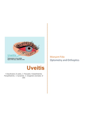
Figure 1 uveitis
Anatomical
classification
Clinical
classification
Pathological
classification
Etiological
classification
(Duke Elder’s)
Classification of uveitis
1. Anterior uveitis
Can be divided as
follow;
1) Iritis_
inflammation
mainly the iris
2) Iridocyclitis _iris
and pars plicata
involved
3) Cyclitis_ pars
plicata is affected
Acute uveitis
Onset is
sudden,
Last for less
than 3 weeks
Granulomatous
uveitis
Infective nature
Inflammation is
insidious in onset
Chronic in nature
with minimum
clinical features
Infective uveitis
2. Intermediate uveitis
Inflammation of pars
plana, peripheral retina
and choroid.
Also called as “pars
planitis”.
Chronic uveitis
Onset is
insidious
Duration is
more than 3
weeks
Non-
granulomatous
uveitis
due to allergic or
immune related
reaction
acute onset
short duration
Allergic uveitis or
immune related
uveitis
3. Posterior uveitis
Inflammation of
choroid(choroiditis)
Associated inflammation
of retina (chorioretinitis)
Recurrent
uveitis
uveitis keeps
reoccurring
periodically
Toxic uveitis
4. Panuveitis
Inflammation of whole
uveal tract
Traumatic uveitis
5. Uveitis
associated with
non-infective
systemic
diseases
6. Idiopathic uveitis
7. Neoplastic
Figure 2 anatomical classification of uveitis
Panuveitis Endophthalmitis Panophthalmitis
Inflammation of all layers
of uvea of eye
Can also affect lens,
retina, optic nerve and
vitreous causing reduced
vision or blindness.
Inflammation of internal
structures of the eye, I;e
choroid, retina and vitreous
Purulent inflammation of all
structures of eye
Including all the three
coats and Tenon’s capsule
as well.
Etiology
1. Idiopathic
After ruling out other
causes
2. Infectious
Tuberculosis
Syphilis
Lyme disease
Leptospirosis
Infectious
endophthalmitis
3. Immune related
Sarcoidosis
Vogt-koyanagi-
Harada syndrome
Sympathetic
ophthalmitis
Behcet syndrome
Etiology
Acute process 1-7 days
following intraocular
surgery such as Cataract
surgery and filtering
operation
Commonly caused by
Bacteria-staphylococcus,
pseudomonas,
pneumococcus,
streptococcus, E. coli,
Fungus -aspergillus
fumigatus, candida
albicans, fusarium,
Etiology
1.Exogenous
Due to infected wounds
Common pathogens are
pneumococcus,
staphylococcus,
pseudomonas,
pneumococcus,
streptococcus, E. coli.
2.Endogenous
Due to metastasis of
infected embolus in retinal
artery and choroidal
vessels.
Clinical Features
 Sudden onset of
unilateral pain,
redness,
photophobia
 Maybe associated
with lacrimation
 Visual acuity is
usually good at
presentation except
in eyes with severe
hypopyon.
 Low IOP
 Fibrinous exudate
Clinical Features
Bacterial
endophthalmitis
 Sudden onset with
severe pain
 Redness
 Visual loss
 Lid oedema,
chemosis, corneal
haze
 Low intraocular
tension(hypotony)
 Fibrinous exudate
or hypopyon seen in
Clinical Features
 Severe pain
 Limitation of eye
movements
 Rise in temp,
headache, vomiting
and rapid failure of
vision
 Red and swollen
lids with marked
conjunctival and
ciliary congestion
 Purulent
conjunctival
 Posterior synechiae
 Miosis
 Aqueous flare and
cells
 Endothelial dusting
anterior chamber
 Associated vitritis
and haze in
vitreous.
 Yellowish reflex
behind lens,
absence of red
reflex, inability to
fundus visualization
with indirect
ophthalmoscope
Fungal endophthalmitis
 Incubation period of
several week
 Mild pain, redness,
transient hypopyon
 Affect anterior
vitreous and
anterior uvea
 Vitreous turns into
granulomatous
mass
discharge,
conjunctival and
ciliary congestion
 Corneal wound
appears necrotic
and hypopyon
present
 Yellow reflex is
seen through pupil
(vitreous abscess)
 Fundus
examination- media
is hazy,
oedematous retina
faintly visible or
invisible.
Synechiae
abnormal adhesions of the iris to other ocular structures.
1. Anterior synechiae is an adhesion of the iris to the posterior cornea due to
abnormal fibro vascular tissue formation.
2. Posterior synechiae is an adhesion of the iris to the anterior lens capsule and/or
vitreous due to abnormal fibrovascular tissue formation or due to organization of
the fibrin rich exudates.
There can also be concurrent anterior and posterior synechiae.
Associated lesions include staphyloma (partial protrusion of the iris into the corneal
stroma), entropion uveae (posterior inversion of the pupillary margin of the iris), and
occlusion of the pupil by an abnormal fibrovascular membrane, and inflammation,
among others.
Figure 3 Synechiae
Types:
Morphologically, posterior synechiae may be segmental, annular or total.
1. Segmental posterior synechiae
adhesions of iris to lens at some points.
2. Annular posterior synechiae
adhesion of the whole rim of the iris to the anterior capsule
3. Total posterior synechiae
adhesion of the total posterior surface of the iris to the anterior
of lens. It is rarely formed in acute plastic type of Uveitis and result in deepening of
anterior chamber.
Annular Posterior
synechiae
Total posterior synechiae Festooned Pupil
Etiology:
 Infective uveitis: such as herpes simplex, herpes zoster, tuberculosis and syphilis
 Allergic (hypersensitivity) uveitis
 Toxic uveitis
 Traumatic uveitis
 Uveitis associated with non-infective systemic diseases
 Posterior synechiae are the most common ocular complications in chronic or
recurrent
 anterior uveitis, such as idiopathic anterior uveitis, and
 iridocyclitis in juvenile idiopathic arthritis, sarcoidosis, intermediate uveitis, lens-
induced
 Intraocular inflammation, especially of the iris and ciliary body.
 Synechiae can also be squeal of many ocular diseases, such as cataract,
increased
 intraocular pressure, compressive or invasive intraocular neoplasms, and
inflammation
 resulting from various causes.
 Idiopathic uveitis
Signs:
 Central iridocorneal synechiae are frequently associated with rubeotic iris vessels
 Pupil is irregular/ festooned pupil
 Synechiae associated with uveitis have signs like Keratic precipitates, anterior
chamber
 cells and flares,
 irregular pupils,
 ciliary injections,
 vitreous cells,
 iris abnormalities,
 fundal changes
Symptoms:
Acute angle closure with the classic constellation of symptoms, including
 ocular pain,
 headaches,
 blurred vision,
 photophobia,
 watering
 halos.
 Reduced vision due to corneal edema or end-stage glaucomatous optic
neuropathy.
 If associated with systemic diseases may have recurrent attacks
CONGENITAL ANOMALIES OF UVEA
1. Heterochromia iridium
One iris may have different color from the other.
Figure 4 Heterochromia iridium
2. Heterochromia iridis
Parts of one iris may have different color
Figure 5 Heterochromia iridis
3. Polycoria
There is more than one pupil.
Figure 6 Polycoria
4. Corectopia
Pupil is not central but displaced to nasal side usually
Figure 7 Corectopia
5. Aniridia
Bilateral condition, iris is absent except for a narrow rim at ciliary border. Often
leads to secondary glaucoma.
Figure 8 Aniridia
6. Persistent pupillary membrane
Incidence: Commonly seen in babies
Etiology: Persistence of part of anterior vascular sheath of lens which normally
disappears before birth.
Clinical features:
o Fine thread stretches across pupil
o Maybe attached to lens capsule
o Pigments are seen on lens surface as fine brown dots
o Not affect vision usually
Figure 9 Persistent pupillary membrane
7. Coloboma
a hole in one of the structures of the eye, such as the iris, retina, choroid, or optic
disc.
Etiology: due to deficient closure of embryonic cleft resulting in abnormal shape of iris.
Clinical features:
Iris
i. Typical_ pear shaped coloboma seen in lower part and slightly inward
Figure 10pear shaped coloboma
ii. Atypical_ defect in iris is seen in any other direction
Choroid and retina
I. Fundus examination show oval or comet-shaped defect with rounded apex
towards disc. Few vessels are seen over surface and edges.
II. Central vision defect
III. Field of vision_ scotoma is present.
Figure 11 retinal and choroidal coloboma
8. Albinism
Defective development of pigment through body
Types
a) Ocular
b) Oculocutaneous
c) Cutaneous
Symptoms
o Defective vision
o Photophobia and dazzling
o Nystagmus
o Strabismus
Signs
o Pink iris
o Fundus examination: Retinal and choroidal vessels seen with great clarity with
glistening white sclera behind them
o Partial albinism is more common. Iris is blue and pigments are absent from
choroid and retina. Macula may look normal.
Figure 12 albinism
9. Cysts
a) Serous cysts_ due to closure of iris crypts.
b) Cyst of posterior epithelium_ must be differentiated from iris bombe.
c) Implantation cyst_ occur after perforating wounds or operations.
Figure 13iris ring cysts

More Related Content

What's hot

neovascular glaucoma
neovascular glaucomaneovascular glaucoma
neovascular glaucoma
SSSIHMS-PG
 
Fungal corneal ulcer
Fungal corneal ulcerFungal corneal ulcer
Fungal corneal ulcer
drkvasantha
 
Assessment of anterior chamber
Assessment of anterior chamberAssessment of anterior chamber
Assessment of anterior chamber
LABISHETTY CHARAN
 
Papilloedema presentation1
Papilloedema presentation1Papilloedema presentation1
Papilloedema presentation1
shovon2026
 
Primary Angle Closure Glaucoma- Saral
Primary Angle Closure Glaucoma- SaralPrimary Angle Closure Glaucoma- Saral
Primary Angle Closure Glaucoma- Saral
Saral Lamichhane
 
Proptosis in ophthalmology
Proptosis  in ophthalmologyProptosis  in ophthalmology
Proptosis in ophthalmology
Dr.Juleena Kunhimohammed
 
Traumatic and complicated cataract
Traumatic and complicated cataractTraumatic and complicated cataract
Traumatic and complicated cataractSamuel Ponraj
 
Primary open angle glaucoma
Primary open angle glaucomaPrimary open angle glaucoma
Primary open angle glaucoma
ketan bhardwaj
 
Phthisis bulbi..elias t
Phthisis bulbi..elias tPhthisis bulbi..elias t
Phthisis bulbi..elias t
Elias Tadesse
 
Lens induced glaucoma - DR ARNAV
Lens induced glaucoma - DR ARNAVLens induced glaucoma - DR ARNAV
Lens induced glaucoma - DR ARNAV
DrArnavSaroya
 
Ptosis
PtosisPtosis
Corneal ulcers
Corneal ulcers Corneal ulcers
Corneal ulcers
sameep94
 
INTRAOCULAR FOREIGN BODY
INTRAOCULAR FOREIGN BODYINTRAOCULAR FOREIGN BODY
INTRAOCULAR FOREIGN BODY
Laxmi Eye Institute
 
Blunt trauma of eye
Blunt trauma of eyeBlunt trauma of eye
Blunt trauma of eye
challenger klvk
 
Iridodialysis repair
Iridodialysis repair  Iridodialysis repair
Iridodialysis repair
ArchanaShetty28
 
Macular function test
Macular function testMacular function test
Macular function test
ankita mahapatra
 
Pseudophakia
PseudophakiaPseudophakia
Pseudophakia
Mujeeb M
 
treatment of non healing corneal ulcer
treatment of non healing corneal ulcertreatment of non healing corneal ulcer
treatment of non healing corneal ulcer
ikramdr01
 

What's hot (20)

neovascular glaucoma
neovascular glaucomaneovascular glaucoma
neovascular glaucoma
 
Fungal corneal ulcer
Fungal corneal ulcerFungal corneal ulcer
Fungal corneal ulcer
 
Assessment of anterior chamber
Assessment of anterior chamberAssessment of anterior chamber
Assessment of anterior chamber
 
Papilloedema presentation1
Papilloedema presentation1Papilloedema presentation1
Papilloedema presentation1
 
Primary Angle Closure Glaucoma- Saral
Primary Angle Closure Glaucoma- SaralPrimary Angle Closure Glaucoma- Saral
Primary Angle Closure Glaucoma- Saral
 
Proptosis in ophthalmology
Proptosis  in ophthalmologyProptosis  in ophthalmology
Proptosis in ophthalmology
 
Traumatic and complicated cataract
Traumatic and complicated cataractTraumatic and complicated cataract
Traumatic and complicated cataract
 
Primary open angle glaucoma
Primary open angle glaucomaPrimary open angle glaucoma
Primary open angle glaucoma
 
Phthisis bulbi..elias t
Phthisis bulbi..elias tPhthisis bulbi..elias t
Phthisis bulbi..elias t
 
Lens induced glaucoma - DR ARNAV
Lens induced glaucoma - DR ARNAVLens induced glaucoma - DR ARNAV
Lens induced glaucoma - DR ARNAV
 
Ptosis
PtosisPtosis
Ptosis
 
Red eye
Red eyeRed eye
Red eye
 
Angle Closure Glaucoma
Angle  Closure  GlaucomaAngle  Closure  Glaucoma
Angle Closure Glaucoma
 
Corneal ulcers
Corneal ulcers Corneal ulcers
Corneal ulcers
 
INTRAOCULAR FOREIGN BODY
INTRAOCULAR FOREIGN BODYINTRAOCULAR FOREIGN BODY
INTRAOCULAR FOREIGN BODY
 
Blunt trauma of eye
Blunt trauma of eyeBlunt trauma of eye
Blunt trauma of eye
 
Iridodialysis repair
Iridodialysis repair  Iridodialysis repair
Iridodialysis repair
 
Macular function test
Macular function testMacular function test
Macular function test
 
Pseudophakia
PseudophakiaPseudophakia
Pseudophakia
 
treatment of non healing corneal ulcer
treatment of non healing corneal ulcertreatment of non healing corneal ulcer
treatment of non healing corneal ulcer
 

Similar to Uveitis (Classification, Panuveitis, Endophthalmitis, Panophthalmitis, Synechiae, Congenital anomalies of uvea)

Acute red eye
Acute red eyeAcute red eye
Acute red eye
medicostest
 
c503c8af-3c4c-4c0a-af67-840c22945426.pptx
c503c8af-3c4c-4c0a-af67-840c22945426.pptxc503c8af-3c4c-4c0a-af67-840c22945426.pptx
c503c8af-3c4c-4c0a-af67-840c22945426.pptx
MainaBidiyasar
 
Ocular manifestations of systemic diseases
Ocular manifestations of systemic diseasesOcular manifestations of systemic diseases
Ocular manifestations of systemic diseases
shovon2026
 
uveitis.pptx
uveitis.pptxuveitis.pptx
uveitis.pptx
geniousg1
 
Clinical approach to uveitis
Clinical approach to uveitisClinical approach to uveitis
Clinical approach to uveitis
SAMEEKSHA AGRAWAL
 
Red eye evaluation
Red eye evaluationRed eye evaluation
Red eye evaluation
Mosese HULKSTAH Tuapati JNR
 
Orbital cellulitis
Orbital cellulitisOrbital cellulitis
Orbital cellulitis
Ankush Weginwar
 
OSCE- Eye
OSCE- Eye OSCE- Eye
OSCE- Eye Dr. Rubz
 
1167_Anterior-Uveitis.ppt (inflammation of iris, cilliary body, choroid
1167_Anterior-Uveitis.ppt (inflammation of iris, cilliary body, choroid1167_Anterior-Uveitis.ppt (inflammation of iris, cilliary body, choroid
1167_Anterior-Uveitis.ppt (inflammation of iris, cilliary body, choroid
anusingh735179
 
Ocular manifestations of systemic diseases
Ocular manifestations of systemic diseasesOcular manifestations of systemic diseases
Ocular manifestations of systemic diseases
shovon2026
 
Uveitis
UveitisUveitis
Uveitis
Sohailislam12
 
optho help.docx
optho help.docxoptho help.docx
optho help.docx
DuniaKhaled1
 
Uveitis
UveitisUveitis
Ophthalmology 5th year, 7th lecture (Dr. Khalid)
Ophthalmology 5th year, 7th lecture (Dr. Khalid)Ophthalmology 5th year, 7th lecture (Dr. Khalid)
Ophthalmology 5th year, 7th lecture (Dr. Khalid)
College of Medicine, Sulaymaniyah
 
Ophthalmology 5th year, 5th & 6th lectures (Dr. Khalid)
Ophthalmology 5th year, 5th & 6th lectures (Dr. Khalid)Ophthalmology 5th year, 5th & 6th lectures (Dr. Khalid)
Ophthalmology 5th year, 5th & 6th lectures (Dr. Khalid)
College of Medicine, Sulaymaniyah
 

Similar to Uveitis (Classification, Panuveitis, Endophthalmitis, Panophthalmitis, Synechiae, Congenital anomalies of uvea) (20)

Acute red eye
Acute red eyeAcute red eye
Acute red eye
 
c503c8af-3c4c-4c0a-af67-840c22945426.pptx
c503c8af-3c4c-4c0a-af67-840c22945426.pptxc503c8af-3c4c-4c0a-af67-840c22945426.pptx
c503c8af-3c4c-4c0a-af67-840c22945426.pptx
 
Ocular manifestations of systemic diseases
Ocular manifestations of systemic diseasesOcular manifestations of systemic diseases
Ocular manifestations of systemic diseases
 
uveitis.pptx
uveitis.pptxuveitis.pptx
uveitis.pptx
 
Clinical approach to uveitis
Clinical approach to uveitisClinical approach to uveitis
Clinical approach to uveitis
 
Red eye evaluation
Red eye evaluationRed eye evaluation
Red eye evaluation
 
Orbital cellulitis
Orbital cellulitisOrbital cellulitis
Orbital cellulitis
 
Eye osce
Eye osceEye osce
Eye osce
 
OSCE- Eye
OSCE- Eye OSCE- Eye
OSCE- Eye
 
1167_Anterior-Uveitis.ppt (inflammation of iris, cilliary body, choroid
1167_Anterior-Uveitis.ppt (inflammation of iris, cilliary body, choroid1167_Anterior-Uveitis.ppt (inflammation of iris, cilliary body, choroid
1167_Anterior-Uveitis.ppt (inflammation of iris, cilliary body, choroid
 
Ocular manifestations of systemic diseases
Ocular manifestations of systemic diseasesOcular manifestations of systemic diseases
Ocular manifestations of systemic diseases
 
Uveitis
UveitisUveitis
Uveitis
 
optho help.docx
optho help.docxoptho help.docx
optho help.docx
 
Uveitis
UveitisUveitis
Uveitis
 
Uveitis 2lectures by dr. khaled
Uveitis 2lectures by dr. khaledUveitis 2lectures by dr. khaled
Uveitis 2lectures by dr. khaled
 
Uveitis 2lectures by dr. khaled
Uveitis 2lectures by dr. khaledUveitis 2lectures by dr. khaled
Uveitis 2lectures by dr. khaled
 
Uveitis 2lectures by dr. khaled
Uveitis 2lectures by dr. khaledUveitis 2lectures by dr. khaled
Uveitis 2lectures by dr. khaled
 
Uveitis 2lectures by dr. khaled
Uveitis 2lectures by dr. khaledUveitis 2lectures by dr. khaled
Uveitis 2lectures by dr. khaled
 
Ophthalmology 5th year, 7th lecture (Dr. Khalid)
Ophthalmology 5th year, 7th lecture (Dr. Khalid)Ophthalmology 5th year, 7th lecture (Dr. Khalid)
Ophthalmology 5th year, 7th lecture (Dr. Khalid)
 
Ophthalmology 5th year, 5th & 6th lectures (Dr. Khalid)
Ophthalmology 5th year, 5th & 6th lectures (Dr. Khalid)Ophthalmology 5th year, 5th & 6th lectures (Dr. Khalid)
Ophthalmology 5th year, 5th & 6th lectures (Dr. Khalid)
 

More from Maryam Fida

Vitreous (Attachments, age changes, vitreous hemorrhage, Vitreous Detachment)
Vitreous (Attachments, age changes, vitreous hemorrhage, Vitreous Detachment)Vitreous (Attachments, age changes, vitreous hemorrhage, Vitreous Detachment)
Vitreous (Attachments, age changes, vitreous hemorrhage, Vitreous Detachment)
Maryam Fida
 
VISUAL FIELD (Classification, neurological and glaucomatous visual fields)
VISUAL FIELD (Classification, neurological and glaucomatous visual fields)VISUAL FIELD (Classification, neurological and glaucomatous visual fields)
VISUAL FIELD (Classification, neurological and glaucomatous visual fields)
Maryam Fida
 
Management of trauma Ropper Hall classification system (Preventive ophthalmol...
Management of trauma Ropper Hall classification system (Preventive ophthalmol...Management of trauma Ropper Hall classification system (Preventive ophthalmol...
Management of trauma Ropper Hall classification system (Preventive ophthalmol...
Maryam Fida
 
Sclera (scleritis and episcleritis, staphyloma)
Sclera (scleritis and episcleritis, staphyloma)Sclera (scleritis and episcleritis, staphyloma)
Sclera (scleritis and episcleritis, staphyloma)
Maryam Fida
 
Retina (Define ,anatomy of retina, examination of retina, classification of ...
Retina (Define ,anatomy of retina, examination of  retina, classification of ...Retina (Define ,anatomy of retina, examination of  retina, classification of ...
Retina (Define ,anatomy of retina, examination of retina, classification of ...
Maryam Fida
 
Retinoblastoma (Preventive measures for retinoblastoma)
Retinoblastoma (Preventive measures for retinoblastoma)Retinoblastoma (Preventive measures for retinoblastoma)
Retinoblastoma (Preventive measures for retinoblastoma)
Maryam Fida
 
RETINOPATHY OF PREMATTURITY (ROP) PREVENTIVE MEASURES
 RETINOPATHY OF PREMATTURITY (ROP)  PREVENTIVE MEASURES RETINOPATHY OF PREMATTURITY (ROP)  PREVENTIVE MEASURES
RETINOPATHY OF PREMATTURITY (ROP) PREVENTIVE MEASURES
Maryam Fida
 
Onchocerciasis
OnchocerciasisOnchocerciasis
Onchocerciasis
Maryam Fida
 
Primary, Secondary and Tertiary Eye Care Services
Primary, Secondary and Tertiary Eye Care ServicesPrimary, Secondary and Tertiary Eye Care Services
Primary, Secondary and Tertiary Eye Care Services
Maryam Fida
 
Optometry and Orthoptics (Laws and Acts)
Optometry and Orthoptics (Laws and Acts)Optometry and Orthoptics (Laws and Acts)
Optometry and Orthoptics (Laws and Acts)
Maryam Fida
 
IOP measurements
IOP measurementsIOP measurements
IOP measurements
Maryam Fida
 
Keratoconus
KeratoconusKeratoconus
Keratoconus
Maryam Fida
 
Corneal Diseases.docx
Corneal Diseases.docxCorneal Diseases.docx
Corneal Diseases.docx
Maryam Fida
 
Glaucoma Preventive Measurements.pptx
Glaucoma Preventive Measurements.pptxGlaucoma Preventive Measurements.pptx
Glaucoma Preventive Measurements.pptx
Maryam Fida
 
COMMUNITY OPHTHALMOLOGY.pptx
COMMUNITY OPHTHALMOLOGY.pptxCOMMUNITY OPHTHALMOLOGY.pptx
COMMUNITY OPHTHALMOLOGY.pptx
Maryam Fida
 
contact lens solution
contact lens solutioncontact lens solution
contact lens solution
Maryam Fida
 
Contact Lens Deposits, Contact lens Aftercare, Overview of care and Maintenance
Contact Lens Deposits, Contact lens Aftercare, Overview of care and MaintenanceContact Lens Deposits, Contact lens Aftercare, Overview of care and Maintenance
Contact Lens Deposits, Contact lens Aftercare, Overview of care and Maintenance
Maryam Fida
 
Contact Lens Intro.ppt
Contact Lens Intro.pptContact Lens Intro.ppt
Contact Lens Intro.ppt
Maryam Fida
 
Intro databases (Table, Record, Field)
Intro databases (Table, Record, Field)Intro databases (Table, Record, Field)
Intro databases (Table, Record, Field)
Maryam Fida
 
Internet, Intranet and Extranet
Internet, Intranet and Extranet Internet, Intranet and Extranet
Internet, Intranet and Extranet
Maryam Fida
 

More from Maryam Fida (20)

Vitreous (Attachments, age changes, vitreous hemorrhage, Vitreous Detachment)
Vitreous (Attachments, age changes, vitreous hemorrhage, Vitreous Detachment)Vitreous (Attachments, age changes, vitreous hemorrhage, Vitreous Detachment)
Vitreous (Attachments, age changes, vitreous hemorrhage, Vitreous Detachment)
 
VISUAL FIELD (Classification, neurological and glaucomatous visual fields)
VISUAL FIELD (Classification, neurological and glaucomatous visual fields)VISUAL FIELD (Classification, neurological and glaucomatous visual fields)
VISUAL FIELD (Classification, neurological and glaucomatous visual fields)
 
Management of trauma Ropper Hall classification system (Preventive ophthalmol...
Management of trauma Ropper Hall classification system (Preventive ophthalmol...Management of trauma Ropper Hall classification system (Preventive ophthalmol...
Management of trauma Ropper Hall classification system (Preventive ophthalmol...
 
Sclera (scleritis and episcleritis, staphyloma)
Sclera (scleritis and episcleritis, staphyloma)Sclera (scleritis and episcleritis, staphyloma)
Sclera (scleritis and episcleritis, staphyloma)
 
Retina (Define ,anatomy of retina, examination of retina, classification of ...
Retina (Define ,anatomy of retina, examination of  retina, classification of ...Retina (Define ,anatomy of retina, examination of  retina, classification of ...
Retina (Define ,anatomy of retina, examination of retina, classification of ...
 
Retinoblastoma (Preventive measures for retinoblastoma)
Retinoblastoma (Preventive measures for retinoblastoma)Retinoblastoma (Preventive measures for retinoblastoma)
Retinoblastoma (Preventive measures for retinoblastoma)
 
RETINOPATHY OF PREMATTURITY (ROP) PREVENTIVE MEASURES
 RETINOPATHY OF PREMATTURITY (ROP)  PREVENTIVE MEASURES RETINOPATHY OF PREMATTURITY (ROP)  PREVENTIVE MEASURES
RETINOPATHY OF PREMATTURITY (ROP) PREVENTIVE MEASURES
 
Onchocerciasis
OnchocerciasisOnchocerciasis
Onchocerciasis
 
Primary, Secondary and Tertiary Eye Care Services
Primary, Secondary and Tertiary Eye Care ServicesPrimary, Secondary and Tertiary Eye Care Services
Primary, Secondary and Tertiary Eye Care Services
 
Optometry and Orthoptics (Laws and Acts)
Optometry and Orthoptics (Laws and Acts)Optometry and Orthoptics (Laws and Acts)
Optometry and Orthoptics (Laws and Acts)
 
IOP measurements
IOP measurementsIOP measurements
IOP measurements
 
Keratoconus
KeratoconusKeratoconus
Keratoconus
 
Corneal Diseases.docx
Corneal Diseases.docxCorneal Diseases.docx
Corneal Diseases.docx
 
Glaucoma Preventive Measurements.pptx
Glaucoma Preventive Measurements.pptxGlaucoma Preventive Measurements.pptx
Glaucoma Preventive Measurements.pptx
 
COMMUNITY OPHTHALMOLOGY.pptx
COMMUNITY OPHTHALMOLOGY.pptxCOMMUNITY OPHTHALMOLOGY.pptx
COMMUNITY OPHTHALMOLOGY.pptx
 
contact lens solution
contact lens solutioncontact lens solution
contact lens solution
 
Contact Lens Deposits, Contact lens Aftercare, Overview of care and Maintenance
Contact Lens Deposits, Contact lens Aftercare, Overview of care and MaintenanceContact Lens Deposits, Contact lens Aftercare, Overview of care and Maintenance
Contact Lens Deposits, Contact lens Aftercare, Overview of care and Maintenance
 
Contact Lens Intro.ppt
Contact Lens Intro.pptContact Lens Intro.ppt
Contact Lens Intro.ppt
 
Intro databases (Table, Record, Field)
Intro databases (Table, Record, Field)Intro databases (Table, Record, Field)
Intro databases (Table, Record, Field)
 
Internet, Intranet and Extranet
Internet, Intranet and Extranet Internet, Intranet and Extranet
Internet, Intranet and Extranet
 

Recently uploaded

Operation Blue Star - Saka Neela Tara
Operation Blue Star   -  Saka Neela TaraOperation Blue Star   -  Saka Neela Tara
Operation Blue Star - Saka Neela Tara
Balvir Singh
 
The Roman Empire A Historical Colossus.pdf
The Roman Empire A Historical Colossus.pdfThe Roman Empire A Historical Colossus.pdf
The Roman Empire A Historical Colossus.pdf
kaushalkr1407
 
How to Create Map Views in the Odoo 17 ERP
How to Create Map Views in the Odoo 17 ERPHow to Create Map Views in the Odoo 17 ERP
How to Create Map Views in the Odoo 17 ERP
Celine George
 
How to Make a Field invisible in Odoo 17
How to Make a Field invisible in Odoo 17How to Make a Field invisible in Odoo 17
How to Make a Field invisible in Odoo 17
Celine George
 
Home assignment II on Spectroscopy 2024 Answers.pdf
Home assignment II on Spectroscopy 2024 Answers.pdfHome assignment II on Spectroscopy 2024 Answers.pdf
Home assignment II on Spectroscopy 2024 Answers.pdf
Tamralipta Mahavidyalaya
 
PART A. Introduction to Costumer Service
PART A. Introduction to Costumer ServicePART A. Introduction to Costumer Service
PART A. Introduction to Costumer Service
PedroFerreira53928
 
Additional Benefits for Employee Website.pdf
Additional Benefits for Employee Website.pdfAdditional Benefits for Employee Website.pdf
Additional Benefits for Employee Website.pdf
joachimlavalley1
 
Ethnobotany and Ethnopharmacology ......
Ethnobotany and Ethnopharmacology ......Ethnobotany and Ethnopharmacology ......
Ethnobotany and Ethnopharmacology ......
Ashokrao Mane college of Pharmacy Peth-Vadgaon
 
TESDA TM1 REVIEWER FOR NATIONAL ASSESSMENT WRITTEN AND ORAL QUESTIONS WITH A...
TESDA TM1 REVIEWER  FOR NATIONAL ASSESSMENT WRITTEN AND ORAL QUESTIONS WITH A...TESDA TM1 REVIEWER  FOR NATIONAL ASSESSMENT WRITTEN AND ORAL QUESTIONS WITH A...
TESDA TM1 REVIEWER FOR NATIONAL ASSESSMENT WRITTEN AND ORAL QUESTIONS WITH A...
EugeneSaldivar
 
Supporting (UKRI) OA monographs at Salford.pptx
Supporting (UKRI) OA monographs at Salford.pptxSupporting (UKRI) OA monographs at Salford.pptx
Supporting (UKRI) OA monographs at Salford.pptx
Jisc
 
CLASS 11 CBSE B.St Project AIDS TO TRADE - INSURANCE
CLASS 11 CBSE B.St Project AIDS TO TRADE - INSURANCECLASS 11 CBSE B.St Project AIDS TO TRADE - INSURANCE
CLASS 11 CBSE B.St Project AIDS TO TRADE - INSURANCE
BhavyaRajput3
 
How to Break the cycle of negative Thoughts
How to Break the cycle of negative ThoughtsHow to Break the cycle of negative Thoughts
How to Break the cycle of negative Thoughts
Col Mukteshwar Prasad
 
Unit 2- Research Aptitude (UGC NET Paper I).pdf
Unit 2- Research Aptitude (UGC NET Paper I).pdfUnit 2- Research Aptitude (UGC NET Paper I).pdf
Unit 2- Research Aptitude (UGC NET Paper I).pdf
Thiyagu K
 
Synthetic Fiber Construction in lab .pptx
Synthetic Fiber Construction in lab .pptxSynthetic Fiber Construction in lab .pptx
Synthetic Fiber Construction in lab .pptx
Pavel ( NSTU)
 
How to Split Bills in the Odoo 17 POS Module
How to Split Bills in the Odoo 17 POS ModuleHow to Split Bills in the Odoo 17 POS Module
How to Split Bills in the Odoo 17 POS Module
Celine George
 
Unit 8 - Information and Communication Technology (Paper I).pdf
Unit 8 - Information and Communication Technology (Paper I).pdfUnit 8 - Information and Communication Technology (Paper I).pdf
Unit 8 - Information and Communication Technology (Paper I).pdf
Thiyagu K
 
1.4 modern child centered education - mahatma gandhi-2.pptx
1.4 modern child centered education - mahatma gandhi-2.pptx1.4 modern child centered education - mahatma gandhi-2.pptx
1.4 modern child centered education - mahatma gandhi-2.pptx
JosvitaDsouza2
 
aaaaaaaaaaaaaaaaaaaaaaaaaaaaaaaaaaaaaaaaa
aaaaaaaaaaaaaaaaaaaaaaaaaaaaaaaaaaaaaaaaaaaaaaaaaaaaaaaaaaaaaaaaaaaaaaaaaaaaaaaaaa
aaaaaaaaaaaaaaaaaaaaaaaaaaaaaaaaaaaaaaaaa
siemaillard
 
Language Across the Curriculm LAC B.Ed.
Language Across the  Curriculm LAC B.Ed.Language Across the  Curriculm LAC B.Ed.
Language Across the Curriculm LAC B.Ed.
Atul Kumar Singh
 
2024.06.01 Introducing a competency framework for languag learning materials ...
2024.06.01 Introducing a competency framework for languag learning materials ...2024.06.01 Introducing a competency framework for languag learning materials ...
2024.06.01 Introducing a competency framework for languag learning materials ...
Sandy Millin
 

Recently uploaded (20)

Operation Blue Star - Saka Neela Tara
Operation Blue Star   -  Saka Neela TaraOperation Blue Star   -  Saka Neela Tara
Operation Blue Star - Saka Neela Tara
 
The Roman Empire A Historical Colossus.pdf
The Roman Empire A Historical Colossus.pdfThe Roman Empire A Historical Colossus.pdf
The Roman Empire A Historical Colossus.pdf
 
How to Create Map Views in the Odoo 17 ERP
How to Create Map Views in the Odoo 17 ERPHow to Create Map Views in the Odoo 17 ERP
How to Create Map Views in the Odoo 17 ERP
 
How to Make a Field invisible in Odoo 17
How to Make a Field invisible in Odoo 17How to Make a Field invisible in Odoo 17
How to Make a Field invisible in Odoo 17
 
Home assignment II on Spectroscopy 2024 Answers.pdf
Home assignment II on Spectroscopy 2024 Answers.pdfHome assignment II on Spectroscopy 2024 Answers.pdf
Home assignment II on Spectroscopy 2024 Answers.pdf
 
PART A. Introduction to Costumer Service
PART A. Introduction to Costumer ServicePART A. Introduction to Costumer Service
PART A. Introduction to Costumer Service
 
Additional Benefits for Employee Website.pdf
Additional Benefits for Employee Website.pdfAdditional Benefits for Employee Website.pdf
Additional Benefits for Employee Website.pdf
 
Ethnobotany and Ethnopharmacology ......
Ethnobotany and Ethnopharmacology ......Ethnobotany and Ethnopharmacology ......
Ethnobotany and Ethnopharmacology ......
 
TESDA TM1 REVIEWER FOR NATIONAL ASSESSMENT WRITTEN AND ORAL QUESTIONS WITH A...
TESDA TM1 REVIEWER  FOR NATIONAL ASSESSMENT WRITTEN AND ORAL QUESTIONS WITH A...TESDA TM1 REVIEWER  FOR NATIONAL ASSESSMENT WRITTEN AND ORAL QUESTIONS WITH A...
TESDA TM1 REVIEWER FOR NATIONAL ASSESSMENT WRITTEN AND ORAL QUESTIONS WITH A...
 
Supporting (UKRI) OA monographs at Salford.pptx
Supporting (UKRI) OA monographs at Salford.pptxSupporting (UKRI) OA monographs at Salford.pptx
Supporting (UKRI) OA monographs at Salford.pptx
 
CLASS 11 CBSE B.St Project AIDS TO TRADE - INSURANCE
CLASS 11 CBSE B.St Project AIDS TO TRADE - INSURANCECLASS 11 CBSE B.St Project AIDS TO TRADE - INSURANCE
CLASS 11 CBSE B.St Project AIDS TO TRADE - INSURANCE
 
How to Break the cycle of negative Thoughts
How to Break the cycle of negative ThoughtsHow to Break the cycle of negative Thoughts
How to Break the cycle of negative Thoughts
 
Unit 2- Research Aptitude (UGC NET Paper I).pdf
Unit 2- Research Aptitude (UGC NET Paper I).pdfUnit 2- Research Aptitude (UGC NET Paper I).pdf
Unit 2- Research Aptitude (UGC NET Paper I).pdf
 
Synthetic Fiber Construction in lab .pptx
Synthetic Fiber Construction in lab .pptxSynthetic Fiber Construction in lab .pptx
Synthetic Fiber Construction in lab .pptx
 
How to Split Bills in the Odoo 17 POS Module
How to Split Bills in the Odoo 17 POS ModuleHow to Split Bills in the Odoo 17 POS Module
How to Split Bills in the Odoo 17 POS Module
 
Unit 8 - Information and Communication Technology (Paper I).pdf
Unit 8 - Information and Communication Technology (Paper I).pdfUnit 8 - Information and Communication Technology (Paper I).pdf
Unit 8 - Information and Communication Technology (Paper I).pdf
 
1.4 modern child centered education - mahatma gandhi-2.pptx
1.4 modern child centered education - mahatma gandhi-2.pptx1.4 modern child centered education - mahatma gandhi-2.pptx
1.4 modern child centered education - mahatma gandhi-2.pptx
 
aaaaaaaaaaaaaaaaaaaaaaaaaaaaaaaaaaaaaaaaa
aaaaaaaaaaaaaaaaaaaaaaaaaaaaaaaaaaaaaaaaaaaaaaaaaaaaaaaaaaaaaaaaaaaaaaaaaaaaaaaaaa
aaaaaaaaaaaaaaaaaaaaaaaaaaaaaaaaaaaaaaaaa
 
Language Across the Curriculm LAC B.Ed.
Language Across the  Curriculm LAC B.Ed.Language Across the  Curriculm LAC B.Ed.
Language Across the Curriculm LAC B.Ed.
 
2024.06.01 Introducing a competency framework for languag learning materials ...
2024.06.01 Introducing a competency framework for languag learning materials ...2024.06.01 Introducing a competency framework for languag learning materials ...
2024.06.01 Introducing a competency framework for languag learning materials ...
 

Uveitis (Classification, Panuveitis, Endophthalmitis, Panophthalmitis, Synechiae, Congenital anomalies of uvea)

  • 1. Uveitis 1.Classification of uveitis, 2. Panuveitis Endophthalmitis Panophthalmitis, 3. Synechiae, 4. Congenital anomalies of uvea Maryam Fida Optometry and Orthoptics
  • 2. Uveitis  Inflammation of uveal tissue.  Associated inflammation of adjacent structures, such as Retina, Vitreous, Sclera and Cornea. Figure 1 uveitis Anatomical classification Clinical classification Pathological classification Etiological classification (Duke Elder’s) Classification of uveitis
  • 3. 1. Anterior uveitis Can be divided as follow; 1) Iritis_ inflammation mainly the iris 2) Iridocyclitis _iris and pars plicata involved 3) Cyclitis_ pars plicata is affected Acute uveitis Onset is sudden, Last for less than 3 weeks Granulomatous uveitis Infective nature Inflammation is insidious in onset Chronic in nature with minimum clinical features Infective uveitis 2. Intermediate uveitis Inflammation of pars plana, peripheral retina and choroid. Also called as “pars planitis”. Chronic uveitis Onset is insidious Duration is more than 3 weeks Non- granulomatous uveitis due to allergic or immune related reaction acute onset short duration Allergic uveitis or immune related uveitis 3. Posterior uveitis Inflammation of choroid(choroiditis) Associated inflammation of retina (chorioretinitis) Recurrent uveitis uveitis keeps reoccurring periodically Toxic uveitis 4. Panuveitis Inflammation of whole uveal tract Traumatic uveitis 5. Uveitis associated with non-infective systemic diseases 6. Idiopathic uveitis 7. Neoplastic
  • 4. Figure 2 anatomical classification of uveitis
  • 5. Panuveitis Endophthalmitis Panophthalmitis Inflammation of all layers of uvea of eye Can also affect lens, retina, optic nerve and vitreous causing reduced vision or blindness. Inflammation of internal structures of the eye, I;e choroid, retina and vitreous Purulent inflammation of all structures of eye Including all the three coats and Tenon’s capsule as well. Etiology 1. Idiopathic After ruling out other causes 2. Infectious Tuberculosis Syphilis Lyme disease Leptospirosis Infectious endophthalmitis 3. Immune related Sarcoidosis Vogt-koyanagi- Harada syndrome Sympathetic ophthalmitis Behcet syndrome Etiology Acute process 1-7 days following intraocular surgery such as Cataract surgery and filtering operation Commonly caused by Bacteria-staphylococcus, pseudomonas, pneumococcus, streptococcus, E. coli, Fungus -aspergillus fumigatus, candida albicans, fusarium, Etiology 1.Exogenous Due to infected wounds Common pathogens are pneumococcus, staphylococcus, pseudomonas, pneumococcus, streptococcus, E. coli. 2.Endogenous Due to metastasis of infected embolus in retinal artery and choroidal vessels. Clinical Features  Sudden onset of unilateral pain, redness, photophobia  Maybe associated with lacrimation  Visual acuity is usually good at presentation except in eyes with severe hypopyon.  Low IOP  Fibrinous exudate Clinical Features Bacterial endophthalmitis  Sudden onset with severe pain  Redness  Visual loss  Lid oedema, chemosis, corneal haze  Low intraocular tension(hypotony)  Fibrinous exudate or hypopyon seen in Clinical Features  Severe pain  Limitation of eye movements  Rise in temp, headache, vomiting and rapid failure of vision  Red and swollen lids with marked conjunctival and ciliary congestion  Purulent conjunctival
  • 6.  Posterior synechiae  Miosis  Aqueous flare and cells  Endothelial dusting anterior chamber  Associated vitritis and haze in vitreous.  Yellowish reflex behind lens, absence of red reflex, inability to fundus visualization with indirect ophthalmoscope Fungal endophthalmitis  Incubation period of several week  Mild pain, redness, transient hypopyon  Affect anterior vitreous and anterior uvea  Vitreous turns into granulomatous mass discharge, conjunctival and ciliary congestion  Corneal wound appears necrotic and hypopyon present  Yellow reflex is seen through pupil (vitreous abscess)  Fundus examination- media is hazy, oedematous retina faintly visible or invisible.
  • 7. Synechiae abnormal adhesions of the iris to other ocular structures. 1. Anterior synechiae is an adhesion of the iris to the posterior cornea due to abnormal fibro vascular tissue formation. 2. Posterior synechiae is an adhesion of the iris to the anterior lens capsule and/or vitreous due to abnormal fibrovascular tissue formation or due to organization of the fibrin rich exudates. There can also be concurrent anterior and posterior synechiae. Associated lesions include staphyloma (partial protrusion of the iris into the corneal stroma), entropion uveae (posterior inversion of the pupillary margin of the iris), and occlusion of the pupil by an abnormal fibrovascular membrane, and inflammation, among others. Figure 3 Synechiae Types: Morphologically, posterior synechiae may be segmental, annular or total. 1. Segmental posterior synechiae adhesions of iris to lens at some points. 2. Annular posterior synechiae adhesion of the whole rim of the iris to the anterior capsule 3. Total posterior synechiae adhesion of the total posterior surface of the iris to the anterior of lens. It is rarely formed in acute plastic type of Uveitis and result in deepening of anterior chamber.
  • 8. Annular Posterior synechiae Total posterior synechiae Festooned Pupil Etiology:  Infective uveitis: such as herpes simplex, herpes zoster, tuberculosis and syphilis  Allergic (hypersensitivity) uveitis  Toxic uveitis  Traumatic uveitis  Uveitis associated with non-infective systemic diseases  Posterior synechiae are the most common ocular complications in chronic or recurrent  anterior uveitis, such as idiopathic anterior uveitis, and  iridocyclitis in juvenile idiopathic arthritis, sarcoidosis, intermediate uveitis, lens- induced  Intraocular inflammation, especially of the iris and ciliary body.  Synechiae can also be squeal of many ocular diseases, such as cataract, increased  intraocular pressure, compressive or invasive intraocular neoplasms, and inflammation  resulting from various causes.  Idiopathic uveitis Signs:  Central iridocorneal synechiae are frequently associated with rubeotic iris vessels  Pupil is irregular/ festooned pupil  Synechiae associated with uveitis have signs like Keratic precipitates, anterior chamber  cells and flares,  irregular pupils,  ciliary injections,  vitreous cells,  iris abnormalities,  fundal changes
  • 9. Symptoms: Acute angle closure with the classic constellation of symptoms, including  ocular pain,  headaches,  blurred vision,  photophobia,  watering  halos.  Reduced vision due to corneal edema or end-stage glaucomatous optic neuropathy.  If associated with systemic diseases may have recurrent attacks CONGENITAL ANOMALIES OF UVEA 1. Heterochromia iridium One iris may have different color from the other. Figure 4 Heterochromia iridium 2. Heterochromia iridis Parts of one iris may have different color Figure 5 Heterochromia iridis 3. Polycoria There is more than one pupil.
  • 10. Figure 6 Polycoria 4. Corectopia Pupil is not central but displaced to nasal side usually Figure 7 Corectopia 5. Aniridia Bilateral condition, iris is absent except for a narrow rim at ciliary border. Often leads to secondary glaucoma. Figure 8 Aniridia 6. Persistent pupillary membrane Incidence: Commonly seen in babies
  • 11. Etiology: Persistence of part of anterior vascular sheath of lens which normally disappears before birth. Clinical features: o Fine thread stretches across pupil o Maybe attached to lens capsule o Pigments are seen on lens surface as fine brown dots o Not affect vision usually Figure 9 Persistent pupillary membrane 7. Coloboma a hole in one of the structures of the eye, such as the iris, retina, choroid, or optic disc. Etiology: due to deficient closure of embryonic cleft resulting in abnormal shape of iris. Clinical features: Iris i. Typical_ pear shaped coloboma seen in lower part and slightly inward Figure 10pear shaped coloboma ii. Atypical_ defect in iris is seen in any other direction
  • 12. Choroid and retina I. Fundus examination show oval or comet-shaped defect with rounded apex towards disc. Few vessels are seen over surface and edges. II. Central vision defect III. Field of vision_ scotoma is present. Figure 11 retinal and choroidal coloboma 8. Albinism Defective development of pigment through body Types a) Ocular b) Oculocutaneous c) Cutaneous Symptoms o Defective vision o Photophobia and dazzling o Nystagmus o Strabismus Signs o Pink iris o Fundus examination: Retinal and choroidal vessels seen with great clarity with glistening white sclera behind them o Partial albinism is more common. Iris is blue and pigments are absent from choroid and retina. Macula may look normal.
  • 13. Figure 12 albinism 9. Cysts a) Serous cysts_ due to closure of iris crypts. b) Cyst of posterior epithelium_ must be differentiated from iris bombe. c) Implantation cyst_ occur after perforating wounds or operations. Figure 13iris ring cysts