SlideShare a Scribd company logo
THE EXCRETORY
SYSTEM
By:
Mrs.Keerthi.K
Asst.Professor
Vijay Marie CON
INTRODUCTION
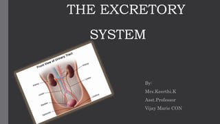
• The urinary system consists of 2 kidneys, 2 ureters one urinary
bladder and one urethra.
• Once the kidneys filter the blood plasma, they return most of the
water and solutes to the blood stream. The remaining water and
solutes constitute URINE which passes through ureters and is
stored in bladder until it is excreted from body through urethra.
• NEPHROLOGY: scientific study of anatomy , physiology and
pathology of kidneys.
• UROLOGY: branch of medicine that deals with male and female
urinary
FUNCTIONS OF KIDNEYS
Regulation of blood ionic composition
Regulation of blood PH
Regulation of blood volume
Regulation of BP
Maintenance of blood osmolarity
Production of hormones
Regulation of blood glucose levels
Excretion of wastes and foreign substances
1.BLOOD IONIC COMPOSITION
• Regulates the levels of several
ions , mostly Na+,ca+2,cl-,Hpo4-
3.REGULATION OF BLOOD VOLUME:
• Kidneys adjust blood volume by
conserving or eliminating water in
urine.
• Increase in blood volume increases
blood pressure and decrease in blood
volume decreases blood pressure.
FUNCTIONS OF KIDNEYS
2.REGULATION OF BLOOD PH:
• They excrete a variable amount of
H+into the urine and conserve Hco3-
which is an important buffer of H+
ion balance.
• Both these activities helps to regulate
PH
4. REGULATION OF BP:
• They secrete renin a hormone which
activates Renin-Angiotensin-
Aldosterone pathway.
• Increased renin causes increased BP
6.PRODUCTION OF HORMONES:
1.CALCITRIOL: active form of vitamin
D that regulates calcium homeostasis.
2. ERYTHROPOIETIN: helps in
production of RBC’s.
FUNCTIONS OF KIDNEYS
5.MAINTAIN BLOOD OSMOLARITY
• By separately regulating loss of
water and loss of solutes in urine.
Kidneys maintain blood osmolarity
to 300 mosm/lit.
7. REGULATION OF BLOOD GLUCOSE
LEVELS:
• Like liver , kidneys can use amino acid
glutamine in gluconeogenesis which
releases glucose into blood to maintain
normal blood glucose levels.
8. EXCRETION OF WASTES AND FOREIGN
SUBSTANCES:
• By forming urine kidneys help excrete wastes. Some
wastes are formed from metabolic reactions in body-
NH4 and urea( from deamination of aminoacids).
• BILIRUBIN: from catabolism of hemoglobin.
• CREATININE: breakdown of creatinine phosphate
in muscle fibers.
• URIC ACID: catabolism of nucleic acids.
• OTHER WASTES: metabolism of diet , drugs and
environmental toxins
FUNCTIONS OF KIDNEYS
• GROSS APPEARANCE: paired, reddish and bean shaped.
• LOCATION: located just above the waist between peritoneum
and posterior wall of abdomen.
• Because their position is posterior to peritoneum of abdominal
cavity – hence they are called RETROPERITONEAL ORGANS.
• Located between levels of last thoracic and 3rd lumbar
vertebrae protected by 11th and 12th pair of ribs.
• Right kidney is slightly lower than the left because of the
presence of liver
ANATOMY OF KIDNEYS
• LENGTH : 10-12cms
• WIDTH : 5-7cms
• THICKNESS: 3cms and size of bar soap
• WEIGHT: 135-150grams
• THE CONCAVE BOARDER: faces the vertebral column. Near
the center of the concave boarder there is an indentation
called HILUM/RENAL HILUS through which ureter emerges
along with blood vessels, lymphatic vessels and nerves.
ANATOMY OF KIDNEYS-EXTERNAL
• Inner layer.
• Smooth
transparent sheet
of dense irregular
connective tissue.
• Barrier against
trauma and helps
to maintain shape
of the kidney.
RENAL
CAPSULE
• Middle layer.
• Mass of fatty
tissue
surrounding the
renal capsule.
• Protects from
trauma and
holds kidney in
place within
abdominal cavity
ADIPOSE
CAPSULE
• Superficial layer
• Thin layer of
dense irregular
connective tissue
that anchors
kidney to
surrounding
structures
RENAL
FASCIA
ANATOMY OF KIDNEYS-EXTERNAL
• A frontal section through kidney reveals 2 distinct
regions:
 RENAL CORTEX- superficial light red area
 RENAL MEDULLA – darker reddish brown inner
region
• The renal medulla consists of several cone shaped
structures- RENAL PYRAMIDS.
 BASE of renal pyramids – faces renal cortex
 APEX – is called RENAL PAPILLA –points towards
renal hilum.
INTERNAL ANATOMY OF KIDNEYS
• RENAL CORTEX: smooth textured area extending
from renal capsule to bases of renal pyramids and
into the spaces between them.
• It is divided into 2 zones:
 OUTER CORTICAL ZONE
 INNER MEDULLARY ZONE
• Portions of renal cortex that extends between renal
pyramids is called RENAL COLUMNS.
INTERNAL ANATOMY OF KIDNEYS
• RENAL LOBE: Renal Pyramid+ overlying area of renal cortex+ half
of each adjacent renal column.
• Together ,the renal cortex and renal pyramids of renal medulla-
RENAL PARENCHYMA or the functional portion of kidney.
• Within the parenchyma , millions of functional units of kidneys
called NEPHRONS are present.
• Filtrate formed from nephrons drains into large papillary ducts and
then into large cup shaped structures called minor and major
calyces.
INTERNAL ANATOMY OF KIDNEYS
• Each kidney has 8-18 minor calyces and 2-3
major calyces.
• Minor calyx receives urine from papillary duct of
one real papilla and delivers it to a major calyx.
• Once the filtrate enters calyces it becomes urine
as further reabsorption doesnot occur
INTERNAL ANATOMY OF KIDNEYS
• Kidneys are abundantly supplied with blood vessels
• In adults renal blood flow is 1200ml/min
• Within the kidney the renal artery divides into several SEGMENTAL
ARTERIES.
• Each segmental artery gives off several branches that enter расенсута b/w
renal pyramids and pass through renal columns as INTERLOBAR
ARTERIES.
• At the base of renal pyramid. the interlobar arteries arch between renal
medulla & content= hence called as ARCUATE –ARTERIES
• Division of arcuate arteries produce a series of INTERLOBULAR
ARTERIES - They pass between lobules into Afferent arterioles.
BLOOD SUPPLY TO KIDNEYS
Each nephron receives one afferent arteriole which
divides into a tangled ,ball shaped capillary network
called GLOMERULUS.
The glomerular capillaries reunite to form efferent
arteriole that carries blood out of glomerulus.
The efferent .A divides to form Peritubular capillaries
BLOOD SUPPLY TO KIDNEYS
Peritubular venules
Interlobular veins
Arcuate veins
Interlobar veins
Segmental veins
BLOOD SUPPLY TO KIDNEYS
• Nephrons are the functional units of kidneys .
• Each nephron has 2 parts:
 RENAL CAPSULE-where blood plasma is filtered
 RENAL TUBULE- into which filtered fluid passes
1.RENAL CAPSULE: there are 2 components
GLOMERULUS- capillary network
GLOMERULAR CAPSULE- double walled epithelial cup that surrounds
capillaries
• Blood plasma is filtered in the glomerular capsule and the filtered fluid passes
into renal tubule
THE NEPHRON
2.RENAL TUBULE: there are 2 main sections
Proximal convoluted tubule (PCT)
Loop of Henle (LOH)
Distal Convulued tubule (DCT)
• PCT denotes a part of tubule attached to
Glomerular capsule .
• Distal denotes the part that is farther away .
• Convoluted means tubule is tightly coiled
THE NEPHRON
• Renal corpuscle and both CT’s lie within the renal cortex, whereas LOH
extends into renal medulla , make a hairpin turn and returns to renal
cortex.
• The DCT of several nephrons empty into a single collecting duct.
• Collecting ducts unite and converge into several hundreds papillary ducts
that drain into minor calyces.
• In a nephron the LOH connects PCT and DCT. The first part of LOH dips
into the renal medulla, where it is called descending limb of LOH.
• It makes a hairpin turn and returns to renal cortex as the ascending limb of
LOH.
THE NEPHRON
CORTICAL NEPHRONS
• About 80-85%.
• Their renal corpuscles lie in outer
portion of renal cortex.
• They have short LOH that lie in
cortex and penetrate only into the
outer region of renal medulla.
JUXTRA-MEDULLARY NEPHRONS
• The other 15-20%.
• Their renal corpuscles lie deep in
the cortex, close to medulla.
• They have long LOH that extends
into the deepest region of
medulla.
• These nephrons have 2 additional
portions:
 Thin ascending LOH
 Thick ascending LOH
THE NEPHRON-TYPES
VISCERAL LAYER:
• modified SSE cells called
PODOCYTES.
• The many foot like projections of
these cells wrap around a single
layer of endothelial cells of
glomerular capillaries and form
inner wall of capsule.
PARIETAL LAYER:
• Consists of SSE and forms outer wall
of capsule.
• Fluid filtered from glomerular
capillaries enters capsular space (the
space between 2 layers of glomerular
capsule)
THE NEPHRON
A single layer of epithelial cells forms the entire wall of the glomerular capsule , renal tubule
and ducts.
However each part has distinctive histological features and reflect its articular functions:
1.GLOMERULAR CAPSULE: 2 layers
• IN PCT: Simple cuboidal cells with prominent brush boarder
of microvilli on their apical surface.
• These microvilli increase surface area of reabsorption and
secretion.
• Descending limb of LOH and 1st part of thin AL of LOH –
simple squamous epithelium.
• Thick ascending limb of LOH- simple cuboidal to low
columnar epithelium.
RENAL TUBULE AND COLLECTING DUCT
• In the renal corpuscle –the columnar tubule cells are crowded and are
called as MACULA DENSA.
• The site where the A/L touches the afferent arteriole – specialized cells
(cuboidal cells are crowded ) called macula densa. Only in macula densa
area the aff.arteriole has JG cells.
• Along side of macula densa the wall of afferent arteriole contains
modified smooth muscle fibers called JUXTRA GLOMERULAR CELLS.
• Together with macula densa + JG cells= JG apparatus- very sensitive to
Na+and Cl- reabsorption
RENAL TUBULE AND COLLECTING DUCT
• There are two types of cells:
1. PRINCIPAL CELLS:
receptors for ADH and
aldosterone
2. INTERCALATED CELLS:
homeostasis of blood PH
DCT
• Each of 2 ureters transports urine from renal pelvis to the bladder.
• Peristalisis contractions of the muscular walls of the ureters push urine
towards the bladder, but the hydrostatic pressure and gravity also
contribute .
• Frequency of peristalitic waves – 5/min.
• LENGTH- 25-30cms
• DIAMETER- 1mm-10mm
• RETROPERITONEAL
URETERS
• There is no anatomical valve at the opening of each
ureter , into bladder , a physiological one is quiet
effective.
• As the bladder fills , pressure with in compresses the
oblique openings into ureters and prevents backflow of
urine.
• If the physiological valve is not functioning then the
microbes travel upto the ureter from bladder to infect
both kidneys.
URETERS
MUCOSA WITH
UNDERLYING
LAMINA PROPRIA
MUSCULARIS ADVENTITIA
• Deepest , lined with
transitional
epithelium and
goblet cells.
• Lamina propria is
the areolar
connective tissue
with collagen,
elastic fibers and
lymphatic tissue
• Middle layer
• INNER-
Longitudinal
• OUTER- circular
helps in peristalsis.
• Transitional
epithelium is able
to stretch- stretches
to accommodate
urine.
• Mucous secreted by
goblet cells
prevents cells of
mucosa to come in
contact with urine.
• Superficial layer-
areolar connective
tissue with blood
vessels, lymphatic
vessels and nerves
that serves
muscularis and
mucosa.
URETERS-LAYERS
• It is hollow , distensible muscular organ situated in pelvic cavity
posterior to the pubic symphysis.
• In males, it is anterior to rectum, in females it is anterior to
vagina and inferior to uterus .
• Folds of peritoneum holds bladder in position.
• SHAPE: spherical –when slightly distended due to accumulation
of urine.
• When it is empty , it collapses . As urine volume increases it
becomes pear shaped and rises into the abdominal cavity.
URINARY BLADDER
• Capacity-700-800ml
• It is smaller in females because the
uterus occupies the space just as
superior to urinary bladder.
URINARY BLADDER
• In the floor of bladder there is small
triangular area called the ‘TRIGONE’
• Posterior corners of trigone- has 2 ureteral
opening and 1 urethral opening called
internal urethral orifice.
• 3 coats makeup the wall of urinary
bladder.
URINARY BLADDER-ANATOMY&HISTOLOGY
1.MUCOSA: deepest made of transitional epithelium +
underlying lamina propria similar to that of ureters.
• RUGAE- are also present to permit the expansion of bladder.
• Surrounding mucosa there is intermediate MUSCULARIS or
DETRUSOR MUSCLE- 3 layers of smooth muscles.
 Inner longitudinal muscle
 Middle circular muscles
 Outer longitudinal muscles
URINARY BLADDER-ANATOMY&HISTOLOGY
• Around the opening to the urethra the circular
fibers form an IU sphincter inferior to it there is
external urethral sphincter composed of skeletal
muscle.
3.ADVENTITIA: most superficial layer
• Areolar connective tissue
• Over superior surface of urinary bladder is SEROSA layer of
visceral peritoneum.
URINARY BLADDER-ANATOMY&HISTOLOGY
• A tubular structure emerging from the neck of
the bladder and opens to the exterior.
• It is the outlet of the bladder and eliminates
urine to outside.
• Present in both males and females but there
are some differences between the two.
URETHRA
MALE URETHRA FEMALE URETHRA
Long Short
Length = 18-20cms Length =4cms
Function: urination
and ejaculation of
semen
Only urination
Course – curved
(double)
Nearly straight –foleys
catheterization is easy.
DIFFERENCES BETWEEN MALE AND FEMALE URETHRA
MALE URETHRA FEMALE URETHRA
Long Short
Length = 18-20cms Length =4cms
Function: urination
and ejaculation of
semen
Only urination
Course – curved
(double)
Nearly straight –foleys
catheterization is easy.
PARTS OF URETHRA

More Related Content

What's hot

HUMAN URINARY SYSTEM - Anatomy & Physiology
HUMAN URINARY SYSTEM - Anatomy & PhysiologyHUMAN URINARY SYSTEM - Anatomy & Physiology
HUMAN URINARY SYSTEM - Anatomy & Physiology
Kameshwaran Sugavanam
 
URINE FORMATION
URINE FORMATION URINE FORMATION
URINE FORMATION
sakshi nayak
 
Kidney – structure and function
Kidney – structure and functionKidney – structure and function
Kidney – structure and function
Carolyn Khoo
 
Function/physiology of genitourinary system
Function/physiology  of genitourinary systemFunction/physiology  of genitourinary system
Function/physiology of genitourinary system
MR. JAGDISH SAMBAD
 
Physiology of Urine Formation
Physiology of Urine Formation Physiology of Urine Formation
Physiology of Urine Formation
Shruti Richa
 
Anatomy of Nephron
Anatomy of NephronAnatomy of Nephron
Anatomy of Nephron
Shruti Richa
 
The urinary system
The urinary systemThe urinary system
The urinary system
Mickelder Kercy
 
Parts, structure and functions of nephron
Parts, structure and functions of nephronParts, structure and functions of nephron
Parts, structure and functions of nephron
AtheenaPandian Enterprises
 
Renal Physiology
Renal PhysiologyRenal Physiology
Renal PhysiologyKern Rocke
 
Human kidney,structure and functions of kidney
Human kidney,structure and functions of kidneyHuman kidney,structure and functions of kidney
Human kidney,structure and functions of kidney
Anand P P
 
Mechanism of formation of urine
Mechanism of formation of urineMechanism of formation of urine
Mechanism of formation of urine
Ambika Jawalkar
 
The urinary system
The urinary systemThe urinary system
The urinary system
Sohel Daria
 
Histology of kidney.pptx
Histology of kidney.pptxHistology of kidney.pptx
Histology of kidney.pptx
UTTARAN MODHUKALYA
 
Nephron (The Guyton and Hall physiology)
Nephron (The Guyton and Hall physiology)Nephron (The Guyton and Hall physiology)
Nephron (The Guyton and Hall physiology)
Maryam Fida
 
GLOMERULAR FILTRATION /TUBULAR REABSORPTION AND SECRETION
GLOMERULAR FILTRATION /TUBULAR REABSORPTION AND SECRETIONGLOMERULAR FILTRATION /TUBULAR REABSORPTION AND SECRETION
GLOMERULAR FILTRATION /TUBULAR REABSORPTION AND SECRETION
keerthi samuel
 
Renal physiology, nephron structure, function,jga. hussein f. sakr
Renal physiology, nephron structure, function,jga. hussein f. sakrRenal physiology, nephron structure, function,jga. hussein f. sakr
Renal physiology, nephron structure, function,jga. hussein f. sakr
Hussein Sakr
 
Anatomy and physiology of Digestive system
Anatomy and physiology of Digestive systemAnatomy and physiology of Digestive system
Anatomy and physiology of Digestive system
Dhruvi Prajapati
 
Renal physiology-1
Renal physiology-1Renal physiology-1
Renal physiology-1
FarragBahbah
 
Renal physiology
Renal  physiologyRenal  physiology
Renal physiology
Nandhu Natchimuthu
 
Kidney structure (anatomy)
Kidney structure (anatomy)Kidney structure (anatomy)
Kidney structure (anatomy)D.A.B.M
 

What's hot (20)

HUMAN URINARY SYSTEM - Anatomy & Physiology
HUMAN URINARY SYSTEM - Anatomy & PhysiologyHUMAN URINARY SYSTEM - Anatomy & Physiology
HUMAN URINARY SYSTEM - Anatomy & Physiology
 
URINE FORMATION
URINE FORMATION URINE FORMATION
URINE FORMATION
 
Kidney – structure and function
Kidney – structure and functionKidney – structure and function
Kidney – structure and function
 
Function/physiology of genitourinary system
Function/physiology  of genitourinary systemFunction/physiology  of genitourinary system
Function/physiology of genitourinary system
 
Physiology of Urine Formation
Physiology of Urine Formation Physiology of Urine Formation
Physiology of Urine Formation
 
Anatomy of Nephron
Anatomy of NephronAnatomy of Nephron
Anatomy of Nephron
 
The urinary system
The urinary systemThe urinary system
The urinary system
 
Parts, structure and functions of nephron
Parts, structure and functions of nephronParts, structure and functions of nephron
Parts, structure and functions of nephron
 
Renal Physiology
Renal PhysiologyRenal Physiology
Renal Physiology
 
Human kidney,structure and functions of kidney
Human kidney,structure and functions of kidneyHuman kidney,structure and functions of kidney
Human kidney,structure and functions of kidney
 
Mechanism of formation of urine
Mechanism of formation of urineMechanism of formation of urine
Mechanism of formation of urine
 
The urinary system
The urinary systemThe urinary system
The urinary system
 
Histology of kidney.pptx
Histology of kidney.pptxHistology of kidney.pptx
Histology of kidney.pptx
 
Nephron (The Guyton and Hall physiology)
Nephron (The Guyton and Hall physiology)Nephron (The Guyton and Hall physiology)
Nephron (The Guyton and Hall physiology)
 
GLOMERULAR FILTRATION /TUBULAR REABSORPTION AND SECRETION
GLOMERULAR FILTRATION /TUBULAR REABSORPTION AND SECRETIONGLOMERULAR FILTRATION /TUBULAR REABSORPTION AND SECRETION
GLOMERULAR FILTRATION /TUBULAR REABSORPTION AND SECRETION
 
Renal physiology, nephron structure, function,jga. hussein f. sakr
Renal physiology, nephron structure, function,jga. hussein f. sakrRenal physiology, nephron structure, function,jga. hussein f. sakr
Renal physiology, nephron structure, function,jga. hussein f. sakr
 
Anatomy and physiology of Digestive system
Anatomy and physiology of Digestive systemAnatomy and physiology of Digestive system
Anatomy and physiology of Digestive system
 
Renal physiology-1
Renal physiology-1Renal physiology-1
Renal physiology-1
 
Renal physiology
Renal  physiologyRenal  physiology
Renal physiology
 
Kidney structure (anatomy)
Kidney structure (anatomy)Kidney structure (anatomy)
Kidney structure (anatomy)
 

Similar to Urinary system

The urinary system
The urinary systemThe urinary system
The urinary system
Nikita Sharma
 
Renal physiology
Renal physiologyRenal physiology
Renal physiology
Kamalsayim
 
URINARY SYSTEM.pptx
URINARY SYSTEM.pptxURINARY SYSTEM.pptx
URINARY SYSTEM.pptx
RameeThj
 
Renal Histology
Renal HistologyRenal Histology
Renal Histology
Jess Little
 
Urinary System
Urinary SystemUrinary System
Urinary System
Mayur Gaikar
 
The urinary system
The urinary systemThe urinary system
The urinary system
EvanceSalvatory
 
Urinary System.pptx
Urinary System.pptxUrinary System.pptx
Urinary System.pptx
Audumbar Mali
 
Washing machine of body
Washing machine of bodyWashing machine of body
Washing machine of body
MubasshirBabar
 
Anatomy of kidneys
Anatomy of  kidneysAnatomy of  kidneys
Anatomy of kidneys
Swaroopa Beulah Perumalla
 
Kidney histology
Kidney histologyKidney histology
Kidney histology
anamika gupta
 
urinary system- pptx.pptx
urinary system- pptx.pptxurinary system- pptx.pptx
urinary system- pptx.pptx
Vivek Bhattji
 
anatomyandphysiologyoftheurinarysystem-1-180326104626.pdf
anatomyandphysiologyoftheurinarysystem-1-180326104626.pdfanatomyandphysiologyoftheurinarysystem-1-180326104626.pdf
anatomyandphysiologyoftheurinarysystem-1-180326104626.pdf
Happychifunda
 
Anatomy and physiology of the Urinary system by Dipali Harkhani
Anatomy and physiology of the Urinary system by Dipali HarkhaniAnatomy and physiology of the Urinary system by Dipali Harkhani
Anatomy and physiology of the Urinary system by Dipali Harkhani
Dipali Harkhani
 
THE KIDNEY.
THE KIDNEY.THE KIDNEY.
THE KIDNEY.
KoukabZargar
 
09 urinary system
09 urinary system09 urinary system
The urinary system 2018
The urinary system 2018The urinary system 2018
The urinary system 2018
falgunasawla
 
Kidney anatomy, physiology and disorders
Kidney anatomy, physiology and disordersKidney anatomy, physiology and disorders
Kidney anatomy, physiology and disordersUniversity of Mauritius
 
Anatomy of kidney By M.Adil
Anatomy of kidney By M.AdilAnatomy of kidney By M.Adil
Anatomy of kidney By M.Adil
Adil Subhani
 
Excretory system
Excretory systemExcretory system
Excretory system
Rohit Paswan
 
KIDNEY
KIDNEY KIDNEY
KIDNEY
KoukabZargar
 

Similar to Urinary system (20)

The urinary system
The urinary systemThe urinary system
The urinary system
 
Renal physiology
Renal physiologyRenal physiology
Renal physiology
 
URINARY SYSTEM.pptx
URINARY SYSTEM.pptxURINARY SYSTEM.pptx
URINARY SYSTEM.pptx
 
Renal Histology
Renal HistologyRenal Histology
Renal Histology
 
Urinary System
Urinary SystemUrinary System
Urinary System
 
The urinary system
The urinary systemThe urinary system
The urinary system
 
Urinary System.pptx
Urinary System.pptxUrinary System.pptx
Urinary System.pptx
 
Washing machine of body
Washing machine of bodyWashing machine of body
Washing machine of body
 
Anatomy of kidneys
Anatomy of  kidneysAnatomy of  kidneys
Anatomy of kidneys
 
Kidney histology
Kidney histologyKidney histology
Kidney histology
 
urinary system- pptx.pptx
urinary system- pptx.pptxurinary system- pptx.pptx
urinary system- pptx.pptx
 
anatomyandphysiologyoftheurinarysystem-1-180326104626.pdf
anatomyandphysiologyoftheurinarysystem-1-180326104626.pdfanatomyandphysiologyoftheurinarysystem-1-180326104626.pdf
anatomyandphysiologyoftheurinarysystem-1-180326104626.pdf
 
Anatomy and physiology of the Urinary system by Dipali Harkhani
Anatomy and physiology of the Urinary system by Dipali HarkhaniAnatomy and physiology of the Urinary system by Dipali Harkhani
Anatomy and physiology of the Urinary system by Dipali Harkhani
 
THE KIDNEY.
THE KIDNEY.THE KIDNEY.
THE KIDNEY.
 
09 urinary system
09 urinary system09 urinary system
09 urinary system
 
The urinary system 2018
The urinary system 2018The urinary system 2018
The urinary system 2018
 
Kidney anatomy, physiology and disorders
Kidney anatomy, physiology and disordersKidney anatomy, physiology and disorders
Kidney anatomy, physiology and disorders
 
Anatomy of kidney By M.Adil
Anatomy of kidney By M.AdilAnatomy of kidney By M.Adil
Anatomy of kidney By M.Adil
 
Excretory system
Excretory systemExcretory system
Excretory system
 
KIDNEY
KIDNEY KIDNEY
KIDNEY
 

More from keerthi samuel

pannel.pptx
pannel.pptxpannel.pptx
pannel.pptx
keerthi samuel
 
EUGENIC MOVEMENT.pptx
EUGENIC MOVEMENT.pptxEUGENIC MOVEMENT.pptx
EUGENIC MOVEMENT.pptx
keerthi samuel
 
urine formation.pptx
urine formation.pptxurine formation.pptx
urine formation.pptx
keerthi samuel
 
Human genome project [autosaved]
Human genome project [autosaved]Human genome project [autosaved]
Human genome project [autosaved]
keerthi samuel
 
Sd,t test
Sd,t testSd,t test
Sd,t test
keerthi samuel
 
Meningitis
MeningitisMeningitis
Meningitis
keerthi samuel
 
Patterns of inheritance
Patterns of inheritancePatterns of inheritance
Patterns of inheritance
keerthi samuel
 
Mendels theory
Mendels theoryMendels theory
Mendels theory
keerthi samuel
 
Dna
DnaDna
Mechanical ventilation
Mechanical ventilationMechanical ventilation
Mechanical ventilation
keerthi samuel
 
Infection control in critical care units
Infection control in critical care unitsInfection control in critical care units
Infection control in critical care units
keerthi samuel
 
CCU
CCUCCU
Correlation
CorrelationCorrelation
Correlation
keerthi samuel
 
Central tendency
Central tendencyCentral tendency
Central tendency
keerthi samuel
 
Chisquare
ChisquareChisquare
Chisquare
keerthi samuel
 
PRESENTATION OF STATISTICAL DATA
PRESENTATION OF STATISTICAL DATAPRESENTATION OF STATISTICAL DATA
PRESENTATION OF STATISTICAL DATA
keerthi samuel
 
Septic arthritis
Septic arthritisSeptic arthritis
Septic arthritis
keerthi samuel
 
LUNG VOLUMES AND CAPACITIES
LUNG VOLUMES AND CAPACITIESLUNG VOLUMES AND CAPACITIES
LUNG VOLUMES AND CAPACITIES
keerthi samuel
 
New microsoft power point presentation
New microsoft power point presentationNew microsoft power point presentation
New microsoft power point presentation
keerthi samuel
 
Gouty arthritis
Gouty arthritisGouty arthritis
Gouty arthritis
keerthi samuel
 

More from keerthi samuel (20)

pannel.pptx
pannel.pptxpannel.pptx
pannel.pptx
 
EUGENIC MOVEMENT.pptx
EUGENIC MOVEMENT.pptxEUGENIC MOVEMENT.pptx
EUGENIC MOVEMENT.pptx
 
urine formation.pptx
urine formation.pptxurine formation.pptx
urine formation.pptx
 
Human genome project [autosaved]
Human genome project [autosaved]Human genome project [autosaved]
Human genome project [autosaved]
 
Sd,t test
Sd,t testSd,t test
Sd,t test
 
Meningitis
MeningitisMeningitis
Meningitis
 
Patterns of inheritance
Patterns of inheritancePatterns of inheritance
Patterns of inheritance
 
Mendels theory
Mendels theoryMendels theory
Mendels theory
 
Dna
DnaDna
Dna
 
Mechanical ventilation
Mechanical ventilationMechanical ventilation
Mechanical ventilation
 
Infection control in critical care units
Infection control in critical care unitsInfection control in critical care units
Infection control in critical care units
 
CCU
CCUCCU
CCU
 
Correlation
CorrelationCorrelation
Correlation
 
Central tendency
Central tendencyCentral tendency
Central tendency
 
Chisquare
ChisquareChisquare
Chisquare
 
PRESENTATION OF STATISTICAL DATA
PRESENTATION OF STATISTICAL DATAPRESENTATION OF STATISTICAL DATA
PRESENTATION OF STATISTICAL DATA
 
Septic arthritis
Septic arthritisSeptic arthritis
Septic arthritis
 
LUNG VOLUMES AND CAPACITIES
LUNG VOLUMES AND CAPACITIESLUNG VOLUMES AND CAPACITIES
LUNG VOLUMES AND CAPACITIES
 
New microsoft power point presentation
New microsoft power point presentationNew microsoft power point presentation
New microsoft power point presentation
 
Gouty arthritis
Gouty arthritisGouty arthritis
Gouty arthritis
 

Recently uploaded

Role of Mukta Pishti in the Management of Hyperthyroidism
Role of Mukta Pishti in the Management of HyperthyroidismRole of Mukta Pishti in the Management of Hyperthyroidism
Role of Mukta Pishti in the Management of Hyperthyroidism
Dr. Jyothirmai Paindla
 
Hemodialysis: Chapter 3, Dialysis Water Unit - Dr.Gawad
Hemodialysis: Chapter 3, Dialysis Water Unit - Dr.GawadHemodialysis: Chapter 3, Dialysis Water Unit - Dr.Gawad
Hemodialysis: Chapter 3, Dialysis Water Unit - Dr.Gawad
NephroTube - Dr.Gawad
 
Novas diretrizes da OMS para os cuidados perinatais de mais qualidade
Novas diretrizes da OMS para os cuidados perinatais de mais qualidadeNovas diretrizes da OMS para os cuidados perinatais de mais qualidade
Novas diretrizes da OMS para os cuidados perinatais de mais qualidade
Prof. Marcus Renato de Carvalho
 
Ozempic: Preoperative Management of Patients on GLP-1 Receptor Agonists
Ozempic: Preoperative Management of Patients on GLP-1 Receptor Agonists  Ozempic: Preoperative Management of Patients on GLP-1 Receptor Agonists
Ozempic: Preoperative Management of Patients on GLP-1 Receptor Agonists
Saeid Safari
 
Gram Stain introduction, principle, Procedure
Gram Stain introduction, principle, ProcedureGram Stain introduction, principle, Procedure
Gram Stain introduction, principle, Procedure
Suraj Goswami
 
Physiology of Special Chemical Sensation of Taste
Physiology of Special Chemical Sensation of TastePhysiology of Special Chemical Sensation of Taste
Physiology of Special Chemical Sensation of Taste
MedicoseAcademics
 
micro teaching on communication m.sc nursing.pdf
micro teaching on communication m.sc nursing.pdfmicro teaching on communication m.sc nursing.pdf
micro teaching on communication m.sc nursing.pdf
Anurag Sharma
 
basicmodesofventilation2022-220313203758.pdf
basicmodesofventilation2022-220313203758.pdfbasicmodesofventilation2022-220313203758.pdf
basicmodesofventilation2022-220313203758.pdf
aljamhori teaching hospital
 
How STIs Influence the Development of Pelvic Inflammatory Disease.pptx
How STIs Influence the Development of Pelvic Inflammatory Disease.pptxHow STIs Influence the Development of Pelvic Inflammatory Disease.pptx
How STIs Influence the Development of Pelvic Inflammatory Disease.pptx
FFragrant
 
Cervical & Brachial Plexus By Dr. RIG.pptx
Cervical & Brachial Plexus By Dr. RIG.pptxCervical & Brachial Plexus By Dr. RIG.pptx
Cervical & Brachial Plexus By Dr. RIG.pptx
Dr. Rabia Inam Gandapore
 
Pharma Pcd Franchise in Jharkhand - Yodley Lifesciences
Pharma Pcd Franchise in Jharkhand - Yodley LifesciencesPharma Pcd Franchise in Jharkhand - Yodley Lifesciences
Pharma Pcd Franchise in Jharkhand - Yodley Lifesciences
Yodley Lifesciences
 
Triangles of Neck and Clinical Correlation by Dr. RIG.pptx
Triangles of Neck and Clinical Correlation by Dr. RIG.pptxTriangles of Neck and Clinical Correlation by Dr. RIG.pptx
Triangles of Neck and Clinical Correlation by Dr. RIG.pptx
Dr. Rabia Inam Gandapore
 
ANATOMY AND PHYSIOLOGY OF URINARY SYSTEM.pptx
ANATOMY AND PHYSIOLOGY OF URINARY SYSTEM.pptxANATOMY AND PHYSIOLOGY OF URINARY SYSTEM.pptx
ANATOMY AND PHYSIOLOGY OF URINARY SYSTEM.pptx
Swetaba Besh
 
KDIGO 2024 guidelines for diabetologists
KDIGO 2024 guidelines for diabetologistsKDIGO 2024 guidelines for diabetologists
KDIGO 2024 guidelines for diabetologists
د.محمود نجيب
 
SURGICAL ANATOMY OF THE RETROPERITONEUM, ADRENALS, KIDNEYS AND URETERS.pptx
SURGICAL ANATOMY OF THE RETROPERITONEUM, ADRENALS, KIDNEYS AND URETERS.pptxSURGICAL ANATOMY OF THE RETROPERITONEUM, ADRENALS, KIDNEYS AND URETERS.pptx
SURGICAL ANATOMY OF THE RETROPERITONEUM, ADRENALS, KIDNEYS AND URETERS.pptx
Bright Chipili
 
Vision-1.pptx, Eye structure, basics of optics
Vision-1.pptx, Eye structure, basics of opticsVision-1.pptx, Eye structure, basics of optics
Vision-1.pptx, Eye structure, basics of optics
Sai Sailesh Kumar Goothy
 
Pharynx and Clinical Correlations BY Dr.Rabia Inam Gandapore.pptx
Pharynx and Clinical Correlations BY Dr.Rabia Inam Gandapore.pptxPharynx and Clinical Correlations BY Dr.Rabia Inam Gandapore.pptx
Pharynx and Clinical Correlations BY Dr.Rabia Inam Gandapore.pptx
Dr. Rabia Inam Gandapore
 
Cardiac Assessment for B.sc Nursing Student.pdf
Cardiac Assessment for B.sc Nursing Student.pdfCardiac Assessment for B.sc Nursing Student.pdf
Cardiac Assessment for B.sc Nursing Student.pdf
shivalingatalekar1
 
Effective-Soaps-for-Fungal-Skin-Infections.pptx
Effective-Soaps-for-Fungal-Skin-Infections.pptxEffective-Soaps-for-Fungal-Skin-Infections.pptx
Effective-Soaps-for-Fungal-Skin-Infections.pptx
SwisschemDerma
 
CDSCO and Phamacovigilance {Regulatory body in India}
CDSCO and Phamacovigilance {Regulatory body in India}CDSCO and Phamacovigilance {Regulatory body in India}
CDSCO and Phamacovigilance {Regulatory body in India}
NEHA GUPTA
 

Recently uploaded (20)

Role of Mukta Pishti in the Management of Hyperthyroidism
Role of Mukta Pishti in the Management of HyperthyroidismRole of Mukta Pishti in the Management of Hyperthyroidism
Role of Mukta Pishti in the Management of Hyperthyroidism
 
Hemodialysis: Chapter 3, Dialysis Water Unit - Dr.Gawad
Hemodialysis: Chapter 3, Dialysis Water Unit - Dr.GawadHemodialysis: Chapter 3, Dialysis Water Unit - Dr.Gawad
Hemodialysis: Chapter 3, Dialysis Water Unit - Dr.Gawad
 
Novas diretrizes da OMS para os cuidados perinatais de mais qualidade
Novas diretrizes da OMS para os cuidados perinatais de mais qualidadeNovas diretrizes da OMS para os cuidados perinatais de mais qualidade
Novas diretrizes da OMS para os cuidados perinatais de mais qualidade
 
Ozempic: Preoperative Management of Patients on GLP-1 Receptor Agonists
Ozempic: Preoperative Management of Patients on GLP-1 Receptor Agonists  Ozempic: Preoperative Management of Patients on GLP-1 Receptor Agonists
Ozempic: Preoperative Management of Patients on GLP-1 Receptor Agonists
 
Gram Stain introduction, principle, Procedure
Gram Stain introduction, principle, ProcedureGram Stain introduction, principle, Procedure
Gram Stain introduction, principle, Procedure
 
Physiology of Special Chemical Sensation of Taste
Physiology of Special Chemical Sensation of TastePhysiology of Special Chemical Sensation of Taste
Physiology of Special Chemical Sensation of Taste
 
micro teaching on communication m.sc nursing.pdf
micro teaching on communication m.sc nursing.pdfmicro teaching on communication m.sc nursing.pdf
micro teaching on communication m.sc nursing.pdf
 
basicmodesofventilation2022-220313203758.pdf
basicmodesofventilation2022-220313203758.pdfbasicmodesofventilation2022-220313203758.pdf
basicmodesofventilation2022-220313203758.pdf
 
How STIs Influence the Development of Pelvic Inflammatory Disease.pptx
How STIs Influence the Development of Pelvic Inflammatory Disease.pptxHow STIs Influence the Development of Pelvic Inflammatory Disease.pptx
How STIs Influence the Development of Pelvic Inflammatory Disease.pptx
 
Cervical & Brachial Plexus By Dr. RIG.pptx
Cervical & Brachial Plexus By Dr. RIG.pptxCervical & Brachial Plexus By Dr. RIG.pptx
Cervical & Brachial Plexus By Dr. RIG.pptx
 
Pharma Pcd Franchise in Jharkhand - Yodley Lifesciences
Pharma Pcd Franchise in Jharkhand - Yodley LifesciencesPharma Pcd Franchise in Jharkhand - Yodley Lifesciences
Pharma Pcd Franchise in Jharkhand - Yodley Lifesciences
 
Triangles of Neck and Clinical Correlation by Dr. RIG.pptx
Triangles of Neck and Clinical Correlation by Dr. RIG.pptxTriangles of Neck and Clinical Correlation by Dr. RIG.pptx
Triangles of Neck and Clinical Correlation by Dr. RIG.pptx
 
ANATOMY AND PHYSIOLOGY OF URINARY SYSTEM.pptx
ANATOMY AND PHYSIOLOGY OF URINARY SYSTEM.pptxANATOMY AND PHYSIOLOGY OF URINARY SYSTEM.pptx
ANATOMY AND PHYSIOLOGY OF URINARY SYSTEM.pptx
 
KDIGO 2024 guidelines for diabetologists
KDIGO 2024 guidelines for diabetologistsKDIGO 2024 guidelines for diabetologists
KDIGO 2024 guidelines for diabetologists
 
SURGICAL ANATOMY OF THE RETROPERITONEUM, ADRENALS, KIDNEYS AND URETERS.pptx
SURGICAL ANATOMY OF THE RETROPERITONEUM, ADRENALS, KIDNEYS AND URETERS.pptxSURGICAL ANATOMY OF THE RETROPERITONEUM, ADRENALS, KIDNEYS AND URETERS.pptx
SURGICAL ANATOMY OF THE RETROPERITONEUM, ADRENALS, KIDNEYS AND URETERS.pptx
 
Vision-1.pptx, Eye structure, basics of optics
Vision-1.pptx, Eye structure, basics of opticsVision-1.pptx, Eye structure, basics of optics
Vision-1.pptx, Eye structure, basics of optics
 
Pharynx and Clinical Correlations BY Dr.Rabia Inam Gandapore.pptx
Pharynx and Clinical Correlations BY Dr.Rabia Inam Gandapore.pptxPharynx and Clinical Correlations BY Dr.Rabia Inam Gandapore.pptx
Pharynx and Clinical Correlations BY Dr.Rabia Inam Gandapore.pptx
 
Cardiac Assessment for B.sc Nursing Student.pdf
Cardiac Assessment for B.sc Nursing Student.pdfCardiac Assessment for B.sc Nursing Student.pdf
Cardiac Assessment for B.sc Nursing Student.pdf
 
Effective-Soaps-for-Fungal-Skin-Infections.pptx
Effective-Soaps-for-Fungal-Skin-Infections.pptxEffective-Soaps-for-Fungal-Skin-Infections.pptx
Effective-Soaps-for-Fungal-Skin-Infections.pptx
 
CDSCO and Phamacovigilance {Regulatory body in India}
CDSCO and Phamacovigilance {Regulatory body in India}CDSCO and Phamacovigilance {Regulatory body in India}
CDSCO and Phamacovigilance {Regulatory body in India}
 

Urinary system

  • 2. INTRODUCTION • The urinary system consists of 2 kidneys, 2 ureters one urinary bladder and one urethra. • Once the kidneys filter the blood plasma, they return most of the water and solutes to the blood stream. The remaining water and solutes constitute URINE which passes through ureters and is stored in bladder until it is excreted from body through urethra. • NEPHROLOGY: scientific study of anatomy , physiology and pathology of kidneys. • UROLOGY: branch of medicine that deals with male and female urinary
  • 3. FUNCTIONS OF KIDNEYS Regulation of blood ionic composition Regulation of blood PH Regulation of blood volume Regulation of BP Maintenance of blood osmolarity Production of hormones Regulation of blood glucose levels Excretion of wastes and foreign substances
  • 4. 1.BLOOD IONIC COMPOSITION • Regulates the levels of several ions , mostly Na+,ca+2,cl-,Hpo4- 3.REGULATION OF BLOOD VOLUME: • Kidneys adjust blood volume by conserving or eliminating water in urine. • Increase in blood volume increases blood pressure and decrease in blood volume decreases blood pressure. FUNCTIONS OF KIDNEYS 2.REGULATION OF BLOOD PH: • They excrete a variable amount of H+into the urine and conserve Hco3- which is an important buffer of H+ ion balance. • Both these activities helps to regulate PH
  • 5. 4. REGULATION OF BP: • They secrete renin a hormone which activates Renin-Angiotensin- Aldosterone pathway. • Increased renin causes increased BP 6.PRODUCTION OF HORMONES: 1.CALCITRIOL: active form of vitamin D that regulates calcium homeostasis. 2. ERYTHROPOIETIN: helps in production of RBC’s. FUNCTIONS OF KIDNEYS 5.MAINTAIN BLOOD OSMOLARITY • By separately regulating loss of water and loss of solutes in urine. Kidneys maintain blood osmolarity to 300 mosm/lit.
  • 6. 7. REGULATION OF BLOOD GLUCOSE LEVELS: • Like liver , kidneys can use amino acid glutamine in gluconeogenesis which releases glucose into blood to maintain normal blood glucose levels. 8. EXCRETION OF WASTES AND FOREIGN SUBSTANCES: • By forming urine kidneys help excrete wastes. Some wastes are formed from metabolic reactions in body- NH4 and urea( from deamination of aminoacids). • BILIRUBIN: from catabolism of hemoglobin. • CREATININE: breakdown of creatinine phosphate in muscle fibers. • URIC ACID: catabolism of nucleic acids. • OTHER WASTES: metabolism of diet , drugs and environmental toxins FUNCTIONS OF KIDNEYS
  • 7. • GROSS APPEARANCE: paired, reddish and bean shaped. • LOCATION: located just above the waist between peritoneum and posterior wall of abdomen. • Because their position is posterior to peritoneum of abdominal cavity – hence they are called RETROPERITONEAL ORGANS. • Located between levels of last thoracic and 3rd lumbar vertebrae protected by 11th and 12th pair of ribs. • Right kidney is slightly lower than the left because of the presence of liver ANATOMY OF KIDNEYS
  • 8. • LENGTH : 10-12cms • WIDTH : 5-7cms • THICKNESS: 3cms and size of bar soap • WEIGHT: 135-150grams • THE CONCAVE BOARDER: faces the vertebral column. Near the center of the concave boarder there is an indentation called HILUM/RENAL HILUS through which ureter emerges along with blood vessels, lymphatic vessels and nerves. ANATOMY OF KIDNEYS-EXTERNAL
  • 9. • Inner layer. • Smooth transparent sheet of dense irregular connective tissue. • Barrier against trauma and helps to maintain shape of the kidney. RENAL CAPSULE • Middle layer. • Mass of fatty tissue surrounding the renal capsule. • Protects from trauma and holds kidney in place within abdominal cavity ADIPOSE CAPSULE • Superficial layer • Thin layer of dense irregular connective tissue that anchors kidney to surrounding structures RENAL FASCIA ANATOMY OF KIDNEYS-EXTERNAL
  • 10. • A frontal section through kidney reveals 2 distinct regions:  RENAL CORTEX- superficial light red area  RENAL MEDULLA – darker reddish brown inner region • The renal medulla consists of several cone shaped structures- RENAL PYRAMIDS.  BASE of renal pyramids – faces renal cortex  APEX – is called RENAL PAPILLA –points towards renal hilum. INTERNAL ANATOMY OF KIDNEYS
  • 11. • RENAL CORTEX: smooth textured area extending from renal capsule to bases of renal pyramids and into the spaces between them. • It is divided into 2 zones:  OUTER CORTICAL ZONE  INNER MEDULLARY ZONE • Portions of renal cortex that extends between renal pyramids is called RENAL COLUMNS. INTERNAL ANATOMY OF KIDNEYS
  • 12. • RENAL LOBE: Renal Pyramid+ overlying area of renal cortex+ half of each adjacent renal column. • Together ,the renal cortex and renal pyramids of renal medulla- RENAL PARENCHYMA or the functional portion of kidney. • Within the parenchyma , millions of functional units of kidneys called NEPHRONS are present. • Filtrate formed from nephrons drains into large papillary ducts and then into large cup shaped structures called minor and major calyces. INTERNAL ANATOMY OF KIDNEYS
  • 13. • Each kidney has 8-18 minor calyces and 2-3 major calyces. • Minor calyx receives urine from papillary duct of one real papilla and delivers it to a major calyx. • Once the filtrate enters calyces it becomes urine as further reabsorption doesnot occur INTERNAL ANATOMY OF KIDNEYS
  • 14. • Kidneys are abundantly supplied with blood vessels • In adults renal blood flow is 1200ml/min • Within the kidney the renal artery divides into several SEGMENTAL ARTERIES. • Each segmental artery gives off several branches that enter расенсута b/w renal pyramids and pass through renal columns as INTERLOBAR ARTERIES. • At the base of renal pyramid. the interlobar arteries arch between renal medulla & content= hence called as ARCUATE –ARTERIES • Division of arcuate arteries produce a series of INTERLOBULAR ARTERIES - They pass between lobules into Afferent arterioles. BLOOD SUPPLY TO KIDNEYS
  • 15. Each nephron receives one afferent arteriole which divides into a tangled ,ball shaped capillary network called GLOMERULUS. The glomerular capillaries reunite to form efferent arteriole that carries blood out of glomerulus. The efferent .A divides to form Peritubular capillaries BLOOD SUPPLY TO KIDNEYS
  • 16. Peritubular venules Interlobular veins Arcuate veins Interlobar veins Segmental veins BLOOD SUPPLY TO KIDNEYS
  • 17. • Nephrons are the functional units of kidneys . • Each nephron has 2 parts:  RENAL CAPSULE-where blood plasma is filtered  RENAL TUBULE- into which filtered fluid passes 1.RENAL CAPSULE: there are 2 components GLOMERULUS- capillary network GLOMERULAR CAPSULE- double walled epithelial cup that surrounds capillaries • Blood plasma is filtered in the glomerular capsule and the filtered fluid passes into renal tubule THE NEPHRON
  • 18. 2.RENAL TUBULE: there are 2 main sections Proximal convoluted tubule (PCT) Loop of Henle (LOH) Distal Convulued tubule (DCT) • PCT denotes a part of tubule attached to Glomerular capsule . • Distal denotes the part that is farther away . • Convoluted means tubule is tightly coiled THE NEPHRON
  • 19. • Renal corpuscle and both CT’s lie within the renal cortex, whereas LOH extends into renal medulla , make a hairpin turn and returns to renal cortex. • The DCT of several nephrons empty into a single collecting duct. • Collecting ducts unite and converge into several hundreds papillary ducts that drain into minor calyces. • In a nephron the LOH connects PCT and DCT. The first part of LOH dips into the renal medulla, where it is called descending limb of LOH. • It makes a hairpin turn and returns to renal cortex as the ascending limb of LOH. THE NEPHRON
  • 20. CORTICAL NEPHRONS • About 80-85%. • Their renal corpuscles lie in outer portion of renal cortex. • They have short LOH that lie in cortex and penetrate only into the outer region of renal medulla. JUXTRA-MEDULLARY NEPHRONS • The other 15-20%. • Their renal corpuscles lie deep in the cortex, close to medulla. • They have long LOH that extends into the deepest region of medulla. • These nephrons have 2 additional portions:  Thin ascending LOH  Thick ascending LOH THE NEPHRON-TYPES
  • 21. VISCERAL LAYER: • modified SSE cells called PODOCYTES. • The many foot like projections of these cells wrap around a single layer of endothelial cells of glomerular capillaries and form inner wall of capsule. PARIETAL LAYER: • Consists of SSE and forms outer wall of capsule. • Fluid filtered from glomerular capillaries enters capsular space (the space between 2 layers of glomerular capsule) THE NEPHRON A single layer of epithelial cells forms the entire wall of the glomerular capsule , renal tubule and ducts. However each part has distinctive histological features and reflect its articular functions: 1.GLOMERULAR CAPSULE: 2 layers
  • 22. • IN PCT: Simple cuboidal cells with prominent brush boarder of microvilli on their apical surface. • These microvilli increase surface area of reabsorption and secretion. • Descending limb of LOH and 1st part of thin AL of LOH – simple squamous epithelium. • Thick ascending limb of LOH- simple cuboidal to low columnar epithelium. RENAL TUBULE AND COLLECTING DUCT
  • 23. • In the renal corpuscle –the columnar tubule cells are crowded and are called as MACULA DENSA. • The site where the A/L touches the afferent arteriole – specialized cells (cuboidal cells are crowded ) called macula densa. Only in macula densa area the aff.arteriole has JG cells. • Along side of macula densa the wall of afferent arteriole contains modified smooth muscle fibers called JUXTRA GLOMERULAR CELLS. • Together with macula densa + JG cells= JG apparatus- very sensitive to Na+and Cl- reabsorption RENAL TUBULE AND COLLECTING DUCT
  • 24. • There are two types of cells: 1. PRINCIPAL CELLS: receptors for ADH and aldosterone 2. INTERCALATED CELLS: homeostasis of blood PH DCT
  • 25. • Each of 2 ureters transports urine from renal pelvis to the bladder. • Peristalisis contractions of the muscular walls of the ureters push urine towards the bladder, but the hydrostatic pressure and gravity also contribute . • Frequency of peristalitic waves – 5/min. • LENGTH- 25-30cms • DIAMETER- 1mm-10mm • RETROPERITONEAL URETERS
  • 26. • There is no anatomical valve at the opening of each ureter , into bladder , a physiological one is quiet effective. • As the bladder fills , pressure with in compresses the oblique openings into ureters and prevents backflow of urine. • If the physiological valve is not functioning then the microbes travel upto the ureter from bladder to infect both kidneys. URETERS
  • 27. MUCOSA WITH UNDERLYING LAMINA PROPRIA MUSCULARIS ADVENTITIA • Deepest , lined with transitional epithelium and goblet cells. • Lamina propria is the areolar connective tissue with collagen, elastic fibers and lymphatic tissue • Middle layer • INNER- Longitudinal • OUTER- circular helps in peristalsis. • Transitional epithelium is able to stretch- stretches to accommodate urine. • Mucous secreted by goblet cells prevents cells of mucosa to come in contact with urine. • Superficial layer- areolar connective tissue with blood vessels, lymphatic vessels and nerves that serves muscularis and mucosa. URETERS-LAYERS
  • 28. • It is hollow , distensible muscular organ situated in pelvic cavity posterior to the pubic symphysis. • In males, it is anterior to rectum, in females it is anterior to vagina and inferior to uterus . • Folds of peritoneum holds bladder in position. • SHAPE: spherical –when slightly distended due to accumulation of urine. • When it is empty , it collapses . As urine volume increases it becomes pear shaped and rises into the abdominal cavity. URINARY BLADDER
  • 29. • Capacity-700-800ml • It is smaller in females because the uterus occupies the space just as superior to urinary bladder. URINARY BLADDER
  • 30. • In the floor of bladder there is small triangular area called the ‘TRIGONE’ • Posterior corners of trigone- has 2 ureteral opening and 1 urethral opening called internal urethral orifice. • 3 coats makeup the wall of urinary bladder. URINARY BLADDER-ANATOMY&HISTOLOGY
  • 31. 1.MUCOSA: deepest made of transitional epithelium + underlying lamina propria similar to that of ureters. • RUGAE- are also present to permit the expansion of bladder. • Surrounding mucosa there is intermediate MUSCULARIS or DETRUSOR MUSCLE- 3 layers of smooth muscles.  Inner longitudinal muscle  Middle circular muscles  Outer longitudinal muscles URINARY BLADDER-ANATOMY&HISTOLOGY
  • 32. • Around the opening to the urethra the circular fibers form an IU sphincter inferior to it there is external urethral sphincter composed of skeletal muscle. 3.ADVENTITIA: most superficial layer • Areolar connective tissue • Over superior surface of urinary bladder is SEROSA layer of visceral peritoneum. URINARY BLADDER-ANATOMY&HISTOLOGY
  • 33. • A tubular structure emerging from the neck of the bladder and opens to the exterior. • It is the outlet of the bladder and eliminates urine to outside. • Present in both males and females but there are some differences between the two. URETHRA
  • 34. MALE URETHRA FEMALE URETHRA Long Short Length = 18-20cms Length =4cms Function: urination and ejaculation of semen Only urination Course – curved (double) Nearly straight –foleys catheterization is easy. DIFFERENCES BETWEEN MALE AND FEMALE URETHRA
  • 35. MALE URETHRA FEMALE URETHRA Long Short Length = 18-20cms Length =4cms Function: urination and ejaculation of semen Only urination Course – curved (double) Nearly straight –foleys catheterization is easy. PARTS OF URETHRA