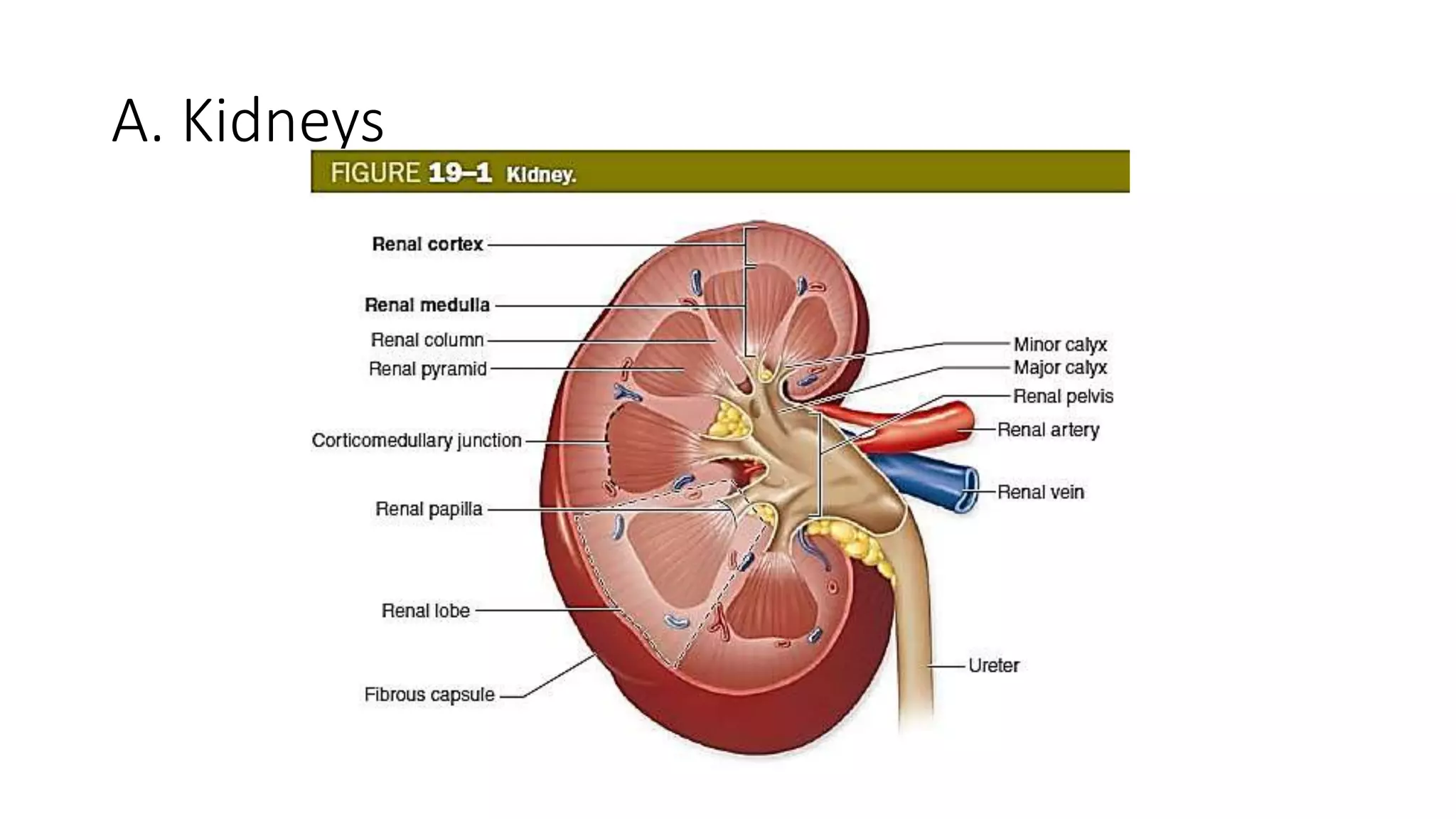
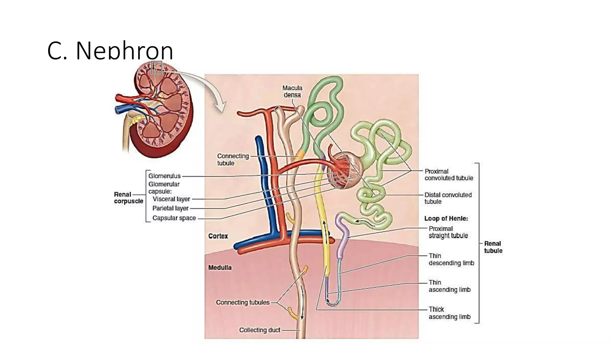
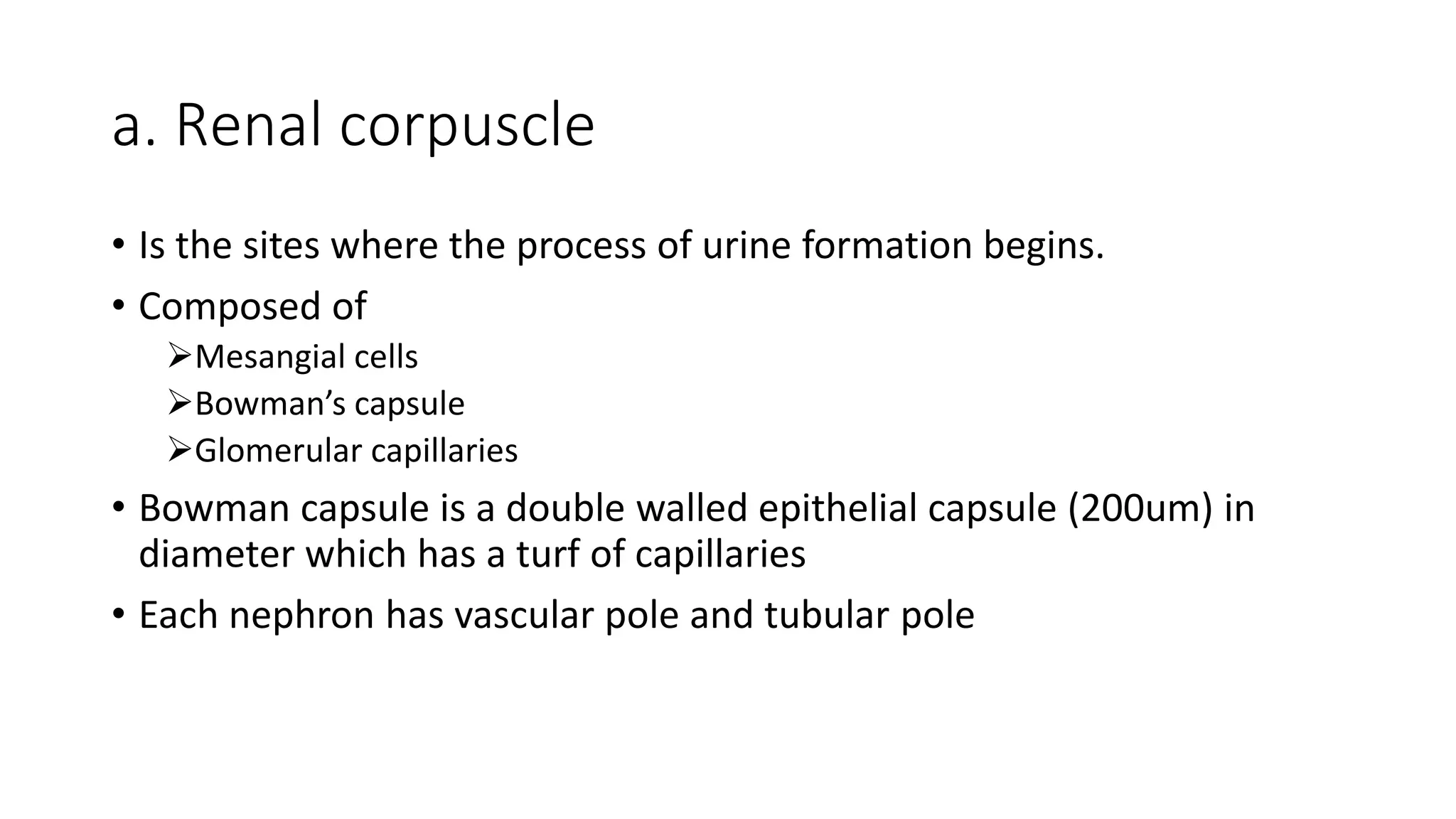
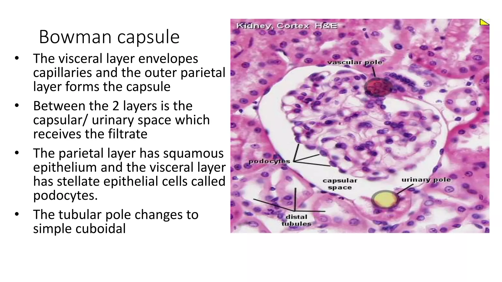
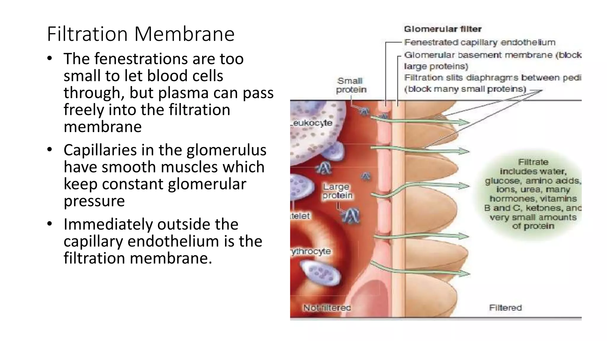
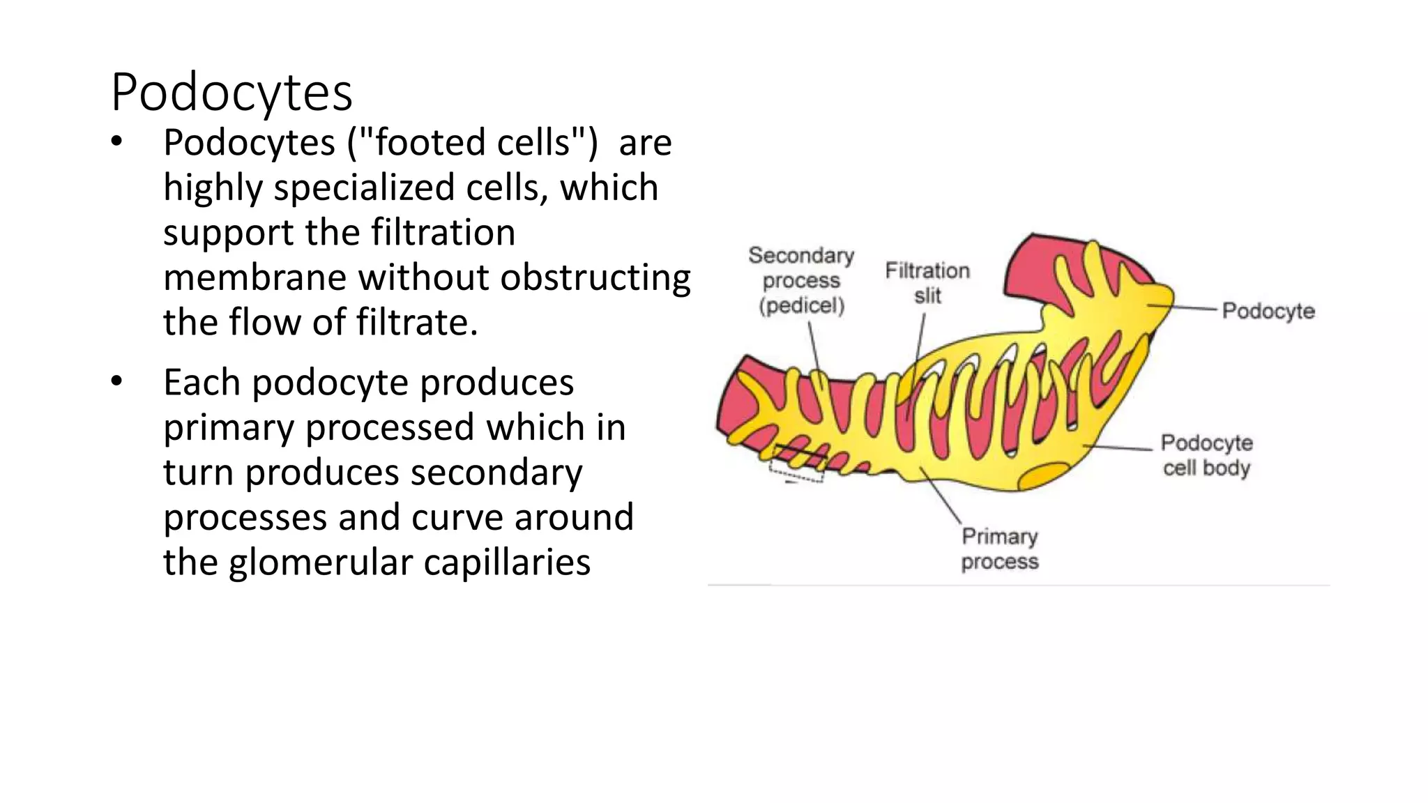
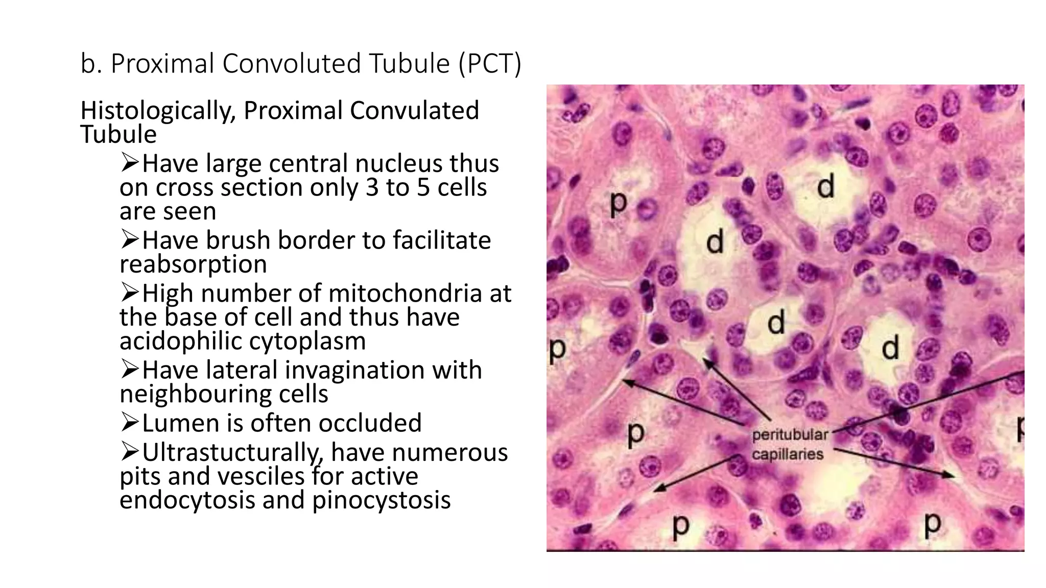
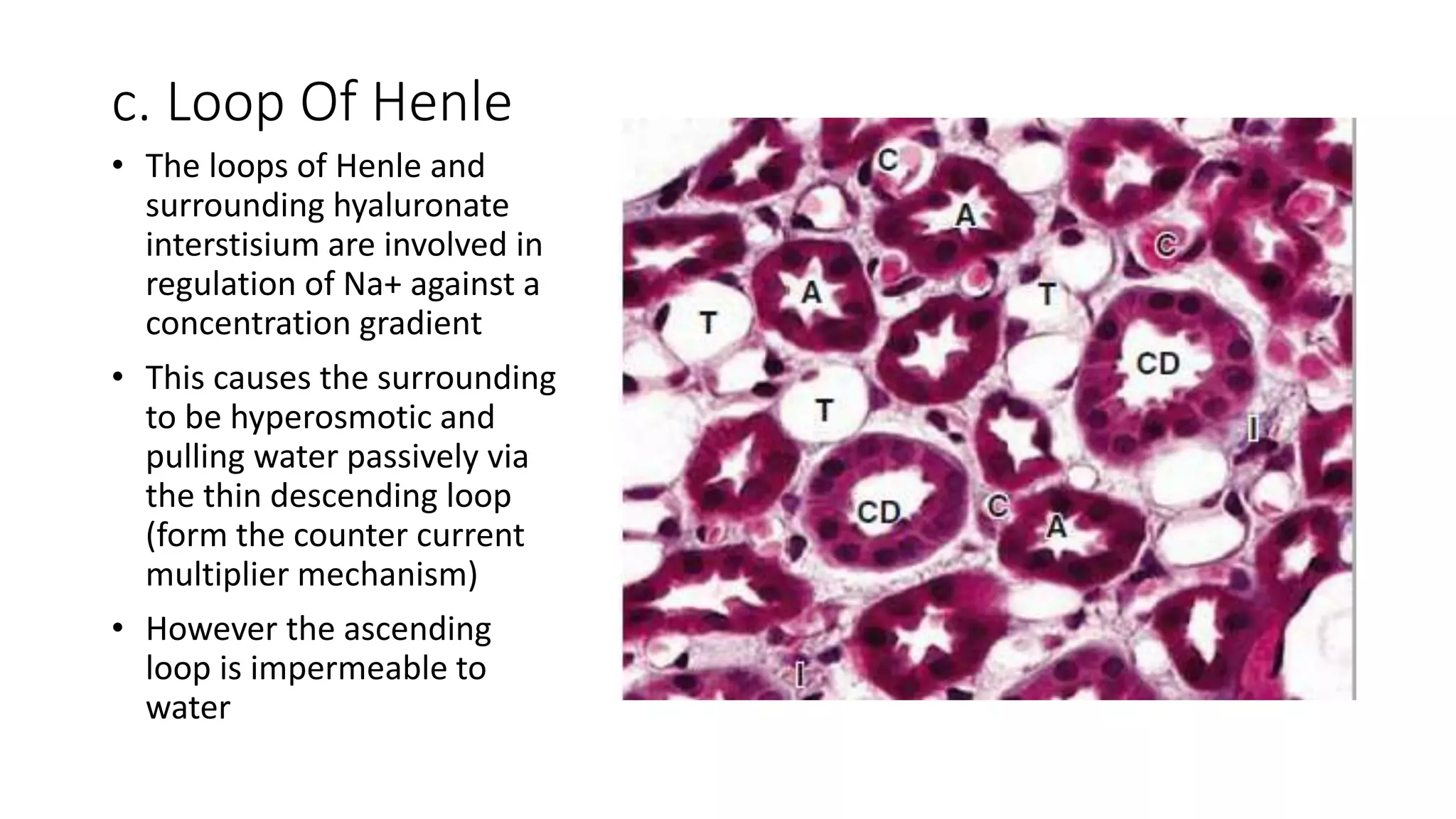
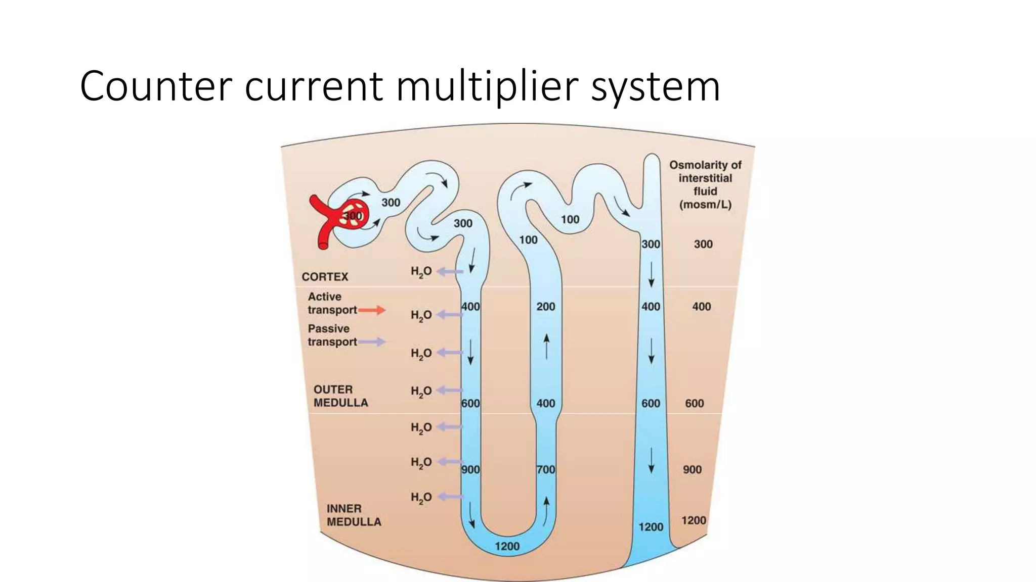
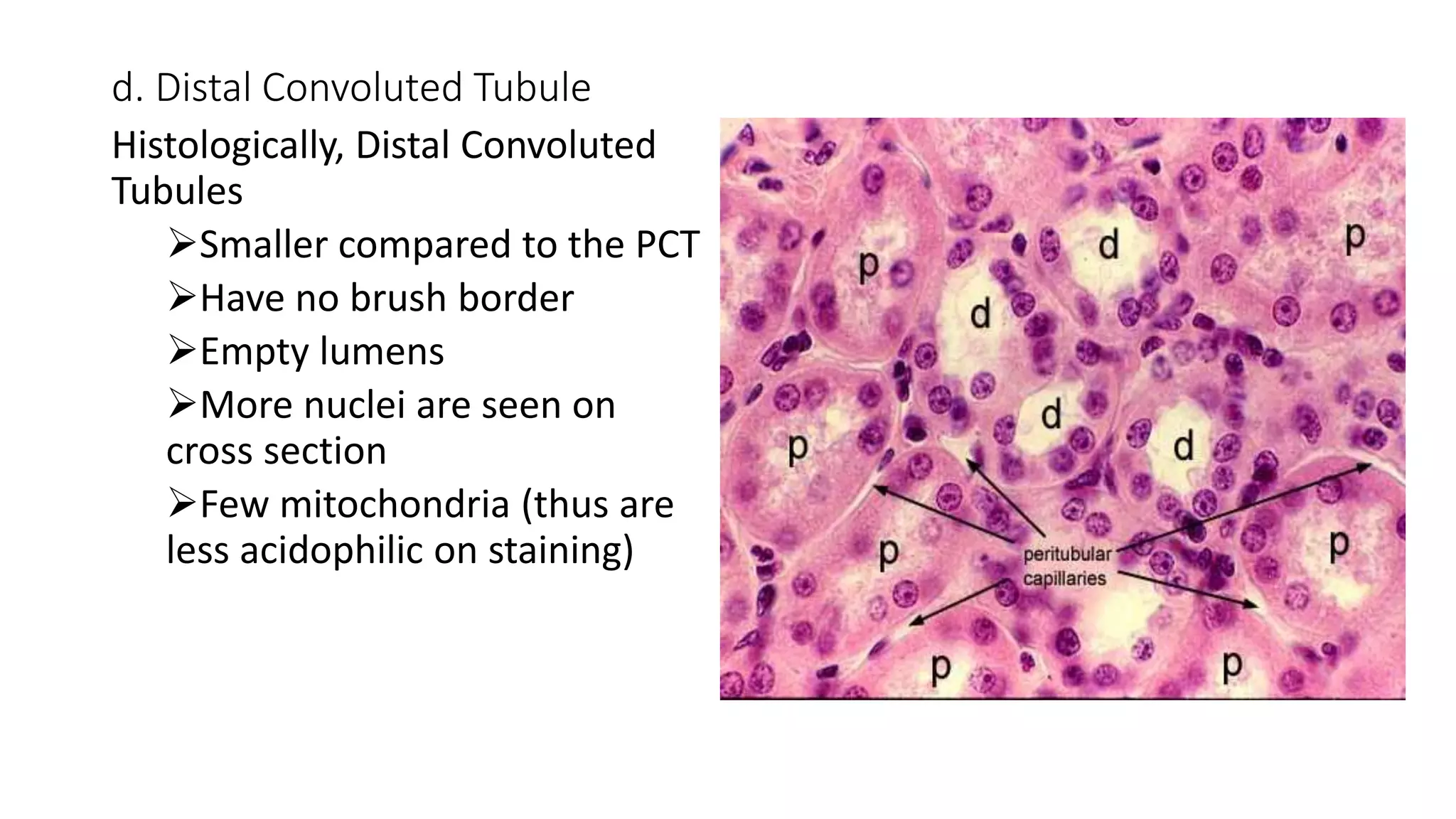
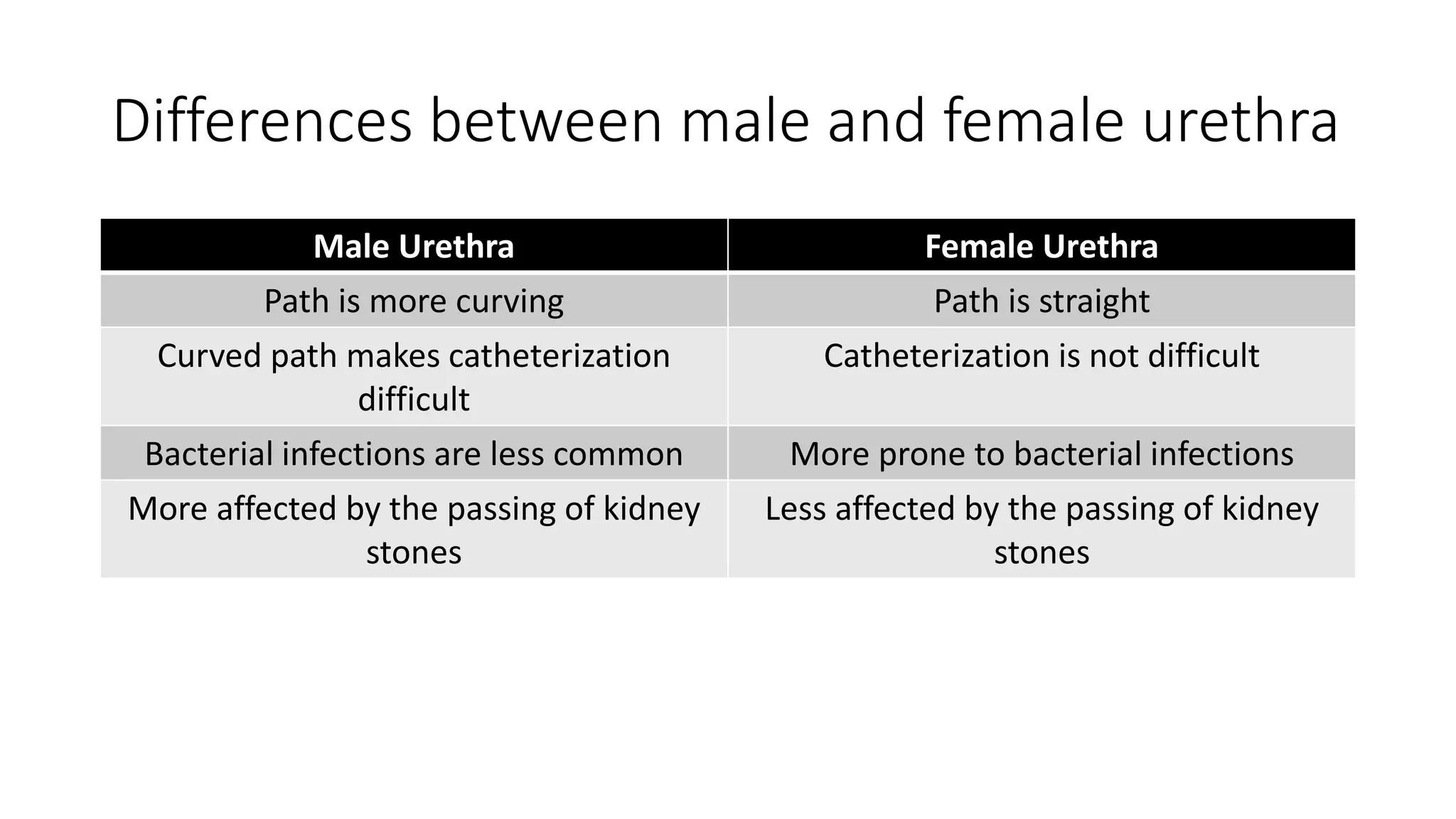
2. OBJECTIVES FOR URINARY SYSTEM
At the end of this presentation, students should be able to:
• Describe components and functions of the Urinary system
• Understand the histology of the kidney, nephron, ureter, urinary
bladder and urethra.
• Describe clinical application of the urinary system components.
3. OUTLINE OF THE URINARY SYSTEM
A. KIDNEYS
B. BLOOD CIRCULATION
C. NEPHRON
• RENAL CORPUSLES & BLOOD FILTRATION
• PROXIMAL CONVOLUTES TUBULTE
• LOOP OF HENLE
• DISTAL CONVOLUTED TUBULE & JUXTAGLOMERULAR APPARATUS
• COLLECTING DUCT
D. URETERS, BLADDER & URETHRA
4. Introduction
• The human body has 2 kidneys and ureters, 1 bladder and 1 urethra
• This system’s primary role is to ensure optimal properties of the
blood, which the kidneys continuously monitor.
• Urine, the excretory product of the kidneys, passes through the
ureters to the bladder for temporary storage and is then released
through the urethra.
5. A. Kidneys
• Each kidney is 12 cm in length 6cm wide
• Has 2 parts:
Cortex
Medulla
• The medulla has 8 to 15 structures called renal pyramids and their
bases are towards the cortex
• Each pyramid and cortical tissues is called the renal lobe
• The tip of the pyramid for the renal papilla and thus form the minor
calyx which join to form the major calyx and then the ureters
7. A. Kidneys
General roles of the kidney:
• Regulation of acid base (with the regulation of mineral ions and
water)
• Excretion of excess water and waste products
• Production of renin (important in maintenance of blood pressure
through cascade of events)
• Production of erytropoetin (responsible for production of RBC)
• Role in gluconeogenesis (make glucose from amino acids)
• Convertion of Vitamin D to its active form (1,25 – dihydroxy Vitamin
D3)
8. Medical Application
• In polycystic kidney disease is where the cortical organization is lost
due to formation of large fluid filled cysts, most arise from epithelial
tissue of the nephrons
9. B. Blood Circulation (microvasculature)
• Blood vessels of the kidneys are named according to their location
• Each renal artery divides into 2 to 3 segmental arteries which branch
further into Interlobar arteries
• Interlobar arteries further branch to Arcuate arteries and run along
the arc of the base of the pyramid
• Arcuate arteries form smaller Interlobular arteries and run into the
cortex
• They form the afferent arterioles which move along and form the
capillary loops around the nephrone
10. B. Blood Circulation
• Blood leaves the glomerular capillaries via efferent arterioles and
branch into Peritubular capillaries
• Note: the Juxtamedullary nephrons form the Vasa Recta (not
peritubular capillaries)
• Blood leaves the kidney in veins that follow the same courses as
arteries and have the same names.
• The cortex receive 10 times more blood then the medulla
12. Medical Application
• Sickle cell nephropathy, one of the most common problems caused by
sickle cell disease, occurs when the affected sickle cell in the vasa
recta. The nephropathy results from renal infarcts, usually within the
renal papillae or pyramids.
13. C. Nephron
• Each kidney has 1 to 4 million nephrons
• Its is the functional unit of the kidney
• There are two types of nephrons.
Cortical nephrons (always lie re in the cortex)
Juxtamedullary nephrons (near the medulla, long loop of
Henle)
14. C. Nephron
• Major division of the nephron
Renal corpuscle
Proximal convoluted tubule
Loop of Henle Renal Tubules
Distal convoluted tubule
Collecting tubule
• Collecting tubule from many nephrons converge to form the renal
papilla and deliver urine to minor calyx then to the major calyx
16. a. Renal corpuscle
• Is the sites where the process of urine formation begins.
• Composed of
Mesangial cells
Bowman’s capsule
Glomerular capillaries
• Bowman capsule is a double walled epithelial capsule (200um) in
diameter which has a turf of capillaries
• Each nephron has vascular pole and tubular pole
17. Bowman capsule
• The visceral layer envelopes
capillaries and the outer parietal
layer forms the capsule
• Between the 2 layers is the
capsular/ urinary space which
receives the filtrate
• The parietal layer has squamous
epithelium and the visceral layer
has stellate epithelial cells called
podocytes.
• The tubular pole changes to
simple cuboidal
18. Mesangial cells
• These have contractile properties
• Functions
Provide physical support to
capillaries
Produce cytokines and
inflammatory markers for
protections and repair
Contract and relax in response to
BP to maintain constant GFR
Phagocytosis of proteins which
adhere to basement membrane
19. The glomerulus
• Is a small knot of capillaries and
supporting structures suspended
within Bowman's capsule.
• Filtration occurs through the
glomerular filter, which has 3 parts
Fenestration of capillary
endothelium, block red cells
Thick basal laminae restrict large
proteins to pass through
Filtration slit diagrams between the
secondary processed of the
podocytes
20. Filtration Membrane
• The fenestrations are too
small to let blood cells
through, but plasma can pass
freely into the filtration
membrane
• Capillaries in the glomerulus
have smooth muscles which
keep constant glomerular
pressure
• Immediately outside the
capillary endothelium is the
filtration membrane.
21. Podocytes
• Podocytes ("footed cells") are
highly specialized cells, which
support the filtration
membrane without obstructing
the flow of filtrate.
• Each podocyte produces
primary processed which in
turn produces secondary
processes and curve around
the glomerular capillaries
22. Podocytes
• Between adjacent pedicels are gaps called filtration slits which permit
free passage of fluid filtrate into Bowman's space.
• The podocytes have lost their ability to divide; perished podocytes
can only be replaced with the help of hypertrophy.
• In typical histological preparations, podocyte nuclei tend to be oval
23.
24. Medical Application
• Diseases such as Glomerulonephritis alters the glomerular filter and
thus can cause proteinurea
• Diabetic glomerulosclerosis, the thickening and loss of function in the
GBM produced as part of the systemic microvascular sclerosis, is the
leading cause of (irreversible) end-stage kidney disease. Treatment
requires either a kidney transplant or regular artificial hemodialysis.
• Hypertension cause hypertrophy of the capillary smooth muscles and
thus loose their ability to contract and relax
• Chronic NSAID use cause vasoconstriction of arterioles thus
decreasing GFR and causing Renal Tubular Necrosis
25. b. Proximal Convoluted Tubule (PCT)
• Have simple cuboidal epithelium
• Are located mostly in the cortex
• Specialised in reabsorption and secretion; half of the water, ions (H+,
HCO3-, PO4-) and all of the organic components (glucose, amino
acids) are reabsorbed
• Trans cellular absorption occurs both passively and actively. i.e. water
move passively across an osmotic gradient
26. b. Proximal Convoluted Tubule (PCT)
• Small proteins are reabsorbed by receptor medicated endocytosis or
degraded on the luminal surface by petidases
• Organic anions, cations (creatinine, urea, bile salts, etc) and drugs not
filtered in the renal corpuscle are secreted into the tubule (because
they are disposed at a higher rate than glomerular filtration alone)
• They also perform hydroxylation of Vitamin D
• Their fibroblastic interstitial cells in the cortex produce erythropoetin
27. b. Proximal Convoluted Tubule (PCT)
Histologically, Proximal Convulated
Tubule
Have large central nucleus thus
on cross section only 3 to 5 cells
are seen
Have brush border to facilitate
reabsorption
High number of mitochondria at
the base of cell and thus have
acidophilic cytoplasm
Have lateral invagination with
neighbouring cells
Lumen is often occluded
Ultrastucturally, have numerous
pits and vesciles for active
endocytosis and pinocystosis
29. Medical Application
• Disease like Fanconi syndrome can affect the Proximal Convoluted
Tubule and in turn affect the reabsorption of ions and lead to
subsequent disorders
• E.g. glucose transport – renal glucosuria, phosphate transport –
hypophosphatemic rickets.
30. c. Loop Of Henle
• It is located in the medulla
• U shaped structure with thin descending loop; thin ascending loop
with diameter of 30um
• Both the thin descending and ascending loop have simple squamous
epithelium
• Both have few organelles and are involved in passive mode of
transport of water and ions
• The thin ascending loop changes into thick ascending loop (TAL)
which is located at the cortico-medullary junction has cuboidal
epithelium and has many mitochondria
31. c. Loop Of Henle
• The loops of Henle and
surrounding hyaluronate
interstisium are involved in
regulation of Na+ against a
concentration gradient
• This causes the surrounding
to be hyperosmotic and
pulling water passively via
the thin descending loop
(form the counter current
multiplier mechanism)
• However the ascending
loop is impermeable to
water
33. Medical Application
• It forms the counter current multiplier system important for water
and Na+ reabsorption
• Bartter and Gitelman syndrome is a rare inherited defect in the TAL of
the loop of Henle problem the NA/K pump. It is characterized by low
serum Na+ K+ levels, increased blood pH, and normal to low BP
34. d. Distal Convoluted Tubule
• Less absorption occurs here compared to PCT
• Have Simple cuboidal epithelium
• Involved in Na+ reabsorption, but here it is regulated by aldosterone
• They also have intercalleted cells for H+ and HCO3- regulation
• When the part of the DCT come closer to the arteriole of the
glomerulus the cells form the macula densa of the PCT. Histologically,
have columnar epithelium
large nucleus and are closely packed together
35. d. Distal Convoluted Tubule
• The juxtaglomerular cells are
present in the afferent arteriole,
histologically have;
Rounded nuclei
Rough Endoplasmic reticulum
Golgi apparatus
Granules which store renin
• This macula densa, the
juxtaglomerular cells of the
afferent arteriole and the
extraglomerular mesangial cells
(lacis cells) form the
Juxtaglomerular apparatus.
36. d. Distal Convoluted Tubule
Histologically, Distal Convoluted
Tubules
Smaller compared to the PCT
Have no brush border
Empty lumens
More nuclei are seen on
cross section
Few mitochondria (thus are
less acidophilic on staining)
37. Medical Application
• Thiazides are hypertensive medications which inhibit the Na/Cl pump
present in the DCT, and thus Na and water being excreted (diuresis)
38. e. Collecting Duct
• Last part of the nephron
• Many collecting tubule combine to form collecting duct
• They carry filtrates into the collecting system than transports it to
minor calyx -> major calyx -> papillary ducts
• They have simple cuboidal epithelium with a diameter of 40um and
they lie in the medulla
• Also a site for final absorption of water and ions
39. e. Collecting Duct
They have 2 types of cells
Principle cells - many
Intercalated cells (also
present in the DCT) – few
40. Principle cells
Histologically, are:
• Pale staining cells with few
organelles and sparse microvilli
• Basal membrane infolding
responsible for Na+ transport,
K+ secretion in response to Anti
Diuretic Hormone (ADH)
• Rich in aquaprorins (are
membrane pore proteins
sensitive to ADH), are in
cytoplasmic vescicles
42. Intercalated cells
• Have more mitochondria
• Have apical folds
• Maintain the H+ and HCO3-
Ultrastructure of the collecting
duct cells
Key:
I – intercalated cells
P – principle cells
43. Medical Application
• Nephrogenic Diabetes Incipidus type 1 and 2, aquaporin molecules
are not stimulated and thus water is not absorbed
• Syndrome Of Inappropriate ADH (SIADH), hypersecretion of ADH
hormone causing over sensitivity of collecting duct and thus, water is
over absorbed causing hypo-osmolarity, hyponatremia.
44.
45. D. Ureters; Bladder; and Urethra
• Urine is transported by the ureters from the renal pelvis to the
urinary bladder where it is stored until emptying by micturition via
the urethra.
• The walls of the ureters are similar to that of the calyces and renal
pelvis, with mucosal, muscular, and adventitial layers and becoming
gradually thicker closer to the bladder.
• The mucosa of these organs is lined by the uniquely stratified
urothelium (transitional epithelium)
46. a. Ureters
Ureter has three layers; Mucosa, muscular, Adventitial layers
Mucosa.
• Consists of transitional epithelium(urothelium) and a lamina propria of
loose-to-dense connective tissue.
• Transitional epithelium that is capable of responding to stretches in the
ureters.
• The transitional epithelium may appear as a columnar epithelia when
relaxed, and squamous epithelia when distended.
47. a. Ureters
Muscular layer.
• The ureter is surrounded by two muscular layers, an inner longitudinal layer
of muscle, and an outer circular or spiral layer of muscle.
Adventitial layer.
• Outer adventitial layer has fibroelastic connective tissue, with blood vessels,
lymphatics and nerves
49. Medical Application
• Transitional cell carcinoma of the ureter. Ureters are the most
common site for primary tumor. The distal ureter is more frequently
affected, presumably due to greater stasis
• Ureter stone is a mineral mass in the ureter, which may or may not
have originated in the kidney and traveled down into the ureter.
• Ureterocele is a congenital abnormality found in the ureter. In this
condition the distal ureter balloons at its opening into the bladder,
forming a sac-like pouch.
50. b. Urinary Bladder
• The mucosa of the bladder is
composed of transitional
epithelium.
• These cells are organized 3 layers
superficial layer umbrella cells
Intermediate region (columnar cells)
Single layer of small basal cells on
thin membrane
• Beneath it is a well-developed
submucosal (laminar propria)
layer formed largely of connective
and elastic tissues.
51. b. Urinary Bladder
• External to the submucosa is the detrusor muscle which is made up of
a mixture of smooth muscle fibers arranged at random in a
longitudinal, circular, and spiral manner.
• The transitional epithelium of the bladder in the undistended state is
five or six cells in thickness; the superficial cells are rounded and
bulge into the lumen. These cells are frequently polyploid or
binucleate.
• When the epithelium is stretched, as when the bladder is full of urine,
the epithelium is only three or four cells in thickness, and the
superfiacial cells become squamous.
52. b. Urinary Bladder
Empty urinary bladder showing multiple layers of
transitional epithelial cells
Full urinary bladder, and thinning of bladder
wall, supporting collagen fibers embedded
between smooth muscle fibers can be seen
53. Medical Application
• Bladder cancer-transitional cell carcinoma. Most common type of
bladder cancer
• Cystitis is an inflammation of the bladder. Inflammation is where part
of your body becomes irritated, red, or swollen. In most cases, the
cause of cystitis is a urinary tract infection (UTI).
54. c. Urethra
• The urethra is a tube that carries the urine from the bladder to the
exterior. In men, sperm also pass through it during ejaculation. In
women, the urethra is exclusively a urinary organ
55. c. Urethra
1. Male Urethra
• Has 3 parts
Prostratic Urethra
o3-4cm long
oPasses through the prostrate
oHas transional epithelium
56. c. Urethra
Membranous Urethra
oShort segment, 1 – 2 cm long
oLined by pseudo stratified columnar epithelium
Spongy Urethra
o15cm long
o Two parts, the bulbous and pendulous urethra
oStratified columnar and pseudo stratified epithelium with stratified squamous
epithelium distally.
57. c. Urethra
2. Female Urethra
• The female urethra is a tube 4.5 cm long, areas of pseudostratified
columnar epithelium in the middle and lined with stratified squamous
epithelium in the distal parts.
• The mid part of the female urethra is surrounded by an external
striated voluntary sphincter.
59. Differences between male and female urethra
Male Urethra Female Urethra
Pass both urine and semen pass Only urine passes
Longer (20cm) Shorter (4cm)
Diameter is 8 - 9 cm Diameter Is 6cm
Opens to the outside at the tip of the penis Opens to the outside anteriorly to the
vaginal opening
Belongs to both urinary and reproductive
system
Belongs only to the urinary system
Serves passage of the ejaculation of semen Female reproductive system is different
Sphincters control movement of urine and
semen
Sphincters only control movement of urine
60. Differences between male and female urethra
Male Urethra Female Urethra
Path is more curving Path is straight
Curved path makes catheterization
difficult
Catheterization is not difficult
Bacterial infections are less common More prone to bacterial infections
More affected by the passing of kidney
stones
Less affected by the passing of kidney
stones
61. Medical Application
• Urethral stricture is a narrowing of the opening of the urethra after
being exposed to STI’s, injury during catheterization
• Urethritis is inflammation of the urethra, sometimes caused by
infection
• Urethral cancer - a rare cancer that happens more often in men
• Epispadias or hypospadias are birth defects where the urethral
opening is on the ventral or dorsal side
62. References
• Junqueira’s basic histology text and Atlas 13th edition
• Histology text book by Bobrysheva I.V and Kachshenko S.A
• www.ncbi.nlm.nih.gove/pmc/articles/PMC3814687/
• www.emedicine.com
• www.Wikipedia.com
• www.eurocytology.eu/en/course/929
• https://www.ncbi.nlm.nih.gov › NCBI › Literature › PubMed Central (PMC)
• https://www.ncbi.nlm.nih.gov/pmc/articles/PMC3905241/
• http://www.news-medical.net/news/20150303/Breakthrough-treatment-
option-for-men-with-benign-prostatic-hyperplasia.aspx